Korean J Gastroenterol Vol. 59 No. 4, 313-316 http://dx.doi.org/10.4166/kjg.2012.59.4.313
CASE REPORT
Korean J Gastroenterol, Vol. 59 No. 4, April 2012 www.kjg.or.kr
두빈-존슨 증후군 환자에서 동반된 급성 A형간염으로 인한 지속성 담즙 정체 1예
라상호, 성세용, 정호연, 차재황, 백순구, 조미연
1, 김문영
연세대학교 원주의과대학 내과학교실, 병리학교실1
A Case of Sustained Cholestasis Caused by Acute A Viral Hepatitis in Dubin-Johnson Syndrome
Sang Ho Ra, Se Yong Sung, Ho Yeon Jung, Jae Hwang Cha, Soon Koo Baik, Mee Yon Cho1 and Moon Young Kim Departments of Internal Medicine and Pathology1, Yonsei University Wonju College of Medicine, Wonju, Korea
Dubin-Johnson syndrome is a rare clinical entity. It shows intermittent symptoms such as chronic or intermittent jaundice, abdominal pain, weakness, nausea, vomiting, anorexia and diarrhea. Symptoms are precipitated or aggravated by pregnancy, alcoholism, surgical procedures and intercurrent disease. Chronic idiopathic jaundice is typical of Dubin-Johnson syndrome and its prognosis is good. We describe a case of prolonged cholestasis for more than 10 months caused by acute A viral hepatitis in a patient with Dubin-Johnson syndrome. It is a first report of cholestasis complicated by acute A viral hepatitis in a patient with Dubin-Johnson syndrome. (Korean J Gastroenterol 2012;59:313-316)
Key Words: Jaundice, chronic idiopathic; Hepatitis A; Cholestasis; Hyperbilirubinemia
Received August 18, 2011. Revised October 24, 2011. Accepted October 24, 2011.
CC This is an open access article distributed under the terms of the Creative Commons Attribution Non-Commercial License (http://creativecommons.org/licenses/
by-nc/3.0) which permits unrestricted non-commercial use, distribution, and reproduction in any medium, provided the original work is properly cited.
교신저자: 김문영, 220-701, 원주시 일산동 162, 연세대학교 원주의과대학 내과학교실
Correspondence to: Moon Young Kim, Department of Internal Medicine, Yonsei University Wonju College of Medicine, 162 Ilsan-dong, Wonju 220-701, Korea. Tel:
+82-33-741-1225, Fax: +82-33-745-1228, E-mail: [email protected] Financial support: None. Conflict of interest: None.
서 론
1954년 Dubin과 Johnson1은 선천성 고빌리루빈혈증을 나 타내는 빌리루빈 대사장애 중 만성황달이 있는 환자에서 간생 검 결과 간세포의 파괴 없이 간세포 내에 갈색 색소과립이 있는 증례들을 분석하여 처음으로 보고하였고 이후 “두빈-존 슨 증후군(Dubin-Johnson syndrome)”으로 명명되었다. 두 빈-존슨 증후군은 상염색체가 우성 또는 열성 유전되는 드문 질환으로, ATP 의존성 담세관막수용체인 multidrug resist- ance protein 2/multi-specific organic anionic transporter protein의 발현 결함이 원인으로 추정되는 포합형 고빌리루 빈혈증을 보인다.2-4 두빈-존슨 증후군은 임상적으로 길버트 증후군, 로터 증후군과 큰 차이가 없으나 혈중 포합형 빌리루
빈의 현저한 증가와 더불어 간세포 내에 특이한 색소과립을 보이는 것이 특징이다.5 간세포 내의 특이한 색소과립은 에피 네프린의 대사산물이 소엽 간세포의 라이소솜에 침착한 것이 며, 결과적으로 간은 전반적으로 검은 색을 띄게 된다.6 만성 적으로 양성 간헐성 황달 증세 이외에는 증상이 없는 경우가 대부분이며, 대증요법으로 양호한 경과를 보인다.7
두빈-존슨 증후군은 처음 보고된 이래 구미, 일본 등에서 많은 예가 보고되었으며 우리나라에도 1963년 이후 보고되고 있지만,8 두빈-존슨 증후군 환자에서 A형간염이 발생한 예후 및 합병증에 대한 보고는 아직 없다. 저자들은 두빈-존슨 증후 군 환자에서 급성 A형간염 이환 후 10개월 이상 지속되는 만 성 담즙정체를 경험하였기에 문헌고찰과 함께 보고한다.
314
라상호 등. 두빈-존슨 증후군 환자에서 동반된 급성 A형간염으로 인한 지속성 담즙 정체 1예The Korean Journal of Gastroenterology
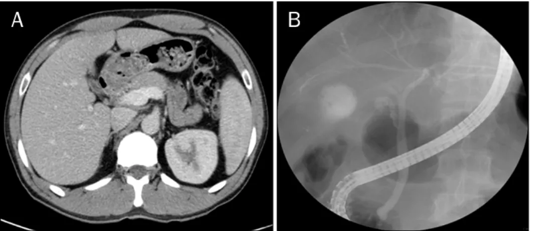
Fig. 2. (A) Liver computed tomography and (B) endoscopic retrograde cho- langiopancreatography showed he- pato-splenomegaly and no signifi- cant abnormal finding in the biliary tract. Those examinations were per- formed in acute phase of acute hepatitis A.
Fig. 1. Microscopically, hepatocytes contained diffuse intracellular deposits of brown pigment (arrows) without evidence of cell damage in liver biopsy in 1995 (H&E, ×200).
증 례
35세 남자가 우상복부 통증 및 발열을 주소로 내원하였다.
환자는 1992년 이후 반복적인 황달로 여러 병원에서 치료받 은 병력이 있었다. 1995년에 황달을 주소로 본원에 내원하여 시행한 혈액검사에서 고빌리루빈혈증을 보였고, 간생검에서 정상적인 간소엽구조를 유지하면서 염증세포 침윤이나 섬유 화가 없고, 간세포 세포질 내에 암갈색의 굵은 과립이 산재되 어 있어(Fig. 1) 두빈-존슨 증후군으로 진단되었다. 이후 주기 적 추적검사에서 3-5 mg/dL 정도의 경증 포합형 고빌리루빈 혈증을 보이며 유지하던 중, 2010년 9월 우상복부 통증, 발열 을 주소로 본원 응급실을 경유하여 입원하였다. 가족력은 특 이사항 없었다.
신체검진에서 내원 당시 활력징후는 혈압 130/92 mmHg, 맥박 61회/분, 호흡 18회/분, 체온 36.7oC였다. 의식은 명료하 였고, 구역질, 전신 쇠약감 및 식욕 부진을 호소하였다. 흉부
청진에서 심음은 규칙적이었으며, 전신 황달 및 공막에 황달 이 관찰되었다. 복부에서 종괴는 촉지되지 않았고, 장음은 약 간 감소되어 있었으며, 우상복부 압통 소견이 관찰되었다. 복 부의 반동압통통은 없었고 복수의 소견도 없었다.
말초혈액검사에서 혈색소 15.3 g/dL, 백혈구 4,200/mm3, 혈소판 239,000/mm3였다. 일반화학검사에서 혈청 총단백 7.3 g/dL, 알부민 4.0 g/dL, AST 11,745 IU/L, ALT 11,779 IU/L, ALP 154 IU/L, GGT 360 IU/L, 총 빌리루빈 14.2 mg/dL, 직접 빌리루빈 12.7 mg/dL, BUN 8.0 mg/dL, 크레 아티닌 0.5 mg/dL, CK 1,024 mg/dL, 젖산탈수소효소 (LDH)>7,500 IU/L였다. 혈액응고검사에서 prothrombin time INR 3.15로 상승되어 있었다. 소변 검사에서 비중 1.020, 요단백 2+, 빌리루빈 2+의 소견을 보여 심한 급성 간염 및 2차적 근육융해증을 시사하였다. 면역혈청검사에서 HBs Ag/Ab (−/+), HCV Ab (−) 소견을 보였고, HAV IgM (+) 소견을 보여 급성 A형간염으로 진단하였다. 급성 A형간 염 진단 시 용혈로 인한 고빌리루빈혈증을 감별하기 위해 시 행한 혈색소, LDH, haptoglobin은 각각 15.3 g/dL, 250 U/L, 43 mg/dL이었고, 3개월 후 관찰 시점에서는 각각 14.4 g/dL, 205 U/L, 153 mg/dL로 모두 정상 범주였다.
영상의학검사에서 두빈-존슨 증후군 진단 당시 초음파와 간스캔에서 간비종대는 관찰되지 않았다. 이번 급성 A형간염 으로 내원하여 시행한 초음파 및 복부전산화단층촬영에서도 간비종대 외에 특이소견은 없었으며, 입원 1개월 경에 시행한 내시경 역행성 담도조영술에서도 담도계의 특이소견은 관찰 되지 않았다(Fig. 2).
1개월 이상의 보존적 치료 후 증상이 호전되었고, AST 61 IU/L, ALT 16 IU/L, ALP 79 IU/L, prothrombin time INR 1.28으로 대부분의 혈액검사 소견이 호전되었으나, 총 빌리루 빈 27.3 mg/dL과 직접 빌리루빈 24.2 mg/dL만 지속적으로 상승되어 있는 담즙정체 소견을 보였다. 이후 환자는 전신상 태가 양호하고 특이 증상이 없어 퇴원하여 주기적으로 외래
Ra SH, et al. A Case of Sustained Cholestasis Caused by Acute A Viral Hepatitis in Dubin-Johnson Syndrome
315
Vol. 59 No. 4, April 2012 Table 1. The Changes of Laboratory Findings
Investigation Days
0 20 50 90 130 190 240 300
Hemoglobin (g/dL) 15.3 12.6 13.0 14.4 13.5 14.3 14.6 14.5
AST (IU/L) 11,745 63 61 51 47 41 43 34
ALT (IU/L) 11,779 69 16 17 21 22 22 21
Total bilirubin (mg/dL) 14.2 26.4 27.3 36.3 26.6 17.3 19.8 18.6
Direct bilirubin (mg/dL) 12.7 23.3 24.2 30.6 22.7 16.2 18.2 17.5
ALP (IU/L) 154 102 79 101 106 103 88 79
GGT (IU/L) 285 177 56 128 60 53 50 51
PT INR 3.15 1.20 1.24 1.12 1.10 1.15 1.10 1.02
Fig. 3. The change of serum total and direct bilirubin levels of the patient.
추적관찰 중인 상태로, 급성 A형간염 발병 300일 이상 지난 최근까지도 prothrombin time INR 1.02, AST 34 IU/L, ALT 21 IU/L는 정상소견이나, 총 빌리루빈 18.6 mg/dL과, 직접 빌리루빈 17.5 mg/dL만 단독으로 상승되어 있어 지속 적인 포합형 고빌리루빈혈증 소견을 보이고 있다(Table 1, Fig. 3).
고 찰
두빈-존슨 증후군은 혈청 AST, ALT, ALP, 알부민, GGT, prothrombin time을 포함한 간기능 검사가 정상이면서 포합 형 고빌리루빈혈증과 함께 소변에서 코프로포피린 I형의 배설 이 80% 이상을 차지하는 특징적인 소견을 보인다. 이번 증례 에서는 소변 코프로포피린 검사를 시행하지 않았으나 간생검 에서 간세포 내에 특이 색소과립을 확인하여 두빈-존슨 증후 군으로 확진되었다.
두빈-존슨 증후군은 양성질환의 경과를 보이고 합병증은 상대적으로 드물어, 임상적으로 경한 황달이 특징적이며, 약 간의 복통이나 무력감이 나타날 수 있다. 생화학검사에서 혈
청 빌리루빈 수치의 증가를 제외하고는 정상 소견을 보이고, 신체진찰에서 황달을 제외하고 정상이나 때때로 간비종대를 보일 수 있다.9 혈청 총빌리루빈의 농도는 대개 2-5 mg/dL이 며, 동반된 질환이 있거나 경구피임약을 복용하는 경우, 혹은 임신 시에 드물게 혈청 총빌리루빈이 20-25 mg/dL까지 상승 하는 경우도 있다.10 일반적으로 포합형 빌리루빈이 더 증가되 는데 비포합형이 더 증가되는 예도 보고되어 있다.1,2 소변검 사에서 수용성 빌리루빈이 나타나며 육안적으로는 짙은 갈색 뇨를 보인다. 소변으로 배설되는 코프로포피린의 총량은 정상 이나, 정상인에서 III형이 75% 이상을 차지하는 데 반해 두빈- 존슨 증후군 환자에서는 일차적인 유전적 결함 또는 코프로포 피린 III cosynthetase의 저하로 I형이 80% 이상을 차지한 다.11 두빈-존슨 증후군은 일반적으로 예후는 좋으며 특별한 치료는 필요치 않고, 기타 다른 합병증도 보고된 바가 없어 정기적인 추적검사도 잘 이루어지지 않고 환자 본인도 질병을 인지하지 못하는 경우가 많다.
급성 A형간염은 소아에서는 대개 불현성 감염에 그치지만 성인에서는 심한 증상을 동반하는 현성 감염이 많고 연령이 높아짐에 따라 그 증상과 합병증이 심각해지며 사망에 이르는 경우도 있다.12 특히, 최근 사회경제적 환경의 변화에 따라 우 리나라 성인의 A형간염 항체 양성률이 낮아지면서 성인기에 현성 급성 A형간염으로 나타나는 경우가 폭발적으로 증가하 고 있어 사회적으로 문제가 되고 있다. 급성 A형간염의 합병 증으로는 Guillain-Barre 증후군, 급성 신부전, 전격성 간부 전, 담낭염, 췌장염, 혈관염, 관절염 등이 있으며13-15 소수의 예에서 재발성 간염, 자가면역성 간염, 지속성 담즙정체 등의 비전형적인 임상 양상이 나타날 수 있다.16-18
급성 A형간염에서 보이는 담즙정체의 경우 소양감, 열, 설 사, 체중 감소를 보일 수 있으며 혈청 빌리루빈이 10 mg/dL 을 초과하는 경우가 많다. 간생검 소견은 문맥역 염증을 동반 한 소엽중심의 담즙정체를 보이고, 심한 담즙정체에서는 만성 활동성 간염에서 관찰되는 문맥 주위의 염증반응과 유사한 소 견을 보인다. 담즙정체 및 이로 인한 황달은 급성 A형간염
316
라상호 등. 두빈-존슨 증후군 환자에서 동반된 급성 A형간염으로 인한 지속성 담즙 정체 1예The Korean Journal of Gastroenterology
환자의 사회ㆍ경제적 활동에 제약을 주는 주원인으로, 빠른 호전을 위한 스테로이드의 사용은 일반적으로 추천되지 않으 며, 보존적 치료만 시행하여도 보통 12주 내에 호전되는 것으 로 알려져 있다.16 그러나, 이번 증례에서와 같이 기존에 두빈- 존슨 증후군같은 선천적인 빌리루빈 대사이상을 가지고 있는 경우, 그 기전은 정확히 알 수 없으나 A형간염에 의해 촉발된 담즙정체가 쉽게 해소되지 않고, 일반적인 경우보다 매우 긴 기간 동안 지속적인 담즙정체를 보이는 경우가 있다. 특히 우 리나라의 경우 학업이나 사회적 활동에 있어서 가장 왕성한 활동을 해야 하는 젊은 층에서 급성 A형간염이 폭발적으로 증가하고 있는데, 이들 중 선천성 빌리루빈 대사이상을 동반 하는 경우 장기간의 지속적인 담즙정체와 황달의 발생으로 인 해 개인은 물론 사회ㆍ경제적으로 막대한 손실이 될 수도 있 다. 따라서 이번 증례에서와 같은 선천적 빌리루빈 대사이상 질병을 가진 취약 환자에 대한 A형간염 항체보유 검사 및 백 신접종 등의 적극적인 예방과 함께 담즙정체 악화, 지속의 기 전 및 해결책에 대한 연구가 필요할 것으로 생각된다. 이러한 의미에서 이번 증례는 향후 선천성 빌리루빈 대사이상 환자의 관리에 있어서 유용한 자료가 될 것으로 생각된다.
REFERENCES
1. Dubin IN, Johnson FB. Chronic idiopathic jaundice with un- identified pigment in liver cells; a new clinicopathologic entity with a report of 12 cases. Medicine (Baltimore) 1954;33:155- 197.
2. Butt HR, Anderson VE, Foulk WT, Baggenstoss AH, Schoenfield LJ, Dickson ER. Studies of chronic idiopathic jaundice (Dubin-Johnson syndrome). II. Evaluation of a large family with the trait. Gastroenterology 1966;51:619-630.
3. Shani M, Seligsohn U, Gilon E, Sheba C, Adam A. Dubin-Johnson syndrome in Israel. I. Clinical, laboratory, and genetic aspects of 101 cases. Q J Med 1970;39:549-567.
4. Toh S, Wada M, Uchiumi T, et al. Genomic structure of the canal-
icular multispecific organic anion-transporter gene (MRP2/cMOAT) and mutations in the ATP-binding-cassette region in Dubin- Johnson syndrome. Am J Hum Genet 1999;64:739-746.
5. Swartz HM, Sarna T, Varma RR. On the natural and excretion of the hepatic pigment in the Dubin-Johnson syndrome. Gastroen- terology 1979;76:958-964.
6. Novikoff AB, Essner E, Quintana N. Golgi Apparatus and Lysosomes. Fed Proc 1964;23:1010-1022.
7. Beker S, Read AE. Familial DubinJohnson syndrome. Gastroen- terology. 1958;35:387-389.
8. Hahn SS, Hahn YC, Hahn YS, Choi KW, Min BC, Kim OJ. A case of Dubin-Johnson syndrome. Taehan Uihak Hyophoe Chi 1963;6:609-614.
9. Berk PD. Bilirubin metabolism and the hereditary hyperbili- rubinemias. Semin Liver Dis 1994;14:321-322.
10. Cohen L, Lewis C, Arias IM. Pregnancy, oral contraceptives, and chronic familial jaundice with predominantly conjugated hyper- bilirubinemia (Dubin-Johnson syndrome). Gastroenterology 1972;62:1182-1190.
11. Koskelo P, Toivonen I, Adlercreutz H. Urinary coproporphyrin iso- mer distribution in the Dubin-Johnson syndrome. Clin Chem 1967;13:1006-1009.
12. Koff RS. Hepatitis A. Lancet 1998;351:1643-1649.
13. Ono S, Chida K, Takasu T. Guillain-Barré syndrome following ful- minant viral hepatitis A. Intern Med 1994;33:799-801.
14. Mourani S, Dobbs SM, Genta RM, Tandon AK, Yoffe B. Hepatitis A virus-associated cholecystitis. Ann Intern Med 1994;120:
398-400.
15. Inman RD, Hodge M, Johnston ME, Wright J, Heathcote J.
Arthritis, vasculitis, and cryoglobulinemia associated with re- lapsing hepatitis A virus infection. Ann Intern Med 1986;105:
700-703.
16. Gordon SC, Reddy KR, Schiff L, Schiff ER. Prolonged intrahepatic cholestasis secondary to acute hepatitis A. Ann Intern Med 1984;101:635-637.
17. Sciot R, Van Damme B, Desmet VJ. Cholestatic features in hep- atitis A. J Hepatol 1986;3:172-181.
18. Schiff ER. Atypical clinical manifestations of hepatitis A. Vaccine 1992;10(Suppl 1):S18-S20.