I N T RO D U CT I O N
Myasthenia gravis (MG) is an autoimmune neu- romuscular junction disease, affecting selectively the ocular muscles or skeletal muscle system. An association between thymic epithelial neoplasms and MG is well known.
1A leiomyosarcoma (LMS) is a relatively rare tumor, most frequently seen in the uterus and gastrointestinal tract, which com- prises 10% of the soft tissue sarcomas found in adults. Most of those tumors occurring in the mediastinum arise from organs in the thoracic cavity such as the atrium, esophagus, and great
vessels, where as an appearance in the soft tissue of the mediastinum is extremely rare.
2 , 3To our knowledge, only a few cases have been reported.
In Stevens-Johnson syndrome (SJS), medications are the offending agents in most patients.
4However, it is rarely combined with MG, medi- astinal LMS, and SJS.
CASE REPORT
A previously healthy 36-year-old housewife was admitted with acute onset headache, right ptosis and general weakness for several days.
She complained intermittent diplopia, dysphagia, and respiratory difficulty. Past, social, and family history was unremarkable. Review of systems revealed shortness of breath, tachypnea, and dull pain in right shoulder. The patient did not take any medication at the time of initial evaluation.
On physical examination, decreased breath
종격 평활근육종 및 Stevens-Johnson증후군과 동반된 중증 근무력증 1예
대구가톨릭대학교 의과대학 신경과학교실
이동국・권영미
A Case of Myasthenia Gravis Combined with Mediastinal Leiomyosarcoma and Stevens-Johnson Syndrome
Dong-Kuck Lee, Young-Mi Kweon
Department of Neurology, School of Medicine, Catholic University of Daegu, Korea
We report a case of 36-year-old woman with myasthenia gravis (MG) combined with mediastinal leiomyosarcoma (LMS) and Stevens-Johnson syndrome (SJS). She was admitted to ICU with the symptoms of acute onset headache, diplopia, ptosis, dysphagia, general weakness, and respiratory difficulty for several days. Physical examination revealed tachypnea, decreased breath sounds and dullness to percussion in right lower chest. Neurologic examination showed ptosis, diplopia, decreased gag reflexes, and generalized proximal weakness. Laboratory studies revealed increased serum acetylcholine receptor antibodies and positive Tensilon test. Chest CT showed a huge mass in the right middle mediastium but no evidence of thymic enlargement. Mediastinal LMS was diagnosed by ultrasound-guided needle biop- sy. The myasthenic symptoms were fluctuated in spite og intravenous immunoglobulin, plasmapheresis, and corticos- teroid. During therapy, SJS developed. She died 4 months after the onset of the myasthenic symptoms despite the chemotherapy for LMS.
Key Words: Myasthenia Gravis, Mediastinal Leiomyosarcoma, Stevens-Johnson Syndrome
Address for correspondence Dong Kuck Lee, M.D.
Department of Neurology, School of Medicine Catholic University of Daegu
3056-6 Daemyung 4 Dong, Namgu, Daegu, 705-718, Korea Tel: +82-53-650-4267 Fax: +82-53-654-9786
E-mail : [email protected]
sounds and dullness to percussion in right lower chest. were detected Neurologic examination revealed bilateral fluctuating ptosis, horizontal and vertical gaze limitation, dysphagia, and weakness of the neck and extremities. Using the Medical Research Council (MRC) scale, motor power was grade 2 in the proximal limbs, and 4 in the distal limbs. Muscles were normotonic and deep tendon reflexes were hypoactive. Plantar stimulation elicited a flexor response bilaterally.
Chest x-ray showed a huge, oval-shaped opac- ity in the lower zone of right lung, which was evaluated as mass lesion. Serum acetylcholine receptor antibody (AchR Ab) titer was increased, and fluorescent antinuclear antibody (FANA) was positive. Tensilon test was positive but repetitive nerve stimulation (RNS) test was normal. She was evaluated with myasthenic crisis and intratho- racic mass.
With respiratory difficulty, she was hospitalized in the intensive care unit for ventilator therapy.
With the intravenous immunoglobulin (IVIG, 0.4 g/kg/day) therapy for 5 days, her ptosis and proximal muscle weakness were much improved.
She could lift her feet off the bed against signifi- cant resistance, and could abduct the arms well.
Foot dorsiflexion and toe extension were normal.
In addition, her dyspnea was improved almost completely and ventilator could be weaned. IVIG and pyridostigmine therapy doesnet impnove pto- sis, proximal weakness and dyspnea consistently.
Chest CT revealed a huge (6.2 ×9.7 cm), lobu-
lated and well-enhanced middle mediastinal mass with dense nodular calcifications, abundant vas- cular components and necrotic area, but no evi- dence of invasion or thymic enlargement (Fig. 1).
In the hospital course, alopecia, oral ulcers involving lips and tongue, and generalized muco- cutaneous maculopapular rashes with vesicular and exfoliative components were developed. These were not improved by antibiotics or antiviral agent such as acyclovir, and not responsive to corticosteroid or antifungal agent over 2 months.
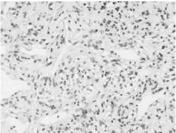
Skin biopsy showed the findings of SJS with staphylococcal and fungal infections (Fig. 2).
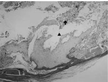
One month after IVIG therapy, she also had plasmapheresis therapy, but her myasthenic symptoms were transiently improved. Follow-up chest CT showed marked increased mass size and enlarged necrotic portions with right pleural effusion. Mediastinal LMS was confirmed by ultrasonogram-guided needle biopsy (Fig. 3).
Chemotherapy was started, but she died 4 months after the onset of myasthenic symptoms.
D I S C U S S I O N
It is widely accepted that MG, a T-cell-depen- dent chronic autoimmune disorder, is induced by sustained production of an antibody to the nico- tinic acetylcholine receptors at the neuromuscular junction. MG is characterized by weakness and fatigability of skeletal, bulbar, and extraocular muscles. During the past 30 years, treatment of
Figure 1. Chest CT shows a huge, well-enhanced mediastinal tumor containing multiple nodular calcifications and abundant vascular components.
Figure 2. Skin biopsy shows dermo-epidermal pustules (▲) containing vacuolar degenerated cells, necrotic keratinocytes at epidermis (△), and perivascular infiltration of lymphocytes in the upper dermis (↑).
MG has advanced significantly and prognosis has greatly improved, with dramatically decreased m o r t a l i t y .
5An association between thymic epithelial neo- plasms and MG is well known. Although approxi- mately 15% of patients with MG have thymoma, 15-80% of patients with thymoma have MG.
6Thymoma has many autoimmune associations, including aplastic anemia and MG. The patho- physiologic relationship between thymoma and its autoimmune sequelae are yet to be fully elucidat- ed, but are thought to be a result of the produc- tion of auto-reactive T cell clones by the thymic epithelium. Ritchie et al.
7report a case in which aplastic anemia did not develop until sometime after remission from thymoma has been induced.
This observation suggests that auto-reactive T cells may be produced by the thymoma but not induce clinical autoimmune disease until a later time, even after eradication of thymoma has been a c h i e v e d .
Extrathymic malignancies associated with thy- moma have been reported at an incidence of 2.6- 27%. The colorectum is the most common site for extrathymic malignancies, followed by the lung, thyroid, prostate, female reproductive organs, breast, kidney, skin (melanoma), and meningies ( m e n i n g i o m a ) .
8There are reports describing an association of MG with thymic and extrathymic lymphoma, suggesting a temporal and therapeutic relationship between MG and lymphoma.
1A b r e y et al.
9report a unique case of extrathymic H o d g k i n ’s lymphoma in a patient with MG in
whom antineoplastic therapy led to a remission of both lymphoma and MG.
The mechanism of the development of extrathymic malignancies associated with thymo- ma might be explained by the immune function of the thymus, whereby its removal could diminish immune surveillance. Immunosuppressed individ- uals are known to have an increased risk of malignancies, however, several series have indi- cated that thymectomy does not influence immunoparameters in patients with thymoma and MG. Moreover, the extrathymic malignancies were often diagnosed before or at the same time as thymoma.
8But Masaoka et al.
1 0suggested that thymoma promotes the development of malignant tumors in other organs, but that thymectomy never enhances the occurrence of malignancy.
Pan et al.
1 1also reported that the association between thymoma and a second malignancy can- not be attributed to thymectomy or radiation therapy. Some reports even suggest that thymec- tomy could reduce the incidence of extrathymic m a l i g n a n c i e s .
1 2Thus, the presence of an abnormal thymus, such as caused by thymoma, might be associated with the development of extrathymic m a l i g n a n c i e s .
And the casual relationship between lym- phoblastic lymphoma and the autoimmune response cannot be demonstrated, it is tempting to speculate that the T-cell defect induced by lymphoma and subsequent chemotherapy might have contributed to the development of autoim- munity. Alternatively, there could have been an immunological disorder such as impaired immune surveillance or impaired immune regulation favoring the occurrence of MG and lymphoma.
1But the precise mechanism between these types of malignancies and MG remains unclear. Our patient was a 36-year-old healthy housewife and did not have a thymoma. So we can not know the pathogenesis of the patient.
MG combined with mediastinal LMS is extreme- ly rare. Because primary mediastinal LMS itself is extremely rare tumor, so the coexistence of the both diseases may be very rare. A LMS most fre- quently seen in the uterus and gastrointestinal tract, which comprises 10% of the soft tissue sar- comas found in adults. The origin of a mediasti- nal LMS arising soft tissue is still controversial, although those have described three possibilities.
Figure 3. Histopathologic study shows bundles of malignant smooth muscle fibers containing spindle-shaped nuclei (H&E,
×100). Immunohistochemical stainings were positive for vimentin and αSMA (alpha smooth muscle actin).
First, an LMS may arise from heterotropic smooth muscle displaced during embryonal development.
The second possibility noted is that an LMS could be a parasitic tumor of the esophagus that has detached from the wall during development. The other potential origin may be from small vessels within the mediastinal soft tissue. However, microscopic observations were unable to indicate the pathogenesis of the tumor.
3Usually death from a malignant mediastinal LMS occurs as a consequence of regional exten- sion and metastasis, but survivals following resection have been reported.
1 3There is no con- sensus with regard to radiotherapy and chemotherapy, but the general consensus on this subject is that the complete resection of these tumor is an important prognostic factor.
2Initially, there was no evidence of metastasis in our patient.
In spite of chemotherapy, the patient developed pleural effusion, and manifested a rapid downhill course in 4 months, indicating the locally aggres- sive nature of this tumor.
Erythema multiforme (EM) is an acute, self- limited dermatosis that may be divided into a minor and major form, that the latter also known as SJS. Herpes simplex virus is the most common agent of infection. Drug-induced EM is usually more extensive than that induced by infectious agents. More than 100 medications have been reported to cause SJS. The most common are trimethoprim/sulfamethoxazole, Fansidar-R, sul- fadoxine plus pyrimethamine, and carbamazepine.
Antibiotics, especially long-acting sulfa drugs and penicillins, other anticonvulsants, antiin- flammatories (NSAIDs), and allopurinol are also common causes.
1 4Our patient presented an extensive generalized EM several days after antibiotics therapy, so we thought it was a drug- induced SJS. We speculated antibiotics might be an etiologic factor in SJS. In addition, her immune system was not good state, so more extensive SJS has been occurred.
EM minor is linked to specific HLA types (HLA- DQ3) and SJS to abnormalities of drug metabo- l i s m .
1 4But our patient did not have an any family history of SJS or other skin diseases.
MG can be accompanied by skin disorders, including pemphigus, vitiligo, and alopecia area- ta, which may be immunological diseases.
Although the association of pemphigus or alope- cia areata with MG is well documented, there have been no reports of the direct association of the three diseases. Chorzelski et al.
1 5reported a case of MG coexisted with Castleman tumor, paraneoplastc pemphigus, and bronchiolitis oblit- erans. Izumi et al.
1 6report a cases of MG with thymoma associated with alopecia areata and pemphigus foliaceus. In regard to the origin of the autoimmune response in the three diseases, the thymoma is implicated as a possible site of origin. They proposed three theories for this association: defective T cell suppression function, cross-reacting between altered thymic antigen and various organs, and general immune dysreg- ulation caused by thymic abnormalities. MG and pemphigus are caused by autoantibodies to AChR and desmoglein, and alopecia areata is rather due to abnormalities of cellular immunity. They spec- ulated that in these case some immunological abnormalities associated with the thymoma may have caused these autoimmune diseases. But our case did not have a thymoma. So we can not know the pathomechanism of the our case which is incidental or paraneoplastc finding or not, but dysfunction or impaired surveillance or regulation of immune system may provide the opportunity of the coexistence of three diseases.
REFERENCES
11. Hasegeliüner A, Abali H, Engin H, Akyol A, Ruacan S, Tan E, Güllü I, Altundag K, Güler N. Myasthenia gravis and lymphoblastic lymphoma involving the thymus: a rare association. Leukemia and lymphoma 2001;42(3):527-31.
12. E r og˘lu A, Kürkçüog˘lu C, Karaog˘ l a n og˘lu N, Gürsan N.
Primary leimyosarcoma of the anterior mediastinum. Eur J Cardio-thoracic Surg 2002;21:943-45.
13. Hirano H, Kizaki T, Sashikata T, Maeda T, Yoshii Y.
Leiomyosarcoma arising from soft tissue tumor of the mediastinum. Med Electron Microsc 2003;36:52-58.
14. C o h e n L M , S k o p i c k i D K , H a r r i s t T J , C l a r k W H . Noninfectious vesiculobullous and vesiculopustular dis- eases. In; Elder D. Lever’s Histopathology of the skin. 8th ed. New York: Lippincott Williams & Wilkins. 1997;239- 40.
15. Konishi T, Yoshiyama Y, Takamori M, Yagi K, Mukai E, Saida T, The Japanese FK506 MG Study Group. Clinical study of FK506 in patients with myasthenia gravis. Muscle Nerve 2003;28:570-4.
16. Koç F, Yerdelen D, Sarica Y. Myasthenia gravis and inva- sive thymoma with multiple intracranial metastases. J
Neuromusc Dis 2003;4:171-3.
17. Ritchie DS, Underhill C, Grigg AP. Aplastic anemia as a late complication of thymoma in remission. Eur J Haematol 2002;68:389-91.
18. Tanakaya K, Konaga E, Takeuchi H, Yasui Y, Takeda A, Yonoki Y, Murakami I. Colon carcinoma after thymecto- my for myasthenia gravis: report of a case. Surg Today 2002;32:896-8.
19. Abrey LE, Askanas V. Association of myasthenia gravis with extrathymic Hodgkin’s lymphoma: complete resolu- tion of myasthenic symptoms following antineoplastic therapy. Neurology 1995;45:1019.
10. Masaoka A, Yamakawa Y, Niwa H, Fukai I, Saito Y, Tokudome S. Thymectomy and malignancy. Eur J Cardiothoracic Surg 1994;8:251-3.
11. Pan CC, Chen PC, Wang LS, Chi KH, Chiang H. Thymoma is associated with an increased risk of second malignancy.
Cancer 2001;92:2406-11.
12. Papatestas AE, Osserman KE, Kark AE. The relationship between the thymus and oncogenesis. A study of the inci- dence of non thymic malignancy in myasthenia gravis. Br J Cancer 1971;25:635-45.
13. Sunderrajan EV, Luger AM, Rosenholtz MJ, Maltby JD.
Leiomyosarcoma in the mediastinum presenting as superi- or vena cava syndrome. Cancer 1984;53:2553-6.
14. Odom RB, James WD, Berger TG. Andrew’s Diseases of the skin. 9th ed. New York: W.B. Saunders Company, 2000;136-50.
15. Chorzelski T, Hashimoto T, Maciejewska B, Amagai M, Anhalt GJ, Jablonska S. Paraneoplastic pemphigus associ- ated with Castleman tumor, myasthenia gravis, and bron- chiolitis obliterans. J Am Acad Dermatol 1 9 9 9 ; 4 1 : 3 9 3 - 4 0 0 . 16. Izumi Y, Kinoshita I, Kita Y, Toriyama F, Tanighuchi H, Motomura M, Yoshimura T. Myasthenia gravis with dif- fuse alopecia areata and pemphigus foliaceus. J Neurol 2002;249:1455-6.