Received December 31, 2018, Revised February 5, 2019, Accepted for publication February 26, 2019
Corresponding author: Seong Jun Seo, Department of Dermatology, Chung- Ang University Hospital, 102 Heukseok-ro, Dongjak-gu, Seoul 06973, Korea.
Tel: 82-2-6299-1525, Fax: 82-2-6299-1718, E-mail: [email protected] ORCID: https://orcid.org/0000-0003-2915-839X
This is an Open Access article distributed under the terms of the Creative Commons Attribution Non-Commercial License (http://creativecommons.
org/licenses/by-nc/4.0) which permits unrestricted non-commercial use, distribution, and reproduction in any medium, provided the original work is properly cited.
Copyright © The Korean Dermatological Association and The Korean Society for Investigative Dermatology
Ann Dermatol Vol. 32, No. 1, 2020 https://doi.org/10.5021/ad.2020.32.1.69
CASE REPORT
Diffuse Systemic Sclerosis in a Patient with Primary Biliary Cirrhosis and Autoimmune Hepatitis Overlap Syndrome: A Case Report
Hye Sung Han, Ga Ram Ahn, Hyung Joon Kim1, Kui Young Park, Kapsok Li, Seong Jun Seo
Departments of Dermatology and 1Internal Medicine, Chung-Ang University College of Medicine, Seoul, Korea
Systemic sclerosis (SSc) is a chronic systemic disease of un- known etiology characterized by vasculopathy, excessive accumulation of extracellular matrix, and fibrosis of the skin and other internal organs. Although its etiology remains elu- sive, approximately one third of SSc patients presents with additional autoimmune disease, which suggests that an auto- immune mechanism is a major component of the underlying pathophysiology. On the other hand, primary biliary cir- rhosis (PBC) and autoimmune hepatitis (AIH) are two main autoimmune liver diseases. A 41-year-old female previously diagnosed with PBC/AIH overlap syndrome presented with multiple, painful brownish to erythematous firm patches on the hands, arms, axillae, neck, abdomen, and thighs. Labora- tory work-up yielded positive results for anti-nuclear anti- body, anti-Ro/Sjögren’s-syndrome-related antigen A auto- antibodies, and perinuclear anti-neutrophil cytoplasmic an- tibodies while punch biopsy of her left hand showed charac- teristics that are consistent with scleroderma. Herein, we re- port the first case of a patient with diffuse cutaneous SSc and concurrent PBC/AIH overlap syndrome and suggest that this coexistence of multiple autoimmune diseases is not a co- incidence but rather that a common autoimmune patho- genesis may exist. (Ann Dermatol 32(1) 69∼73, 2020)
-Keywords-
Autoimmune diseases, Hepatitis, autoimmune, Liver cir- rhosis, biliary, Scleroderma, diffuse, Scleroderma, systemic
INTRODUCTION
Systemic sclerosis (SSc) is a rare and chronic multisystem disease characterized by fibrosis of the skin and internal organs, especially the gastrointestinal tract. Although hep- atobiliary involvement in SSc has been historically consid- ered insignificant, recent investigations have revealed that autoimmune liver diseases (AILDs) are the most common form of liver diseases associated with SSc. Primary biliary cirrhosis (PBC), a chronic cholestatic liver disease, is an AILD that is most frequently observed in SSc patients.
Autoimmune hepatitis (AIH) is another AILD that has been associated with SSc, which is characterized by interface hepatitis with lymphocyte and plasma cell infiltrates. Al- though uncommon, these two AILDs can coexist in one patient and the term PBC/AIH overlap syndrome (OS) is used to describe this phenomenon. Furthermore, a small number of PBC/AIH OS patients present with one or more additional extrahepatic autoimmune diseases. Regarding SSc, three cases of PBC/AIH OS with SSc have been pre- viously reported in the literature. All three cases exhibited the limited cutaneous form of SSc (limited cutaneous sys- temic sclerosis [lcSSc])1-3. Herein, we report the first case of a patient presenting with PBC/AIH OS with diffuse cuta- neous SSc (diffuse cutaneous systemic sclerosis [dcSSc]) which suggests the presence of shared genetic and im- munologic susceptibility factors in AILDs and SSc, irre- spective of the cutaneous subtype of SSc.
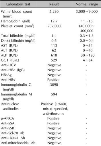
Table 1. Initial laboratory characteristics of the patient
Laboratory test Result Normal range
White blood count (mm3)
5,280 3,000∼9,000
Hemoglobin (g/dl) 12.7 11∼15
Platelet count (mm3) 207,000 140,000∼
400,000
Total bilirubin (mg/dl) 1.4 0.3∼1.3
Direct bilirubin (mg/dl) 0.6 0.0∼0.4
AST (IU/L) 113 0∼34
ALT (IU/L) 62 0∼40
ALP (IU/L) 618 30∼120
GGT (IU/L) 529 4∼34
Anti-HCV Negative -
Anti-HBc (IgG) Negative -
HBsAg Negative -
Anti-HBs Positive -
Immunoglobulin G (mg/dl)
3098 -
Immunoglobulin M (mg/dl)
594 -
Antinuclear antibodies
Positive (1:640), mixed speckled, anti-ribosome
-
p-ANCA Positive -
Anti-SSA Positive -
Anti-SSB Negative -
Anti-Scl-70 Ab Negative -
Anti-LKM-1 Ab Negative -
Anti-mitochondrial Ab Negative -
AST: aspartate aminotransferase, ALT: alanine aminotransferase, ALP: alkaline phosphatase, GGT: gamma glutamyltransferase, Anti-HCV: antibodies against hepatitis C virus, anti-HBc: anti- body to hepatitis B core antigen, HbsAg: hepatitis B virus surface antigen, Anti-HBs: anti-hepatitis B surface antibody, p-ANCA:
perinuclear anti-neutrophil cytoplasmic antibodies, Anti-SSA:
anti-Sjögren’s-syndrome-related antigen A, Anti-SSB: anti-Sjög- ren’s-syndrome-related antigen B, Anti-Scl-70 Ab: anti-topoiso- merase I antibody, Anti-LKM-1 Ab: anti-liver-kidney microsomal antibody, -: not available.
Fig. 1. Liver specimen showed a moderate portal inflammation with abundant lymphoplasma cells and mild bile duct damage with lymphocytic cholangitis (hematoxylin and eosin, ×200).
CASE REPORT
A 41-year-old female presented with multiple, painful brownish to erythematous firm patches on the both hands, arms, axillae, thighs, neck, and abdomen for 2 years. Two months prior to her visit to our department, she had been referred to our hospital from a local clinic for abnormal liver function test results and was diagnosed as PBC/AIH OS by a hepatologist in the hospital. The initial laboratory work-up obtained from the department of internal medi- cine was as follows; hematological data showed normal white blood cell counts (5,280/mm3), hemoglobin (12.7 g/
dl), and platelet counts (207,000/mm3); blood biochemi-
cal data revealed elevated total bilirubin (1.4 mg/dl), direct bilirubin (0.6 mg/dl), aspartate aminotransferase (113 IU/L), alanine aminotransferase (62 IU/L), alkaline phosphatase (618 IU/L), γ-guanosine triphosphate (529 IU/L), IgG (3,098 mg/dl), and IgM (594 mg/dl). Anti-nuclear antibody (ANA titer 1:640; mixed speckled), perinuclear anti-neu- trophil cytoplasmic antibodies, and anti-Ro/Sjögren’s-syn- drome-related antigen A autoantibodies were positive (Table 1). Liver biopsy showed moderate portal inflam- mation with lymphoplasma cells, moderate lobular activ- ity, mild fibrosis, and bile duct with lymphocytic chol- angitis (Fig. 1). She fulfilled the diagnostic criteria for PBC as well as AIH proposed by the American Association for the Study of Liver Disease and was diagnosed as PBC/AIH OS4,5. She was then referred to our department for a thor- ough investigation of her skin lesions.
The skin lesions initially developed 2 years ago starting from her fingers, which progressively spread to the hands, forearms, axillae, and trunk. Diffusely puffy hands with shiny skin suggesting impending skin thickening were ob- served (Fig. 2A). An indurated subcutaneous nodule was found on her left thumb (Fig. 2B). Widespread discolored indurated patches surrounded by hyperpigmented areas were observed on the abdomen and back (Fig. 2C, D). Dif- fuse hard patches with salt-and-pepper hypopigmentation were seen on both axillae (Fig. 2E, F). She complained of pain and tightness of the skin lesions. Physical examina- tion revealed sclerodactyly and Raynaud’s phenomenon.
With the impression of scleroderma, a punch biopsy of her left hand was performed. The skin biopsy revealed a
“square” appearance with thickened, closely packed colla- gen bundles in the reticular dermis (Fig. 3). These histo- logical findings were consistent with scleroderma. By ful- filling the 2013 American College of Rheumatology/
European League Against Rheumatism criteria for SSc, a fi-
Fig. 3. Finger specimen. (A) A ‘squ- are appearance’ in a punch biopsy (hematoxylin and eosin [H&E], ×40).
(B) Collagenous fibrosis in the upper and reticular dermis (H&E, ×100).
Fig. 2. Clinical photographs of the patient on initial visit. (A) Hands, dif- fusely puffy hands with shiny skin.
(B) Left thumb, indurated subcuta- neous nodule. (C) Abdomen, wide- spread discolored indurated patches surrounded by hyperpigmented areas. (D) Back, linear band of firm plaques and pigmentary changes.
(E, F) Axillae, salt-and-pepper hypo- pigmentation and diffuse hyperpig- mentation.
nal diagnosis of SSc was made6. We received the patient’s consent form about publishing all photographic materials.
DISCUSSION
Historically, hepatobiliary involvement in SSc has not been considered characteristic; however, recent investigations have revealed a higher prevalence of liver disturbances in SSc patients ranging from 37% to 52%7. Interestingly, pri- mary AILDs accounted for 77% of all liver diseases in SSc patients with PBC as the main cause (57%) followed by AIH (16%)7. Previously, some researchers argued that hep- atobiliary involvement in SSc occurs through chance and is possibly related to coexisting liver steatosis, viral in- fections, or drug-induced toxicity from SSc treatment8. However, we suggest that there may exist a shared auto- immune basis in AILDs and SSc etiology. Our patient sup- ports this argument since she presented with SSc and an OS of two AILDs. Her laboratory findings were positive
for multiple autoantibodies and PBC/AIH OS was diag- nosed before the discovery of SSc or any therapeutic inter- ventions regarding SSc. There was no evidence for other causes of liver dysfunction, such as alcohol abuse, drugs, or viral infection, which could idiosyncratically cause hep- atitis, including those that can mimic PBC or AIH.
PBC, AIH, and SSc are disorders involving a complex in- teraction between genetic and environmental factors; en- vironmental trigger in a background of genetic defects in immune regulation induces persistent inflammation and breakdown of self-tolerance. Regarding the genetics un- derlying SSc and AILDs, many common genetic loci of PBC and SSc have been identified, including several hu- man leukocyte antigen (HLA) regions (HLA-DRB1, DQA1, and DQB1) and non-HLA regions (interferon regulatory factor 5 and signal transducer and activator of transcrip- tion 4)9,10. With this genetic background, several triggering events, such as viruses, herbs, or the use of antibiotics in AIH, urinary tract infections, vaginal infections, and ciga-
rette smoking in PBC, and viral or bacterial infections and environmental toxins in SSc, initiate autoimmune attack11-13. Apart from the genetic and environmental factors, the adaptive immunity also seems to play a pivotal role since the dysregulation of Th17 and Tregs have been commonly noted in SSc, PBC and AIH14-16. Interleukin-6, an import- ant mediator in SSc which is also elevated in the hepato- cytes of AILDs, may polarize lymphocytes, favoring Th17 differentiation or macrophages17,18.
To our knowledge, this is the first report of an association between dcSSc and PBC/AIH OS. In the literature, all three previous cases of PBC/AIH OS and SSc were in the limited cutaneous form1-3. Based on these findings, some authors have suggested that the lcSSc subset may have a distinct pathogenesis with a greater autoimmune back- ground7. However, our patient’s condition shows that dcSSc can also coexist with PBC/AIH OS and that a common au- toimmune basis may exist between AILDs and SSc—irre- spective of the SSc cutaneous subtype. In conclusion, we suggest that a common autoimmune mechanism may un- derlie AILDs and SSc in etiology and that patients with PBC/AIH OS should be closely monitored for the risk of developing SSc and vice versa. Finally, this common auto- immune basis should be further investigated for better un- derstanding of the disease and development of novel ther- apeutic options.
CONFLICTS OF INTEREST
The authors have nothing to disclose.
ORCID
Hye Sung Han, https://orcid.org/0000-0002-3556-0740 Ga Ram Ahn, https://orcid.org/0000-0002-5696-4699 Hyung Joon Kim, https://orcid.org/0000-0002-1165-948X Kui Young Park, https://orcid.org/0000-0001-5965-1754 Kapsok Li, https://orcid.org/0000-0002-1333-1680 Seong Jun Seo, https://orcid.org/0000-0003-2915-839X
REFERENCES
1. Efe C, Ozaslan E, Nasiroglu N, Tunca H, Purnak T, Altiparmak E. The development of autoimmune hepatitis and primary biliary cirrhosis overlap syndrome during the course of con- nective tissue diseases: report of three cases and review of the literature. Dig Dis Sci 2010;55:2417-2421.
2. Toyoda M, Yokomori H, Kaneko F, Yoshida H, Hoshi K, Takeuchi H, et al. Primary biliary cirrhosis-autoimmune he- patitis overlap syndrome concomitant with systemic sclerosis, immune thrombocytopenic purpura. Intern Med 2009;48:
2019-2023.
3. West M, Jasin HE, Medhekar S. The development of connective tissue diseases in patients with autoimmune hepatitis: a case series. Semin Arthritis Rheum 2006;35:344-348.
4. Lindor KD, Gershwin ME, Poupon R, Kaplan M, Bergasa NV, Heathcote EJ; American Association for Study of Liver Dis- eases. Primary biliary cirrhosis. Hepatology 2009;50:291-308.
5. Manns MP, Czaja AJ, Gorham JD, Krawitt EL, Mieli-Vergani G, Vergani D, et al.; American Association for the Study of Liver Diseases. Diagnosis and management of autoimmune hepatitis. Hepatology 2010;51:2193-2213.
6. van den Hoogen F, Khanna D, Fransen J, Johnson SR, Baron M, Tyndall A, et al. 2013 classification criteria for systemic sclerosis: an American College of Rheumatology/European League against Rheumatism collaborative initiative. Arthritis Rheum 2013;65:2737-2747.
7. Marí-Alfonso B, Simeón-Aznar CP, Guillén-Del Castillo A, Rubio-Rivas M, Trapiella-Martínez L, Todolí-Parra JA, et al.;
RESCLE Investigators, Systemic Autoimmune Diseases Study Group (GEAS). Hepatobiliary involvement in systemic sclerosis and the cutaneous subsets: characteristics and survival of patients from the Spanish RESCLE Registry. Semin Arthritis Rheum 2018;47:849-857.
8. Takahashi A, Abe K, Yokokawa J, Iwadate H, Kobayashi H, Watanabe H, et al. Clinical features of liver dysfunction in collagen diseases. Hepatol Res 2010;40:1092-1097.
9. Gorlova O, Martin JE, Rueda B, Koeleman BP, Ying J, Teruel M, et al.; Spanish Scleroderma Group. Identification of novel genetic markers associated with clinical phenotypes of sys- temic sclerosis through a genome-wide association strategy.
PLoS Genet 2011;7:e1002178.
10. Agarwal SK, Reveille JD. The genetics of scleroderma (sys- temic sclerosis). Curr Opin Rheumatol 2010;22:133-138.
11. Ngu JH, Gearry RB, Frampton CM, Stedman CA. Autoim- mune hepatitis: the role of environmental risk factors: a population-based study. Hepatol Int 2013;7:869-875.
12. Gershwin ME, Selmi C, Worman HJ, Gold EB, Watnik M, Utts J, et al.; USA PBC Epidemiology Group. Risk factors and comorbidities in primary biliary cirrhosis: a controlled interview-based study of 1032 patients. Hepatology 2005;
42:1194-1202.
13. Arnson Y, Amital H, Guiducci S, Matucci-Cerinic M, Valentini G, Barzilai O, et al. The role of infections in the immuno- pathogensis of systemic sclerosis--evidence from serological studies. Ann N Y Acad Sci 2009;1173:627-632.
14. Fenoglio D, Battaglia F, Parodi A, Stringara S, Negrini S, Panico N, et al. Alteration of Th17 and Treg cell subpopulations co-exist in patients affected with systemic sclerosis. Clin Im- munol 2011;139:249-257.
15. Lan RY, Cheng C, Lian ZX, Tsuneyama K, Yang GX, Moritoki Y, et al. Liver-targeted and peripheral blood alterations of regulatory T cells in primary biliary cirrhosis.
Hepatology 2006;43:729-737.
16. Longhi MS, Hussain MJ, Kwok WW, Mieli-Vergani G, Ma Y, Vergani D. Autoantigen-specific regulatory T cells, a potential tool for immune-tolerance reconstitution in type-2 autoimmune hepatitis. Hepatology 2011;53:536-547.
17. Khanna D, Denton CP, Jahreis A, van Laar JM, Frech TM,
Anderson ME, et al. Safety and efficacy of subcutaneous tocilizumab in adults with systemic sclerosis (faSScinate): a phase 2, randomised, controlled trial. Lancet 2016;387:2630- 2640.
18. Zhao L, Tang Y, You Z, Wang Q, Liang S, Han X, et al. Inter- leukin-17 contributes to the pathogenesis of autoimmune hepatitis through inducing hepatic interleukin-6 expression.
PLoS One 2011;6:e18909.
![Fig. 3. Finger specimen. (A) A ‘squ- ‘squ-are appearance’ in a punch biopsy (hematoxylin and eosin [H&E], ×40).](https://thumb-ap.123doks.com/thumbv2/123dokinfo/5167199.104832/3.892.76.602.154.595/fig-finger-specimen-appearance-punch-biopsy-hematoxylin-eosin.webp)
