서 론
뇌 의 퇴행성 질환 중의 하나인 알쯔하이머병에 대한 연구가 선진국에서 노령인구가 점차로 증가 함에 따라 비가역적으로 치매가 진행 되며, 특히 노령인구에 많이 나타나 는 이 질환에 대하여 주의를 집중하 게 되었다.
알쯔하이머병(Alzheimer disease) 으로 알려지고 있는 노인성 치매 질 환은 노인인구가 급증하고 있는 현 재, 최대의 노화 질환일 뿐만 아니라 21세기에는 인류가 당면할 최대의 보건문제로 등장하고 있다. 이미 미 국에서는 400만명 이상의 환자에 사 망률의 제4위를 점하고 있으며, 2000년대 중반까지는 1,500만명의 환 자가 발생되리라 예견되고 있다. 현재 서구사회에서는 65세 이상 인구의 약 10%, 85세 이상 인구의 약 40~50%
에서 알쯔하이머병이 발생되고 있으
Pathogenesis of Alzheimer’s Dementia
Abstract
A
lzhelmer's disease (AD) is the most common cause of dementia that arises on a neuropathological background of amyloid plaques containing βamylold (Aβ) derived from amyloid precursor protein (APP) and tau-rich neurofibrillary tangles. To date, the cause and progression of familial or sporadic AD have not been fully elucidated. About 10% of all cases of AD occur as autosomal dominant inherited forms of early-onset AD, which are caused by mutations in the genes encoding APP, presenilin-1 and presenilin-2. Proteolytic processing of APP by β-γ-secretase and caspase generates Aβand carboxyl-terminal fragments of APP (APP-CTFs), which have been implicated in the pathogenesis of AD. The presenilins function as one of the γ-secretases. Aβwhich is the main component of the amyloid plaques found, is known to exert neurotoxicity by accumulating free radicals, disturbing calcium homeostasis, evoking inflammatory response and activating signaling pathways. The CTFs have been found in AD patients' brain and reported to exhibit much greater neurotoxicity than Aβ. Furthermore CTFs are known to impair calcium homeostasis and learning and memory, triggering a strong inflammatory reaction through MAPKs- and NF-κB-dependent astrocytosis and iNOS induction. Recently, it was reported that CTF translocated into the nucleus and in turn, affected transcription of genes including glycogen synthase kinase-3β which results in the induction of tau-rich neurofibrillary tangles and subsequently cell death. One of the hallmarks of AD, neurofibrillary tangles (NFT), is formed by insoluble intracellular polymers of hyperphosphorylated tau that is believed to cause apoptosis by disrupting cytoskeletal and axonal transport. This review covers the processing of APP, toxic mechanisms of Aβand CTFs of APP, presenilin and also tau in relation to the pathogenesis of AD.
Keywords : Pathogenesis; AD 핵 심 용 어 : 치매; 병인
서 유 헌
서울의대 약리학교실 Yoo--Hun Suh, MD
Department of Pharmacology, Seoul National University College of Medicine E-mail : yhsuh@snu.ac.kr
J Korean Med Assoc 2006; 49(8): 717 - 30
며, 우리나라와 일본에서도 혈관성 치매에 비해 해마다 알쯔하이머병 발생빈도가 증가하고 있어 크나 큰 사회문 제를 야기하고 있다. 우리나라에서도 앞으로 평균수명이 증가할수록 이 병이 큰 사회적 문제를 야기하리라고 예상 되고 있으며, 인간이 태초부터 갈망하던 장수에 가장 큰 도전이 되고 있다.
치매라는 것은 소위 정신 기능의 전반적인 장애가 일어 나는 증후군을 말하며, 치매의 원인은 다양하다.
알쯔하이머병은 그 단독으로나 혈관계 질환과 병합되 어 치매의 흔한 원인이 되고 있다. 심한 치매에 의하여 입 원한 환자들을 대상으로 한 사후연구(postmortem study)에 의하면 약 50%가 알쯔하이머병, 20%는 다발 성 경색 질환(multinfarct disease), 그리고 15~20%가 두 질환을 모두 앓고 있다고 한다.
정신 기능의 평가를 위해 신경과나 정신과에 입원한 환자를 대상으로 한 임상연구에 의하면 치매를 일으키는 다른 원인들을 배제해가면서 진단하였을 때 알쯔하이머 병이나 원인불명으로 진단되는 것이 연구에 따라 전체 환자의 24~57%라 한다. 이러한 연구들은 선정된 집단 을 대상으로 하였기 때문에 일상적인 집단에서 치매의 원인으로 알쯔하이머병이 어느 정도 기여하는지에 대해 서는 정확한 지표를 제시해 주지 못한다. 하지만 이러한 연구들이 제시해 주는 바는 병원에서 평가를 받아야 할 정도로 심한 치매나 나중에 입원치료를 받아야 되는 심 한 치매의 중요한 원인의 하나가 알쯔하이머병이라는 것 이다.
알쯔하이머병(AD)은 뇌 조직을 현미경으로 검사함 으로써만 정확하게 진단할 수 있다. 이 질환은 Alois Alzheimer(1907)의 이름을 딴 것인데, 그는 기억장 애, 지남력상실, 그리고 고등기능의 전반적인 와해를 보이는 치매로 사망한 중년여성의 뇌에서 3가지 특징
적인 병리학적 소견, 즉 노인반(senile plaque)과 신경 섬유 덩어리(neurofibrillary tangle)와 아밀로이드 혈관을 발견하였다(1). 이 환자는 51세 밖에 되지 않았 지만 환자의 질환과 나이를 먹게 되면 흔하게 나타나는 질환 사이의 임상적, 신경병리학적 유사성을 알쯔하이 머로 하여금 두 가지 상태가 동일질환이라는 확신을 갖 게 하였다.
결국 세포의 amyloid와 관련된 비정상적인 축삭돌기 말단으로 이루어진 신경반(neuritic plaque)과 세포 내 의 paired helical filament로 이루어진 신경섬유 덩어리 (neurofibrillary tangle)가 늙기 전 환자와 늙은 환자 모 두에서 발견되었다(2~4). 이러한 신경반(plaque)과 신 경섬유 덩어리는 AD에 특이적인 것은 아니다. 하지만 일 반적으로 다른 질환에서는 병리학적 변화가 알쯔하이머 병에서만큼 심하지 않으며, 병리학적 변화의 분포도 차이 가 있다. AD에서는 대뇌의 측두엽, 전두엽, 두정엽의 일 차연상역(primary association area)이 특히 영향을 받 으며, 일차시각, 체성감각역 및 운동역은 거의 손상을 입 지 않는다. 해마와 편도 핵이 심하게 영향을 받는다. 부가 적인 병리학적 변화로서 해마의 추체세포의 세포 내 소포 들로 이루어진 과립공포성 변성(granulovacuolar dege- neration)이 정상 노인에서 보다 훨씬 빈발한다. 젊은 환 자와 노인 환자에서 나타나는 알쯔하이머병의 질적인 차 이에 대해서는 확실한 것이 없기는 하지만, 임상적으로 볼 때 젊은 환자의 알쯔하이머병이 훨씬 빨리 진행되는 것 같다(2~4).
알쯔하이머병의 흔한 증상으로는 비교적 선택적으로, 가까운 과거의 일을 기억하는 데 있어서의 장애가 있다.
이 질환이 점점 진행됨에 따라 뇌의 고등기능 전반에서
장애가 생기고 나중에는 운동장애, 요실금 및 변실금도
나타난다(2~4).
아밀로이드 전구단백질 (Amyloid Precursor Protein, APP)
1. 아밀로이드 전구단백질의 구조와 대사
최근에 유전공학적 기법이 획기적인 공헌을 하고 있는 분야가 노인성 치매 분야이다. 이 노인성 치매의 중요한 병변은 아밀로이드 β단백질이 세포 내와 외, 그리고 혈관 에 침착하는 것이다(세포 내에 신경섬유 덩어리, 세포 외 에 신경반, 혈관에 혈관아밀로이드: Vascular amyloid) (2~4). 그 결과 뇌기능의 광범위한 장애가 오게 된다. 여 러 학자들이 뇌세포에 침착하는 이 아밀로이드 단백질을 환자의 뇌에서 추출, 정제하여 그 일부의 아미노산 배열
을 밝혔으며 이 아미노산 배열에 대한 oligonucleotide를
만들어 cDNA 유전자 분리를 시도하여 성공하였다
(Figure 1). 이 아밀로이드 단백질이 βpleated sheet 구
조를 가지고 있어서 βamyloid 단백이라 부르게 되었으
며, 대개 40~43개의 아미노산으로 구성된다. 이 단백질
은 큰 분자량의 전구단백질(APP
695, APP
751, APP
770)에서
잘라져서 형성된다(2~4). Figure 1에 보이는 것처럼 아
밀로이드 β단백질은 막에 1/3이, 막 바깥 부분에 2/3가
위치하고 있다. 현재 쿠니츠(kunitz)형의 프로테아제 억
제부[protease inhibitor domain (PI)]가 없는 전구단백
질은 APP
695(695개 아미노산 가짐)이며, PI를 가지고 있
는 전구단백질은 751, 770개의 아미노산을 가지고 있는
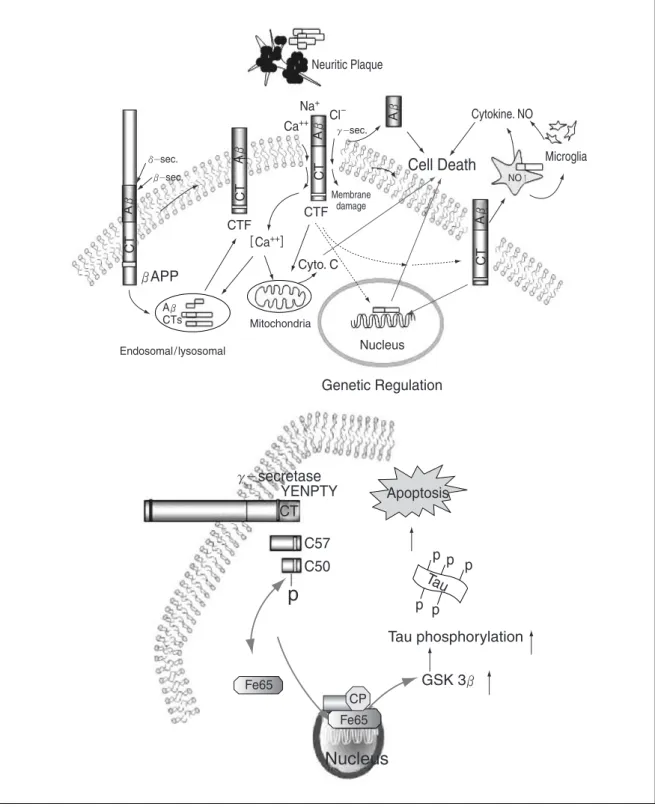
Figure 1. Structure and processing of APPFigure 2. Hypothesis of an etiological role of amyloidogenic CTF of APP in AD
APP
751, APP
770이다. 이 PI의 생체 내 의의에 대해서는 잘 모르고 있으나 아마도 세포성장을 촉진하거나 안정한 시냅스 형성(stable synaptogenesis)을 야기하는 것 같 다(2~4).
이 전구단백질은 여러 말초조직에서도 발현되고 있으 나 β단백질 침착은 뇌에서만 일어나고 있다. 알쯔하이머 병에서는 KPI domain을 가지고 있는 전구단백질의 생성 이 높아진다는 사실이 보고되고 있다(2, 4). 아마 KPI domain이 형성된 β단백질 대사를 억제하는 것 같다.
막단백질인 아밀로이드 전구 단백질은 내형질세망 (ER)과 골지체(Golgi)에서 α , β , γ세크레타제(secre-
tase)에 의해 잘라진다(Figure 1)(2~4).
α세크레타제는 Aβ부위의 가운데 16번 Lys과 17번 Leu 사이를 잘라 수용성인 α-APPs와 83개의 아미노산 으로 구성된 C단 단백질(CT
83)을 만들며 β세크레타제는 A β의 N단 시작부위를 잘라 수용성인 β-APPs (100~120kda)와 99개의 아미노산으로 구성된 C단 단 백질(CT
99)을 만든다.
이 C단 단백질(CT
83, CT
99)이 γ -세크레타제에 의해 다시 잘리면 3KD(P3) 혹은 4-KD 아밀로이드베타(Aβ ) 와 C단 단백질인 CT
57-59(아밀로이드 세포 내 부위, Amyloid intracellular domain, AICD)가 만들어진다.
Table 1. Neurotoxic mechanisms of Aβ
Toxic Mechanisms Effects References
Alteration of ion homeostasis
Potentiation of calcium channels or for- mation of calcium permeable chan- nels
Enhancement of glutamate-mediated excitotoxicity
Inhibition of Na+/Ca2+exchanger Selective Inhibition of potassium channel Impairment of Na+/K+ATPase
Generation of reactive oxygen species Production of free radical
Activation of inflammatory response Activation of microglia/astrocytes
Activation of signaling pathway Activation of MAPK Activation of JNK
Activation of calpain I and Cdk5 Activation of Src family tyrosine kinase
[Ca2+]
[Ca2+] [Ca2+] [Ca2+] [Ca2+]
Lipid peroxidation, DNA oxidation and Protein
oxidation
Release of lipid
Release of cytokines and chemo- kines
No generation
tau phosphorylation induction of Fas ligand tau phosphorylation
tyrosine phosphorylation of cyto- skeletal proteins(tau, microtu- ble-associated protein 2c)
Mattson et al., 1992; Vitek et al., 1994; Lin et al., 1999; Zhu et al., 2000; Green and Peers, 2001;
Lin et al., 2001 Mattson et al,1992 Wu et al.,1997 Good et al.,1996 Mark et al,1995
Behl et al., 1994; Butterfield et al., 1994;
Mattson et al., 1995; Varadarajan et al., 2000;
Monji et al., 2001 Michikawa et al., 2001 Tan et al.,1999; Lue et al., 2001 Tran et al., 2001
Rapoport and Ferreira, 2000 Morishima et al., 2001
Patrick et al.,1999; Lee et al., 2000 Williamson et al., 2002
그리고 ε세크레타제에 의해서 CT
50이 만들어진다. 대다 수의 베타펩티드는 40개의 아미노산으로 구성되어 있는 Aβ
40이며 소량이 응집력이 좋으며 신경반에서 주로 발견 되는 42개의 아미노산으로 구성되어 있는 Aβ
42이다.
β 와 γ절단부위 근처에서 일어나는 돌연변이 때는 Aβ 와 C단 단백질들의 생성이 증가하며 Aβ
42와 CT
50-99형성 사이에는 깊은 관계가 있다(5).
또한 캐스파제(Caspase) 3, 6, 2 효소가 C단 단백질을 잘라 31개 아미노산으로 구성된 C단 단백질(CT
31)을 생 성한다(6, 7).
아밀로이드베타 펩티드(APP695의 597~639 아미노 산)의 1~17 사이는 세포막 바깥에 존재하며 헤파린 결 합부는 N단 쪽에 있으며 인간과 쥐의 아미노산 서열은 3군데만 다르다(5번째 아미노산이 인간은 R, 쥐는 G, 10번째는 인간이 Y, 쥐가 F, 13번째는 인간이 H, 쥐가 R이다).
생성된 Aβ 의 대다수와 일부의 C단 단백질들은 세포 외 로 유리되는 것 같다(2, 3). APP의 세포질 내 C단 부
위에 있는 NPXY 부위는 단백질 을 세포 내로 이동시켜 Aβ 와 C단 단백질을 형성하는 데 중요할 뿐 아니라 세포질 내의 적응단백질 (adaptor protein)이 결합하여 C단 단백질을 핵 내로 이동시켜 중요 유전자의 전사 조절에 깊게 관여 하고 있는 것으로 보고되고 있다 (Figure 2)(8, 9).
현재 연구의 초점은 전구단백질 에 β 와 γ -secretase가 작용해서 어 떤 조절기전에 의해 독성 β단백질 과 C단 단백질이 과도하게 형성되 어 뇌에 침착되는지에 모아지고 있다.
알츠하이머 치매 병인에서의 아밀로이드 가설 (Amyloid Cascade Hypothesis)
1. 베타 아밀로이드(Aβ)가설
많은 치매 연구자들은 베타아밀로이드 독성에 주로 연 구 초점을 맞추고 있다.
베타펩티드는 앞에서 보는 바와 같이 APP 대사에 의 해 생성되는 정상적인 대사물이며 혈장과 뇌 척수액에서 검출된다(10~500pM 농도). 또한 수용성 펩티드가 베타 병풍구조(βplated sheet formation)를 형성하면 섬유체 (fibril)를 형성하여 쉽게 침전되어 독성을 형성하며 이것 이 알츠하이머 치매(AD)의 중요한 병인으로 생각되고 있다(2~4).
그러나 최근에는 섬유체보다 중합체(oligomer) 형성이 치매에서의 병변과 더 잘 일치한다는 보고가 많다(10).
또한 아밀로이드반이 형성되기 이전에 시냅스와 인지
Figure 3. The tau and tangle hypothesis기능 장애가 나타난다는 보고도 많다(4). 따라서 아밀로 이드 침착이 일어나기 전에 시냅스의 변화가 먼저 일어날 가능성도 있기 때문에 여기에도 연구가 진행되고 있다.
Aβ
40는 주로 유리되는 형태이며 Aβ
42는 아미로이드반 에 침착되는 형태이다.
이것으로 볼 때 프로테아제에 저항하는 섬유체가 중 합하여 만들어지는 과정이 치매 발병의 중요 단계인 것 같다(10).
베타펩티드독성은 반응성 산소라디칼형성, Ca
++항상 성의 장애, 염증반응과 세포 내 시그날전도 활성화 등의 여러 기전으로 나타나는 것 같다(Table 1)(11~32).
즉 베타펩티드는 ① 자유라디칼 형성을 증가시키고,
② 염증반응을 야기시키고, ③ 흥분독성을 증가시키고,
④ 전압의존 Ca
++채널을 통한 Ca
++내향 이동을 증가
시키고, ⑤ 세포막에 양이온 특이 이온채널을 형성하고,
⑥ NMDA 수용체의 Mg
++차단을 감소시켜 Ca
2++이 동을 증가시키고, ⑦ K
+채널과 Na
+/ Ca
2+교환을 억제 시킨다.
2. C단 단백질 가설
16-22KDa 크기의 여러 종류의 C단 단백질들이 말 초 혈소판이나 거대 핵세포의 세포막 부분이나(33~
35) 환자의 림프구 세포질(36~38)은 물론 신경반이나 뇌의 혈관벽, 그리고 백질(39) 회백질, 시신경에서도 검 출된다(40~44).
Table 2와 Figure 2에서 보는 것처럼 C단 단백질의 독성은 배양세포(45~47)와 뇌실 내 투여할 때(48, 49) 베타펩티드보다 10배 이상 강하며 채널효과도 개구리 난
Table 2. Summary of various effects of Aand CTFAβ CTF Reference
Neurotoxicity Cultured cells in vivo i.c.v.
Neuritic plaques of transgenic mice Channel effect
Xenopus oocytes Purkinje cells Lipid bilayer Intracellular Ca2+
Block of LTP in hippocampus Learning and memory impairment Cholinergic deficit
Free radical generation NO generation MAPK signaling NF-B
Nuclear translocation
Inflammatory cytokines & chemokines Gliosis & astrocytosis
+ + +++
- - + + + + +
+ + + - + +
+10 ++++
+++
+++
+++
++
+++
+++
+++
+++
+++
+++
+++
+++
+++
+++
45, 46, 47 48, 49 44, 50, 51, 52
53, 54, 55 56 57, 58 55, 57, 59, 60 44, 61, 62
44, 48, 49, 50, 51, 63 49
64, 65 50, 64, 65, 66 64, 65, 66 8,9, 7~69, 70, 71 65, 66
48, 66, 65
모세포(53~55), 신경세포(56), 인공지질막(57, 58)에서 베타펩티드보다 훨씬 크다.
C단 단백질에 의한 세포막 채널 활성화는 이온에 대하 여 비선택성을 보이며 강한 내향전류를 유발하며 세포 내 Ca
++농도를 증가시킨다(55, 57, 59, 60). 이러한 비선 택적 채널이나 세포막 구멍 형성은 Perforin 등과 비슷 하다.
베타펩티드보다 해마에서 LTP 억제효과(44, 61, 62) 학습과 기억력 손상 효과(44, 48~50, 51, 63), 아세틸콜 린계 장애효과(49)도 더 크게 나타난다.
자유산소라디칼 형성(64, 65)이나 염증반응(65, 66)도 더 강하게 유발시킨다.
최근 여러 종류의 C단 단백질들[CT
99, AICD(CT
57-59, CT
50), CT
30]이 세포질 내의 적응단백질(adaptor pro- tein)의 일종인 FE65와 YENPTY 부위에서 결합한 후 핵 내로 들어가게 되며 전사인자인 CP2와 다시 결합하여 삼중중합체(ternary complex)를 형성한 후 타우를 과인 산화시키는 GSK-3 β 유전자의 전사조절촉진부 (Promoter)를 활성화시켜 타우를 과인산 시킨다(Figure 2, 3). 타우의 과인산화로 세포사가 증가되고 신경섬유 덩어리(Neurofibrillary tangle)가 형성되어 치매가 발병 하게 된다(8, 9, 72)(Figure 2, 3).
C단 단백질들의 FE65와 결합에 668번째 트레오닌의 인산화가 중요한 조절 작용을 한다(9).
APP에서 유래된 C단 단백질 뿐만 아니라 APLP2(베 타펩티드 부가 없는 APP 유사단백질)의 C단 단백질들 도 같은 효과를 미친다(72). 이러한 사실로 볼 때 아밀 로이도 전구단백질(APP)과 유사단백질인 APLP2가 상호보완적 작용을 하여 치매 발병원인에 기여하는 것 같다(72).
APP와 APLP2 유전자를 같이 제거해주면 살지 못하
나, APP와 APLP1 유전자의 제거는 생존이 가능한 것 으로 보아서 APLP2의 생체 내 기능이 더욱 중요한 것 같다(72).
프리세닐린(Presenilins)
AD 중 약 10~20%가 유전성 알츠하이머 치매이며 대 다수가 프리세닐린 유전자의 돌연변이로 발생한다. 또한 상염색체 우성으로 유전되며 30세 이전에도 발병한다.
1995년 이래 PS1과 PS2 유전자에 100개 이상의 돌연 변이가 보고되고 있다(2, 73).
프리세닐린 1과 2(PS1과 PS2)는 각각 467개와 448개 의 아미노산으로 구성된 펩티드이며 7~9개의 막을 가로 지르는 부위를 가지고 있다(2, 4, 74). PS1과 PS2는 67%의 아미노산 통일성을 가지며 막 부위에 특히 돌연 변이가 많이 모여 있고 주로 뉴런에 분포하고 있다(74).
프리세닐린은 28~30KDa 크기의 Neks부와 18KDa 크 기의 C단부가 프리세닐린아제(Presenilinase)효소에 의 해 잘라져서 만들어지며 서로 결합하여 고분자량의 이성 복합체(hetero dimer)를 형성한다.
프리세닐린은 γ-세크레타제 복합체(Nicastrin, PEN2 APH-1)의 하나로 C단 단백질에서 베타펩티드 생성에 중심 역할을 한다(2, 4).
타우 단백질과 아밀로이드 단백질의 관계
치매에서 가장 중요한 2가지 병변 중 하나인 신경섬유
덩어리(Neurofibrillary tangle, NFT)는 타우의 과인산
화에 의해서 생성되나 돌연변이 APP와 돌연변이 PS 유
전자 과발현 형질전환 마우스에서는 이 병변이 발견되지
않는다(75, 76).
돌연변이 타우(P301L) 유전자와 돌연변이 APP 유전 자 과발현 마우스에서는 신경반과 신경섬유 덩어리가 발 견되며(77), 돌연변이 타우 유전자(P301L) 과발현 마우 스에 베타아밀로이드를 투여하면 신경섬유덩어리(NFT) 형성이 5배 정도 증가한다(78).
최근 본 연구팀에서는 여러 종류의 C단 단백질들 (CT
99, AICD, CT
31)을 과발현시킬 때 이들이 핵 내로 들 어가서 GSK-3β유전자를 활성화시켜 과인산화 타우의 발현을 증가시키고 세포사를 유발한다는 사실을 처음으 로 밝혔다(Figure 2, 3)(8, 9, 72).
이런 사실로 볼 때 C단 단백질들은 신경반, 세포막, 세 포질에서 검출될 뿐만 아니라 Figure 2와 3에서 보는 바 와 같은 여러 기전으로 세포사를 유발시키고 핵 속에서 전사 기전을 통해 GSK-3β등의 중요 유전자를 활성화 시켜 타우의 과인산화를 촉진시킴으로써 치매 발병을 야기한다는 사실을 알 수 있다. 베타단백질 뿐만 아니라 C단 단백질들의 생성, 핵 내 이동 등에 직접 타겟하는 약물 개발이 앞으로 치매 예방 및 치료에 유용하리라 생 각된다.
참 고 문 헌
1. Alzheimer A. Uber eine eigenartige Erkrankung der Hirnrinde, Allg Zeitschr Psychiatr Psychiatr-Gerichtl Med 1907; 146 - 8 2. Suh YH, Checler F. Amyloid Precursor Protein, Presenilins, and
α-synuclein: Molecular Pathogenesis and Pharmacological Applications in Alzheimer’s disease. Pharmacol Rev 2002; 54:
469 - 525
3. Suh YH. An etiological role of amyloidogenic carboxyl-ter- minal fragments of the beta-amyloid precursor protein in Alzheimer’s disease. J Neurochem 1997; 68: 1781 - 91
4. Selkoe DJ. Alzheimer’s disease: genes, proteins, and therapy.
Physiol Rev 2001; 81: 741 - 66
5. Sato T, Dohmae N, Qi Y, Kakuda N, Misonou H, Mitsumori R, et al. Potential link between amyloid beta-protein 42 and Cterminal fragment gamma 49-99 of beta-amyloid pre- cursor protein. J Biol Chem 2003; 278: 24294 - 301
6. Lu DC, Rabizadeh S, Chandra S, Shayya RF, Ellerby LM, Bredesen DE, et al. A second cytotoxic proteolytic peptide derived from amyloid beta-protein precursor. Nat Med 2000;
6: 397 - 404
7. Su JH, Zhao M, Anderson AJ, Srinivasan A, Cotman CW.
Activated caspase-3 expression in Alzheimer’s and aged control brain: correlation with Alzheimer pathology. Brain Res 2001; 898: 350 - 7
8. Kim HS, Kim EM, Lee JP, Park CH, Kim S, Suh YH, et al. C-
terminal fragments of Amyloid Precursor Protein Exert Neuro- toxicity by Inducing Glycogen Synthase Kinase-3β‚ Expre- ssion. FASEB J 2003; 17: 1951 - 3
9. Chang KA, Kim HS, Ha TY, Ha JW, Shin KY, Suh YH, et al.
Phosphorylation of APP at Thr668 regulates the nuclear translocation of AICD and induces neurodegeneration. Mole- cular and cellular biology 2006; 26: 4327 - 38
10. Neve RL, McPhie DL, Chen Y. Alzheimer’s disease: a dys- function of the amyloid precursor protein(1). Brain Res 2000;
886: 54 - 66
11. Walsh DM, Klyubin I, Fadeeva JV, Cullen WK, Anwyl R, Selkoe DJ, et al. Naturally secreted oligomers of amyloid beta protein potently inhibit hippocampal long-term potentiation in vivo. Nature 2002; 416: 535 - 9
12. Vitek MP, Bhattacharya K, Glendening JM, Stopa E, Vlassara H, Cerami A, et al. Advanced glycation end products con-
tribute to amyloidosis in Alzheimer disease. Proc Natl Acad Sci USA 1994; 91: 4766 - 70
13. Lin H, Zhu YJ, Lal R. Amyloid beta protein (1~40) forms calcium-permeable, Zn2+-sensitive channel in reconstituted lipid vesicles. Biochemistry 1999; 38: 11189 - 96
14. Zhu YJ, Lin H, Lal R Fresh, nonfibrillar. Amyloid beta protein(1- 40) induces rapid cellular degeneration in aged human fi- broblasts: evidence for AbetaP-channel-mediated cellular toxicity. FASEB J 2000; 14: 1244 - 54
15. Green KN, Peers C. Amyloid beta peptides mediate hypoxic augmentation of Ca(2+) channels. J Neurochem 2001; 77:
953 - 6
16. Lin H, Bhatia R, Lal R. Amyloid beta protein forms ion chan- nels: implications for Alzheimer’s disease pathophysiology.
FASEB J 2001; 15: 2433 - 44
17. Wu A, Derrico CA, Hatem L, Colvin RA. Alzheimer’s amyloid-
beta peptide inhibits sodium / calcium exchange measured in rat and human brain plasma membrane vesicles. Neuro- science 1997; 80: 675 - 84
18. Good TA, Smith DO, Murphy RM. Beta-amyloid peptide blocks the fast-inactivating K+current in rat hippocampal neurons. Biophys J 1996; 70: 296 - 304
19. Mark RJ, Hensley K, Butterfield DA, Mattson MP. Amyloid b-peptide impairs ion-motive ATPase activities: evidence for a role in loss of neuronal Ca2+homeostasis and cell death. J Neurosci 1995; 15: 6239 - 49
20. Behl C, Davis JB, Lesley R, Chubert D. Hydrogen Peroxide Mediates Amyloid Protein Toxicity. Cell 2000; 77: 817 - 27 21. Butterfield DA, Hensley K, Harris M, Mattson MP, Carney J.
Beta-amyloid peptide free radical fragments initiate sy- naptosomal lipoperoxidation in a sequence-specific fashion:
implications to Alzheimer’s disease. Biochem Biophys Res Commun 1994; 200: 710 - 5
22. Mattson MP, Lovell MA, Furukawa K, Markesbery WR.
Neurotrophic factors attenuate glutamate-induced accu- mulation of peroxides, elevation of intracellular calcium concentration, and neurotoxicity and increase antioxidant enzyme activities in hippocampal neurons. J Neurochem 1995; 65: 1740 - 51
23. Varadarajan S, Yatin S, Aksenova M, Butterfield AD. Alz- heimer’s amyloid beta peptide-associated free radical oxi- dative stress and neurotoxicity. Journal of Structural Biology 2000; 130: 184 - 208
24. Monji A, Utsumi H, Ueda T, Imoto T, Yoshida I, Tashiro, et al.
The relationship between the aggregational state of the amyloid-beta peptides and free radical generation by the peptides. J Neurochem 2001; 77: 1425 - 32
25. Michikawa M, Gong JS, Fan QW, Sawamura N, Yanagisawa K. A novel action of alzheimer’s amyloid beta-protein (Abeta):
oligomeric Abeta promotes lipid release. J Neurosci 2001; 21:
7226 - 35
26. Tan J, Town T, Paris D, Mori T, Suo Z, Mullan M, et al. Mi- croglial activation resulting from CD40-CD40L interaction after beta-amyloid stimulation. Science 1999; 286: 2352 - 5 27. Lue LF, Rydel R, Brigham EF, Yang LB, Hampel H, Rogers J,
et al. Inflammatory repertoire of Alzheimer’s disease and nondemented elderly microglia in vitro. Glia 2001; 35: 72 - 9 28. Rapoport M, Ferreira A. PD98059 prevents neurite dege-
neration induced by fibrillar beta-amyloid in mature hippo- campal neurons. J Neurochem 2000; 74: 125 - 33
29. Morishima Y, Gotoh Y, Zieg J, Barrett T, Takano H, Greenberg ME, et al. Beta-amyloid induces neuronal apoptosis via a
mechanism that involves the c-Jun N-terminal kinase path- way and the induction of Fas ligand. J Neurosci 2001; 2: 7551 - 60
30. Patrick GN, Zukerberg L, Nikolic M, de la Monte S, Dikkes P, Tsai LH. Conversion of p35 to p25 deregulates Cdk5 activity and promotes neurodegeneration. Nature 1999; 402: 615 - 22 31. Lee MS, Kwon YT, Li M, Peng J, Friedlander RM, Tsai LH.
Neurotoxicity induces cleavage of p35 to p25 by calpain.
Nature 2000; 405: 360 - 4
32. Williamson R, Scales T, Clark BR, Gibb G, Reynolds CH, Anderton BH, et al. Rapid tyrosine phosphorylation of neuronal proteins including tau and focal adhesion kinase in response to amyloid-beta peptide exposure: involvement of Src family protein kinases. J Neurosci 2002; 22: 10 - 20
33. Ghiso J, Rostagno A, Gardella JE, Liem L, Gorevic PD, Frangione B. A 109-amino-acid C-terminal fragment of Alzheimer’s-disease amyloid precursor protein contains a sequence, -RHDS-, that promotes cell adhesion. Biochem J 1992; 288: 1053 - 9
34. Gardella JE, Gorgone GA, Candela L, Ghiso J, Castano EM, Frangione B, et al. High-level expression and in vitro muta- genesis of a fibrillogenic 109-amino-acid C-terminal frag- ment of Alzheimer’s-disease amyloid precursor protein.
Biochem J 1993; 294: 667 - 74
35. Li QX, Evin G, Small DH, Multhaup G, Beyreuther K, Masters CL. Proteolytic processing of Alzheimer’s disease beta A4 amyloid precursor protein in human platelets. J Biol Chem 1995; 270: 14140 - 7
36. Matsumoto A, Fujiwara Y. Abnormal and deficient processing of beta-amyloid precursor protein in familial Alzheimer’s di- sease lymphoblastoid cells. Biochem Biophys Res Commun
1991; 175: 361 - 5
37. Matsumoto A, Fujiwara Y. Aberrant proteolysis of the beta-
amyloid precursor protein in familial Alzheimer’s disease lymphoblastoid cells. Eur J Biochem 1993; 217: 21 - 7 38. Matsumoto A, Matsumoto R. Familial Alzheimer’s disease
cells abnormally accumulate beta-amyloid-harbouring pep- tides preferentially in cytosol but not in extracellular fluid. Eur J Biochem 1994; 225: 1055 - 62
39. Tokuda T, Tanaka K, Kametani F, Ikeda S, Yanagisawa N.
Secretory cleavage of beta-amyloid precursor protein in the cerebral white matter produces amyloidogenic carboxyl-
terminal fragments. Neurosci Lett 1995; 186: 149 - 52 40. Nordstedt C, Gandy SE, Alafuzoff I, Caporaso GL, Iverfeldt K,
Grebb JA, et al. Alzheimer beta/A4 amyloid precursor protein in human brain: aging-associated increases in holoprotein and in a proteolytic fragment. Proc Natl Acad Sci USA 1991;
88: 8910 - 4
41. Estus S, Golde TE, Kunishita T, Blades D, Lowery D, Eisen M, et al. Potentially amyloidogenic, carboxyl-terminal derivatives of the amyloid protein precursor. Science 1992; 255: 726 - 8 42. Tamaoka A, Kalaria RN, Lieberburg I, Selkoe DJ. Identification
of a stable fragment of the Alzheimer amyloid precursor con- taining the beta-protein in brain microvessels. Proc Natl Acad Sci USA 1992; 89: 1345 - 9
43. Amaratunga A, Fine RE. Generation of amyloidogenic C-
terminal fragments during rapid axonal transport in vivo of betaamyloid precursor protein in the optic nerve. J Biol Chem 1995; 270: 17268 - 72
44. Nalbantoglu J, Tirado-Santiago G, Lahsaini A, Poirier J, Goncalves O, Verge G, et al. Impaired learning and LTP in mice expressing the carboxy terminus of the Alzheimer
amyloid precursor protein. Nature 1997; 387: 500 - 5 45. Tanzi RE, Bertram L. New frontiers in Alzheimer’s disease
genetics. Neuron 2001; 32: 181 - 4
46. Lee JP, Chang KA, Kim HS, Kim SS, Jeong SJ, Suh YH. APP carboxyl-terminal fragment without or with abeta domain equally induces cytotoxicity in differentiated PC12 cells and cortical neurons. J Neurosci Res 2000; 60: 565 - 70
47. Marcon G, Giaccone G, Canciani B, Cajola L, Rossi G, De Gioia L, et al. A betaPP peptide carboxyl-terminal to Abeta is neurotoxic. Am J Pathol 1999; 154: 1001 - 7
48. Song DK, Won MH, Jung JS, Lee JC, Kang TC, Suh HW, et al.
Behavioral and neuropathologic changes induced by central injection of carboxyl-terminal fragment of beta-amyloid precursor protein in mice. J Neurochem 1998; 71: 875 - 8 49. Choi SH, Park CH, Koo JW, Seo JH, Kim HS, Jeong SJ, et al.
Memory impairment and cholinergic dysfunction by centrally administered Abeta and carboxyl-terminal fragment of Alz- heimer’s APP in mice. FASEB J 2001; 15: 1816 - 8
50. Lambourne SL, Sellers LA, Bush TG, Choudhury SK, Emson PC, Suh YH, et al. Increased tau phosphorylation on mito- genactivated protein kinase consensus sites and cognitive decline in transgenic models for Alzheimer’s disease and FTDP-17: evidence for distinct molecular processes under- lying tau abnormalities. Molecular Cell Biol 2005; 25: 278 - 93 51. Kammesheidt A, Boyce FM, Spanoyannis AF, Cummings BJ,
Ortegon M, Cotman C, et al. Deposition of beta /A4 immuno- reactivity and neuronal pathology in transgenic mice expres- sing the carboxyl-terminal fragment of the Alzheimer amy- loid precursor in the brain. Proc Natl Acad Sci USA 1992; 89:
10857 - 61
52. Ando K, Iijima KI, Elliott JI, Kirino Y, Suzuki T. Phosphory-
lation- dependent regulation of the interaction of amyloid precursor protein with Fe65 affects the production of beta- amyloid. J Biol Chem 2001; 276: 40353 - 61
53. Fraser SP, Suh YH, Djamgoz MBA. Ionic effects of the Alz- heimer’s disease -amyloid precursor protein and its meta- bolic fragments. Trends Neurosci 1997; 20: 67 - 72
54. Fraser SP, Suh YH, Chong YH, Djamgoz MB. Membrane currents induced in Xenopus oocytes by the C-terminal fragment of the beta-amyloid precursor protein. J Neuro- chem 1996; 66: 2034 - 40
55. Kim JH, Rah JC, Fraser SP, Chang KA, Djamgoz MB, Suh YH.
Carboxyl-terminal peptide of beta-amyloid precursor pro- tein blocks inositol 1, 4, 5-trisphosphate-sensitive Ca2+
release in Xenopus laevis oocytes. J Biol Chem 2002; 277:
20256 - 63
56. Hartell NA, Suh YH. Peptide fragments of beta-amyloid precursor protein: effects on parallel fiber-Purkinje cell sy- naptic transmission in rat cerebellum. J Neurochem 2000; 74:
1112 - 21
57. Kim HS, Lee JH, Suh YH. C-terminal fragment of Alzheimer’s amyloid precursor protein inhibits sodium/calcium exchanger activity in SK-N-SH cell. Neuroreport 1999; 10: 113 - 6 58. Arispe N, Rojas E, Pollard HB. Alzheimer disease amyloid beta
protein forms calcium channels in bilayer membranes: block- ade by tromethamine and aluminum. Proc Natl Acad Sci USA 1993; 90: 567 - 71
59. McKeon-O’Malley C, Wells J, Fine R, Ullman MD, Volicer L.
PC12 cells transfected with a C-terminal fragment of the amyloid precursor protein (APP C-100), exhibit enhanced sensitivity to the calcium ionophore A23187, and diminished sensitivity to hydrogen peroxide. Brain Res Mol Brain Res
1999; 72: 103 - 7
60. Kim HS, Park CH, Cha SH, Lee JH, Lee S, Kim Y, et al. Car- boxyl-terminal fragment of Alzheimer’s APP destabilizes calcium homeostasis and renders neuronal cells vulnerable to excitotoxicity. FASEB J 2000; 14: 1508 - 17
61. Cullen WK, Suh YH, Anwyl R, Rowan MJ. Block of LTP in rat hippocampus in vivo by beta-amyloid precursor protein fragments. Neuroreport 1997; 8: 3213 - 7
62. Kim JH, Anwyl R, Suh YH, Djamgoz MB, Rowan MJ. Use- dependent effects of amyloidogenic fragments of (beta)-
amyloid precursor protein on synaptic plasticity in rat hippo- campus in vivo. J Neurosci 2001; 21: 1327 - 33
63. Sato M, Kawarabayashi T, Shoji M, Kobayashi T, Tada N, Matsubara E, et al. Neurodegeneration and gliosis in trans- genic mice overexpressing a carboxy-terminal fragment of Alzheimer amyloid-beta protein precursor. Dement Geriatr Cogn Disord 1997; 8: 296 - 307
64. Bach JH, Chae HS, Rah JC, Lee MW, Park CH, Choi SH, et al.
C-terminal fragment of amyloid precursor protein induces astrocytosis. J Neurochem 2001; 78: 109 - 20
65. Rah JC, Kim HS, Kim SS, Bach JH, Kim YS, Park CH, et al.
Effects of carboxyl-terminal fragment of Alzheimer’s amyloid precursor protein and amyloid beta-peptide on the pro- duction of cytokines and nitric oxide in glial cells. FASEB J 2001; 15: 1463 - 5
66. Chong YH, Sung JH, Shin SA, Chung JH, Suh YH. Effects of the -amyloid and carboxy-terminal fragment of Alzheimer’s amyloid precursor protein on the production of the tumor necrosis factor-and matrix metalloproteinase-9 by human monocytic THP-1. J Biol Chem 2001; 276: 23511 - 7 67. DeGiorgio LA, DeGiorgio N, Milner TA, Conti B, Volpe BT.
Neurotoxic APP C-terminal and beta-amyloid domains colocalize in the nuclei of substantia nigra pars reticulata neurons undergoing delayed degeneration. Brain Res 2000;
874: 137 - 46
68. Cao X, Sudhof TC. A transcriptionally [correction of trans- criptively] active complex of APP with Fe65 and histone acetyltransferase Tip60. Science 2001; 293: 115 - 20 69. Kimberly WT, Zheng JB, Guenette SY, Selkoe DJ. The intra-
cellular domain of the beta-amyloid precursor protein is stabilized by Fe65 and translocates to the nucleus in a notch- like manner. J Biol Chem 2001; 276: 40288 - 92
70. Gao Y, Pimplikar SW. The gamma -secretase-cleaved Cterminal fragment of amyloid precursor protein mediates signaling to the nucleus. Proc Natl Acad Sci USA 2001; 98:
14979 - 84
71. Cupers P, Bentahir M, Craessaerts K, Orlans I, Vanderstichele H, Saftig P, et al. The discrepancy between presenilin sub- cellular localization and -secretase processing of amyloid precursor protein. J Cell Biol 2001; 154: 731 - 40
72. Xu Y, Kim HS, Joo Y, Choi Y, Chang KA, Suh YH, et al. In- tracellular Domains of Amyloid Precursor-Like Protein 2 Interact with CP2 Transcription Factor in the Nucleus and Induce Glycogen Synthase Kinase-3β‚ Expression. Cell Death and Differentiation. 2006; doi: 10.1038/sj.cdd.4401928 73. Hardy J. Amyloid, the presenilins and Alzheimer’s disease.
Trends Neurosci 1997; 20: 154 - 9
74. Prihar G, Fuldner RA, Perez-Tur J, Lincoln S, Duff K, Adams MD, et al. Structure and alternative splicing of the presenilin-
2 gene. Neuroreport 1996; 7: 1680 - 4
75. Borchelt DR, Ratovitski T, Van Lare J, Lee MK, Gonzales V, Sisodia SS, et al. Accelerated amyloid deposition in the brains
of transgenic mice coexpressing mutant presenilin 1 and amyloid precursor proteins. Neuron 1997; 19: 939 - 45 76. Holcomb L, Gordon MN, McGowan E, Yu X, Benkovic S, Duff
K, et al. Accerlated Alzheimer-type phenotype in transgenic micecarrying both mutant amyloid precursor protein and pre- senilin 1 transgenes. Nature Medicine 1998; 4: 97 - 100 77. Lewis J, Dickson DW, Lin WL, Chisholm L, Corral A, Mc-
Gowan E, et al. Enhanced neurofibrillary degeneration in transgenic mice expressing mutant tau and APP. Science 2001; 293: 1487 - 91
78. Gotz J, Chen F, van Dorpe J, Nitsch RM. Formation of neuro- fibrillary tangles in P301l tau transgenic mice induced by Abeta 42 fibrils. Science 2001; 293: 1491 - 5
Peer Reviewer Commentary
본 논문은 최근 10년간 세계적으로 고령인구의 증가 추세와 더불어 급격히 증가 일로에 있는 알츠하이머병의 발병기 전들에 대한 서론적 소개와 함께 최근 제시되고 있는 여러 가설들에 대하여도 실험적 근거를 바탕으로 간략히 기술 하고 있다. 또한 베타 아밀로이드(A β)와 C단 단백질에 의한 직접적인 독성 이외에도 C단 단백질의 핵내 유입에 의 한 유전자 전사 활성화 과정으로 GSK-3β활성화가 촉진되어 Tau 과인산화가 초래되면서, 이로 인한 신경독성 초 래 및 NFT 형성 가능성이 본 필자에 의해 처음 제시되고 있다. 한편, Aβfibril이나 AβPlaque가 형성되기 전에 수 용성인 Aββoligomer가 synaptic cleft에 쉽게 접근할 수 있어 시넵스 기능 장애를 유도하여 치매 초기병변인 기 억력 감퇴 현상을 초래할 수 있다는 가설이 최근 제시되고 있는 시점이다. 아울러 Tau 과인산화 뿐만 아니라 Tau 절단산물들(Cleaved Tau)에 의한 신경독성 야기 및 NFT 형성 가능성이 최근 연구의 초점이 되고 있다. 따라서 CT 단백질과 이러한 현상들과의 상호 연계성에 대한 지속적인 연구를 수행함으로써, 본 필자에 의해 제시된 A β및 C단 단백질 가설을 치매발병의 예방 및 지연, 치료를 위한 유용한 약물 개발에 매우 중요한 타겟으로 적용할 수 있을 것 으로 사료된다.
정 영 해 (이화의대 미생물학교실)