ABSTRACT
Primary pleomorphic liposarcoma of the breast is rare, and only a few cases in the literature have reported imaging findings. Herein, we report a rare case of primary pleomorphic liposarcoma of the breast in a 38-year-old woman and describe the imaging findings including mammography, ultrasonography, computed tomography, magnetic resonance imaging, and
18F-Fluorodeoxyglucose-positron emission tomography. Although most fat- containing breast masses are benign, malignancy can occur. Magnetic resonance imaging can be helpful for further evaluation of breast masses.
Keywords:
Breast; Liposarcoma; Magnetic resonance imaging; Mammography;
Ultrasonography
INTRODUCTION
Liposarcoma is one of the rarest malignancies of the breast comprising only 0.003% of all malignant breast tumors [1]. Among liposarcomas, the pleomorphic subtype is the least common, and only a few cases have reported imaging findings [1-5]. Herein, we describe a case of primary pleomorphic liposarcoma of the breast in a 38-year-old woman including various imaging findings.
CASE REPORT
This study adhered to the Declaration of Helsinki and was approved by the Institutional Review Board of Inje University Haeundae Paik Hospital (HP IRB 2020-02-006-001).
Informed consent was waived due to the retrospective nature of the study.
A 38-year-old woman presented with a growing palpable mass in her left breast. The mass was biopsied at an outside hospital, and pleomorphic liposarcoma was considered as a possible diagnosis. Following the biopsy, the patient was referred to our hospital. She had no remarkable medical or family history. In the physical examination, a 5 × 4 cm solitary mass was palpable in the left upper outer breast.
Case Report
Received: Feb 15, 2020 Accepted: Apr 9, 2020 Correspondence to Hyun Kyung Jung
Department of Radiology, Inje University Haeundae Paik Hospital, 875 Haeun-daero, Haeundae-gu, Busan 48108, Korea.
E-mail: drsjung@gmail.com
© 2020 Korean Breast Cancer Society This is an Open Access article distributed under the terms of the Creative Commons Attribution Non-Commercial License (https://
creativecommons.org/licenses/by-nc/4.0/) which permits unrestricted non-commercial use, distribution, and reproduction in any medium, provided the original work is properly cited.
ORCID iDs Sung Jae Jo
https://orcid.org/0000-0001-8190-7744 Hyun Kyung Jung
https://orcid.org/0000-0001-6086-7771 Kyung Han Nam
https://orcid.org/0000-0002-0041-7637 Conflicts of Interest
The authors declare that they have no competing interests.
Author Contributions
Supervision: Nam KH; Writing - original draft:
Jo SJ; Writing - review & editing: Jung HK.
Sung Jae Jo
1, Hyun Kyung Jung
1, Kyung Han Nam
21Department of Radiology, Inje University Haeundae Paik Hospital, Busan, Korea
2Department of Pathology, Inje University Haeundae Paik Hospital, Busan, Korea
Recurrent Primary Pleomorphic
Liposarcoma of the Breast: A Case
Report with Imaging Findings
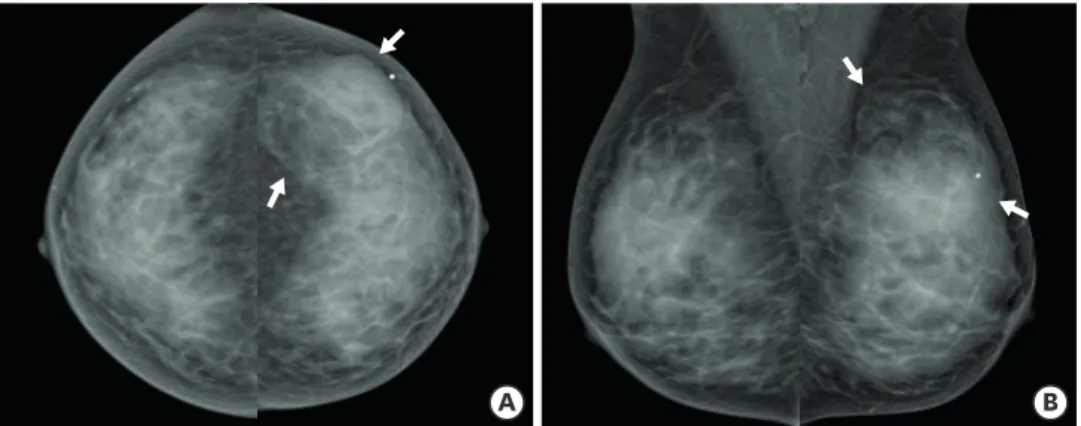
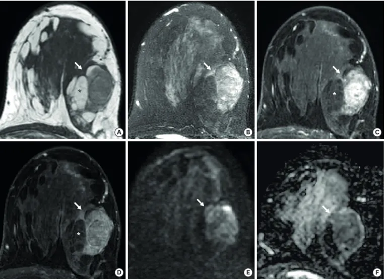
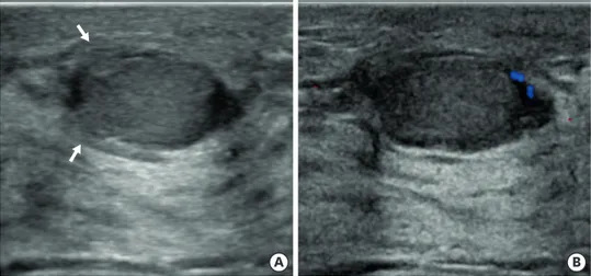
Mammography performed at the outside hospital depicted an oval-shaped fat-containing mass with obscured margin in the left upper outer breast (Figure 1), whereas ultrasonography (US) depicted an oval-shaped heterogeneous hyperechoic mass with indistinct margin and internal and rim vascularity (Figure 2). Dynamic contrast-enhanced magnetic resonance imaging (MRI) of the breast showed a heterogeneously enhanced oval-shaped mass with an irregular margin and kinetic features of rapid initial and delayed wash-out enhancement (Figure 3). Non-fat-suppressed T1-weighted image (T1WI) of the mass showed areas of hyperintensity that did not exhibit enhancement on post-contrast T1WI. The diffusion- weighted image (DWI) and apparent diffusion coefficient (ADC) map revealed a focal area of restricted diffusion in the enhanced parts of the solid mass. Contrast-enhanced chest computed tomography (CT) showed an oval-shaped heterogeneously enhanced mass with a non-enhanced fat component (Figure 4).
18F-fluorodeoxyglucose (FDG)-positron emission tomography (PET) showed focal FDG-avid uptake in the left breast with a maximum standardized uptake value of 3.0. Metastatic work-up was negative on chest CT, abdomen- pelvis CT, and PET. The patient underwent left lumpectomy without axillary dissection at a different hospital. Histologic examination confirmed that the mass was a pleomorphic liposarcoma, 5.5 × 3.8 × 3.4 cm in size with a clear resection margin. Chemotherapy or radiation therapy was not performed after surgery.
Primary Pleomorphic Liposarcoma of the Breast
A B
Figure 1. Craniocaudal (A) and mediolateral oblique (B) mammography showed an oval-shaped fat-containing mass with an obscured margin in the left outer breast (arrows).
A B
Figure 2. Transverse ultrasonography (A) showed an oval-shaped heterogeneous hyperechoic mass with an indistinct margin (arrows). Color Doppler image of the mass (B) showed internal and peripheral vascularity.
* *
*
A B
D E
C
F
Figure 3. On axial T1-weighted (A) and T2-weighted image (B), the mass (arrows) showed heterogeneous signal intensity. Non-fat-suppressed T1WI (A) revealed hyperintense areas (asterisks) that did not show enhancement on post-contrast T1WI (C and D). Dynamic contrast-enhanced magnetic resonance imaging showed an oval-shaped irregular heterogeneously enhanced mass with a rapid enhancement in the early phase (C), followed by a delayed wash-out enhancement in the late phase (D). A focal area of the mass showed restricted diffusion on the diffusion-weighted image (E) and apparent diffusion coefficient map (F).
T1WI, T1-weighted image.
A B
Figure 4. Contrast-enhanced axial (A) and coronal chest computed tomography (B) showed an oval-shaped heterogeneously enhanced mass with a non-enhancing fat component (arrows).
Twelve months after the initial diagnosis, a mass was again palpable in her left breast.
Following a lumpectomy, the mass was diagnosed as a recurrence of pleomorphic
liposarcoma at a different hospital. Seven months after this recurrence, she again presented with a palpable mass in the left breast. US revealed two oval-shaped isoechoic masses with indistinct margin (Figure 5). Breast MRI revealed a circumscribed oval mass with heterogeneous enhancement and kinetic features of rapid initial and delayed wash-out enhancement (Figure 6). A focal area of restricted diffusion was seen on the DWI and ADC map. Unlike the first mass that was detected in the patient, the other 2 masses exhibited an intermediate signal intensity on T1WI.
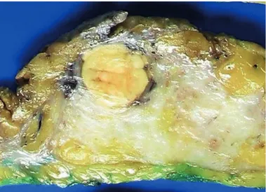
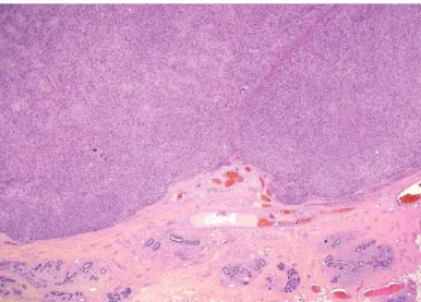
Modified radical mastectomy was performed, and the two masses were confirmed to be recurrent pleomorphic liposarcomas, with sizes of 2.5 × 2.1 × 1.9 cm and 1.1 × 1.0 × 0.8 cm, respectively. Through extensive sampling of the two masses, malignant phyllodes tumors with liposarcomatous differentiation were excluded. Macroscopically, the masses were both well-circumscribed with lobulated margin. The cut surfaces were rubbery and yellowish- white in color (Figure 7). On 1.25× low power magnification, the mass presented with a well-circumscribed lobulated margin (Figure 8). Histologically, the tumors were composed of large pleomorphic cells with round to bizarre nuclei and eosinophilic to vacuolated cytoplasm (Figure 9). In addition, scattered multivacuolated lipoblasts were observed.
DISCUSSION
Primary breast sarcomas are rare malignancies arising from the mesenchymal tissue of the mammary gland, and it accounts for less than 1% of all malignant breast tumors [2,6].
Liposarcomas are rare, and the pleomorphic subtype is the least common and accounts for only 5%–15% of all liposarcomas [7]. It usually presents as a painless, slow-growing mass but may have diverse clinical manifestations [8].
Due to its rarity, imaging findings of primary liposarcoma of the breast have rarely been reported [9,10]. Previous imaging findings of this tumor have been limited to mammography and US, with mammography generally depicting high-density masses with irregular margin and US depicting solid or complex cystic and solid masses [1-5]. In the present case, it
Primary Pleomorphic Liposarcoma of the BreastA B
Figure 5. Transverse ultrasonography (A) showed an oval-shaped isoechoic mass with indistinct margin (arrows).
Color Doppler image of the mass (B) showed peripheral vascularity.
A B
D E
C
F
Figure 6. On axial T1-weighted (A) and T2-weighted image (B), the 2 masses (arrows) showed intermediate signal intensity. Dynamic contrast-enhanced magnetic resonance imaging showed an oval heterogeneously enhanced mass with rapid enhancement in the early phase (C), followed by a delayed wash-out enhancement in the late phase (D). The masses showed restricted diffusion on the diffusion-weighted image (E) and apparent diffusion coefficient map (F).
Figure 7. Mastectomy specimen showed well-circumscribed masses with a yellowish-white cut surface.
appeared as an oval-shaped fat-containing mass on mammography, and a hyperechoic mass on US, thus, potentially mimicking a benign mass. MRI depicted an oval-shaped heterogeneously enhanced mass with initial rapid enhancement followed by a delayed wash- out pattern on the kinetic image and a focal diffusion restricted to the enhanced solid portion of the mass on DWI. To our knowledge, this is the first case report describing MRI features of this type of tumor.
Although there is no definite consensus on the treatment for primary breast sarcomas, surgical resection is generally considered as the gold standard. Yin et al. [11] recommended breast-conserving surgery if complete resection with negative margins can be achieved and adjuvant radiation therapy if the tumor is over 5 cm in size.
Local recurrence of breast liposarcoma has been associated with a pleomorphic subtype and infiltrative margins [12]. In the present case, the 38-year-old woman underwent surgery without adjuvant chemotherapy or radiotherapy at the time of initial diagnosis, even though the tumor was over 5 cm in size. Although the surgical margin was negative, she had two recurrences at the same operation site. Therefore, multimodal therapies are recommended in patients with poor prognosis.
Primary Pleomorphic Liposarcoma of the Breast
Figure 8. Mass was well-circumscribed with a lobulated margin (hematoxylin and eosin, magnification ×1.25).
A B
Figure 9. Microphotograph (A) showed pleomorphic cells with eosinophilic to vacuolated cytoplasm (H&E, magnification ×200). Microphotograph (B) showed multivacuolated lipoblasts (H&E, magnification ×400).
H&E, hematoxylin and eosin.
In conclusion, we have reported various imaging findings of primary pleomorphic liposarcoma of the breast including mammography, US, CT, MRI, and PET. Radiologists should be aware that even if a breast mass exhibits a fat-containing density on mammography and hyperechogenicity on US, it may not be benign. Therefore, MRI can be a useful
additional imaging tool for the evaluation of breast masses.
REFERENCES
1. Mardi K, Gupta N. Primary pleomorphic liposarcoma of breast: a rare case report. Indian J Pathol Microbiol 2011;54:124-6.
PUBMED | CROSSREF
2. Üzüm N, Celasin H, Ataoğlu O, Koçak S. Pleomorphic liposarcoma of the breast misdiagnosed as carcinoma in a tru-cut biopsy. J Breast Health 2010;6:87-90.
3. Nandipati KC, Nerkar H, Satterfield J, Velagapudi M, Ruder U, Sung KJ. Pleomorphic liposarcoma of the breast mimicking breast abscess in a 19-year-old postpartum female: a case report and review of the literature. Breast J 2010;16:537-40.
PUBMED | CROSSREF
4. Whitsell T, Marcovis K, Ruhs S, Andres M, Beck S. High-grade pleomorphic liposarcoma of the breast.
Grand Rounds 2011;11:87-91.
CROSSREF
5. Mukherjee A, Nath J, Dey D, Chakravorty S, Sinha S, Chatterjee T. A rare case report of primary pure pleomorphic liposarcoma of breast with cytological and histopathological findings. J Cancer Sci Clin Oncol 2017;4:102.
CROSSREF
6. Adem C, Reynolds C, Ingle JN, Nascimento AG. Primary breast sarcoma: clinicopathologic series from the Mayo Clinic and review of the literature. Br J Cancer 2004;91:237-41.
PUBMED | CROSSREF
7. Murphey MD, Arcara LK, Fanburg-Smith J. From the archives of the AFIP: imaging of musculoskeletal liposarcoma with radiologic-pathologic correlation. Radiographics 2005;25:1371-95.
PUBMED | CROSSREF
8. Briski LM, Jorns JM. Primary breast atypical lipomatous tumor/well-differentiated liposarcoma and dedifferentiated liposarcoma. Arch Pathol Lab Med 2018;142:268-74.
PUBMED | CROSSREF
9. Smith TB, Gilcrease MZ, Santiago L, Hunt KK, Yang WT. Imaging features of primary breast sarcoma.
AJR Am J Roentgenol 2012;198:W386-93.
PUBMED | CROSSREF
10. Wienbeck S, Meyer HJ, Herzog A, Nemat S, Teifke A, Heindel W, et al. Imaging findings of primary breast sarcoma: results of a first multicenter study. Eur J Radiol 2017;88:1-7.
PUBMED | CROSSREF
11. Yin M, Mackley HB, Drabick JJ, Harvey HA. Primary female breast sarcoma: clinicopathological features, treatment and prognosis. Sci Rep 2016;6:31497.
PUBMED | CROSSREF
12. Austin RM, Dupree WB. Liposarcoma of the breast: a clinicopathologic study of 20 cases. Hum Pathol 1986;17:906-13.
PUBMED | CROSSREF
