43
책임저자 : 김흥철, 강원도 춘천시 교동 153
200-060, 춘천성심병원 영상의학과 Tel: 033-240-5158, Fax: 033-242-7085 E-mail: [email protected]
게재승인일:2008년 2월 27일
갑상샘의 미만성 경화아형 유두상암의 1예 보고: 초음파와 컴퓨터 단층촬영 영상
춘천성심병원 영상의학과, 1외과, 2병리과
김흥철ㆍ남궁숙ㆍ홍명선ㆍ황임경ㆍ김한준1ㆍ최영희2
A Case Report on Diffuse Sclerosing Papillary Carcinoma of the Thyroid: The Ultrasound and CT Images
Heung Cheol Kim, M.D., Sook Namkung, M.D., Myong Son Hong, M.D., Im Kyung Hwang, M.D., Han Joon Kim, M.D.
1 and Young Hee Choi, M.D.2We report here on a case of diffuse sclerosing papillary carcinoma (DSPC), which is a subtype of papillary carcino- ma of thyroid, in a 27-year-old female. The ultrasound im- ages showed diffuse enlargement of the thyroid lobes and this was associated with underlying diffuse scattered micro- calcifications and a heterogeneous hypoechoic background parenchyma that was without any masses. The CT image showeddiffuse scattered dot-like hyperdensities with bi- lateral multiple metastastic lymph nodes. Because making the cytological diagnosis of DSPC is still challenging, the radiological findings that show this disease’s characteristic features may be important clues for making the correct diagnosis. (Korean J Endocrine Surg 2008;8:43-47)
Key Words: Thyroid, Papillary carcinoma, Diffuses scleros-
ing variant, Ultrasonography, CT
중심 단어:갑상샘, 유두상암, 미만성 경화아형, 초음 파, 컴퓨터 단층촬영
Departments of Radiology, 1Surgery, 2Pathology, Chunchon Sacred Heart Hospital, Chuncheon, Korea
서 론
갑상샘의 유두상암(papillary carcinoma)은 전체 일차성 갑 상샘암의 70∼80%를 차지하며,(1,2) 이중 대부분은 고전형
(classic type), 소포형(follicular type), 피막형(encapsulated type) 등이 차지하지만, 이 외에 다른 여러 아형들이 존재하 며 최근 보고들에 의하면 이들은 예후에 영향을 주는 고전 형 유두상암과는 다른 생물학적 성질들을 갖는다. 그 중 미 만성 경화 유두상암(diffuse sclerosing papillary cancer, DSPC) 은 매우 드문 아형이며 임상적으로 매우 공격적인 특성을 갖는다. 그러므로 이 질환의 조기진단은 갑상샘암 환자들 의 치료 방법을 선택하고 예후를 결정짓는데 매우 중요하 다. 그러나 이 질환은 진단이 어렵고 임상적으로 갑상샘염 과 혼동하기 쉬우므로 진단이 지연될 수 있다.(3) 저자들은 일반적인 유두상암과는 다른 특징적인 방사선학적 소견을 보였던 DSPC 1예를 임상소견 및 수술, 조직소견과 함께 보 고한다.
증 례
27세 여자 환자가 내원 약 6개월 전에 우연히 발견되어 서서히 크기가 증가하는 양측 갑상샘의 종괴를 주소로 내 원하였다. 환자는 과거력과 가족력에서 특이 소견 없었다.
이학적 검사에서 갑상샘 양엽에 경계가 그려지지 않는 연 한 무통성 종괴가 관찰되었고 압통은 없었으며, 인접한 우 측 흉쇄유돌근의 내연을 따라 커진 림프절 들이 여러 개 촉지 되었다. 갑상샘 기능에 대한 검사 결과는 TSH 1.70 uIU/ml (정상: 0.35∼4.94), T4 8.1ug/dl (정상: 4.87∼11.73) free T4 1.14 ng/dl (정상: 0.89∼1.76), T3 107.2 ng/dl (정상:
58∼159)으로 정상 범위에 속했다. 초음파는 HDI5000 (Phi- lips-ATL, Bothell, WA, USA)의 선형 탐촉자(5∼12 MHz)를 사용하였다. 초음파 검사에서 갑상샘 양엽에 미만성 종대 를 보였고 전반적으로 불균질한 저음영에코가 보였으며, 우엽에는 약 0.5 cm 크기를 보이는 저에코음영의 고립성 소 결절이 관찰되었다. 갑상샘 우엽의 대부분에 미세석회화를 시사하는 점상 고에코음영(punctate hyperechogenicity)들이 미만성으로 분포하였고, 고립성 소결절 이외에 다른 경계 가 그려지는 종괴는 관찰되지 않았다(Fig. 1A, B). 갑상샘 우엽과 인접한 부위에 분엽성 외연을 보이는 최대 직경이 1 cm 이상으로 커진 림프절들이 다발성으로 관찰되었고 각 각은 편심성 피질 두께 증가(eccentric cortical thickening), 증
가된 주변부의 혈관분포(increased peripheral vascular dis- tribution) 및 림프절문부의 정상 고에코음영 소실 등의 악성 림프절을 시사하는 소견들이 보였고, 갑상샘 우엽과 마찬 가지로 내부에 미세석회화과 관찰되었다(Fig. 1C). CT 검사 에서는 미세석회화가 갑상샘 우엽에 전반적으로, 갑상샘 좌엽에는 일부에서 흩어져 관찰되었고, 갑상샘 우엽에 조 영증강이 덜 되는 고립성 소결절이 보였다(Fig. 2). 동반되 어 비균질한 조영증강을 보이는 림프절들이 우측 level 3, 4에서 다발성으로 관찰되었다(Fig. 2B). 초음파 유도하에 갑 상샘 우엽에 있는 저에코음영의 소결절과 다발성 점상 고 에코음영 부위, 커진 우측 level 3 경부 림프절에서 각각 미 세바늘흡인세포검사를 시행하였다. 갑상샘 우엽의 고립성 저에코음영 결절에서 시행한 미세바늘흡인세포검사결과에 서 유두상암으로 진단되었고 점상 고에코음영부위에서는 림프구들이 흩어져 있는 소견을 보여 갑상샘염으로 진단하 였다. 우측 경부 림프절은 갑상샘 유두상암의 전이성 림프 절로 진단되었다. 환자는 전신마취하에 양측 갑상샘전절제 술과 우측 변형근치경부절제술을 시행하였고, 동결절편에 서 종양의 침윤을 보였던 좌측 부갑상샘 및 인접한 림프절
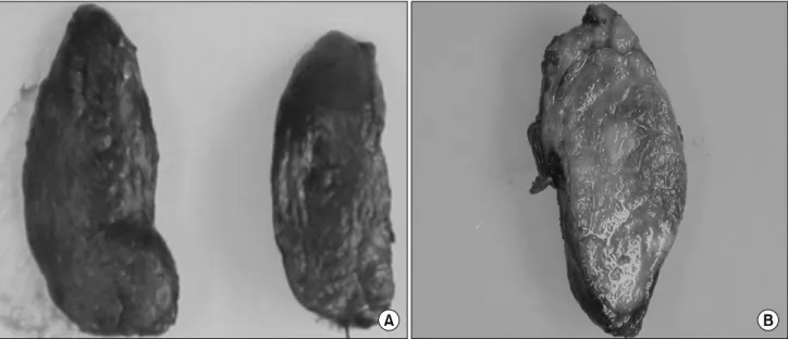
을 포함한 갑상샘 좌엽의 주변조직을 같이 제거하였다. 제 거된 갑상샘 우엽은 5×3×2 cm 크기를 보였고 갑상샘 좌엽 은 3×3.5×2.5 cm 크기였다(Fig. 3A). 수술 중 동결절편을 위 해 보내온 갑상샘 우엽에서는 불명확한 경계를 보이는 0.5 cm 크기의 결절이 관찰되었고, 단면은 하시모토 갑상샘염 을 연상케 하는 약간 단단한 구조물이었다(Fig. 3B). 조직검 사에서 갑상샘 우엽의 전반에 걸쳐 유두상암이 관찰되었고 다수의 모레종체(psammoma body)가 보였으며 그 사이로 섬유화, 소량의 편평상피세포화, 림프관내에 작은 암종 등 이 관찰되었다(Fig. 4). 갑상샘 좌엽은 일부에 흩어져 있는 작은 유두암종들이 관찰되었고, 갑상샘 양엽에서 림프여포 (lymphoid follicle)들과 림프구들이 미만성으로 분포하였다 (Fig. 4). 조직검사에서 갑상샘 우엽에 미만성, 좌엽은 국소 성 침범을 보이는 유두상암의 미만성 경화아형(diffuse scle- rosing subtype)으로 진단되었고, 좌측 부갑상샘을 포함한 양 측 갑상샘의 주변 조직으로의 종양의 침범이 있었다. 우측 경부 림프절(lateral group)은 제거된 33개의 림프절 중 8개 에서 전이의 소견을 보였고, 중심림프절(central group)은 제 거된 7개의 림프절 중 4개의 림프절에서 암전이의 소견을 Fig. 1. Sonographic findings of thyroid DSPC. (A, B) Axial (A) and
longitudinal (B) scans show heterogeneous hypoechoic pa- renchymas and abundant diffuse scattered psammoma bodies as punctate strong echos in thyroid parenchymas. A ill de- fined small hypoechoic and solid nodule is noted (white ar- row). (C) Longitudinal sonogram show multiple nodal meta- stases with eccentric cortical thickenings, loss of central hy- perechogencity and foci of calcification (white arrow).
Fig. 2. Axial CT images of thyroid DSPC. (A) Pre-contrast CT image shows diffuse punctate calcific densities in both thyroid lobes, findings indicative of microcalcifications (white arrow). (B) Axial contrast-enhanced CT image shows heterogeneous enhanced both thyroid parenchymas and multiple enhanced metastatic lymph nodes with ill defined margins in right neck (white arrow).
A poorly enhanced small nodule is noted in right thyroid lobe (arrowhead).
Fig. 3. Gross specimen of both thyroid DSPC. (A) This thyroid shows firm parenchyma. But no obvious mass is found. (B) The cut surface shows ill defined gray and white areas without formation of any masses. The background parenchyma is firm and some- what white appearance.
보였다. 환자는 수술 후 고용량 방사성 요오드 요법 치료 를 받았고, 좌측 경부림프절에 대해서는 초음파 검사 및 미세바늘흡인세포검사를 통한 주의 깊은 추적 검사를 통 해 전이가 발견되면 변형근치경부절제술도 시행 할 예정 이다.
고 찰
갑상샘 유두상암은 여러 아형들로 분류되며 일부 아형들 은 갑상샘 조직 이외로 전이를 하며 수술 시 불완전 절제의 가능성이 높은 매우 공격적인 생물학적 양상을 보인다.(2,4,5) 갑상샘 유두상암의 아형 중 DSPC 아형은 매우 드문 갑상샘
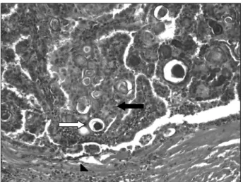
Fig. 4. Histopathologic appearance of the DSPC (H&E stain,
×200). Numerous psammoma bodies (white arrow) with neoplastic cells are noted (black arrow). Fibrous stroma with heavy lymphocytic infiltration is noted (arrowhead).
의 악성종양으로 전체 유두상암의 0.75∼5.3%를 차지하 며,(4,5) 일부에서는 0.02∼0.03% 등의 매우 낮은 빈도로 보 고되고 있다.(3,6) 이 아형은 여자에게 호발 하지만 고전형 유두상암과 비교하여 좀 더 젊은 연령에 나타나며,(3,5-7) 종괴가 더 크고, 림프절 전이를 포함하여 갑상샘 이외의 조 직으로 전이를 더 잘한다.(5,6) 일부 보고에서는 폐와 뇌에 도 전이가 보고되는 등 더욱 공격적인 양상을 보이며, 치료 후 재발이 흔한 것으로 알려져 있다.(5,8,9) 또한 anti-thyro- globulin autoantibody가 양성인 경우가 흔하며 갑상샘의 양 측 혹은 편측성 미만성 종대를 보이기 때문에 하시모토 갑 상샘염과 구별이 어렵고, 조기 발견 및 수술 전 진단이 지연 될 수 있다.(4,7,10,11) DSPC 아형은 고전형 유두상암과 비 교하여 예후가 더 나쁜 것으로 알려져 있지만,(6) 일부에서 는 비슷한 결과를 보였던 경우 등이 있는데,(3,4) 이는 고전 형 유두상암보다 더욱 공격적인 치료의 결과로 생각되며, 대부분은 양측 갑상샘 전절제술, 변형근치경부절제술, 수술 후 131I 방사선 요법 등을 시행한다.(3,5)
유두상암의 아형들 중 tall cell, columnar cell, DSPC 아형 들은 좀 더 공격적 성향을 가지며, 고전형 유두상암과는 다 른 임상적 양상과 예후를 보이므로, 수술 전 진단은 환자의 치료방법 결정 및 예후에 매우 중요하다.(8,12) 갑상샘 종양 의 진단에 있어서 초음파 유도하의 수술 전 미세바늘흡인 세포검사는 경제적인 면과 접근의 용이성, 높은 정확도 등 으로 현재 유두상암을 진단하는데 매우 유용한 방법으로 사용 되고 있다.(13,14) 일반적으로 고전형 유두상암은 세포 학적 형태와 조직학적 소견 간에 높은 연관관계를 보이므 로 미세바늘흡인세포검사가 수술 전 진단방법으로 보편화 되어 있다.(2,13) 그러나 세포학적 검사를 통해 유두상암을 다양한 아형으로 분류 할 수 있는 능력에 대해서는 저자들
마다 이견이 있으며, 검체습득방법이나 검사자 요인, 유두 상암의 아형에 따라 다양한 결과들을 보일 수 있다.(1,8,15) DSPC 아형의 세포와 조직소견간의 연관관계에 대한 연구 에서 특정 아형으로 분류 자체가 불가능했던 경우와, DSPC 아형의 조직학적 특징인 섬유화 소견은 미세바늘흡인세포 검사의 펴 바른 표본에서는 발견 할 수 없기 때문에 DSPC 아형을 세포학적으로 진단하기에 어려웠던 경험들이 보고 되어 있다.(2,15,16) DSPC 아형의 조직병리소견은 미만성 침범, 경화, 풍부한 모레종체, 편평상피화생, 광범위한 림프 관 침투 등이며, 특징적 소견인 갑상샘 내에 넓게 분포하는 모레종체는 방사선학적으로도 중요한 진단적 요소이다.
(10,11,17) 과거에는 모레종체에 의한 미세석회화는 연조직 일반 방사선 촬영을 이용하여 관찰하였지만,(3) 현재는 보 편화된 고해상 초음파를 통해 방사선 피폭 없이 쉽게 진단 할 수 있다. 고전형 유두상암에 대한 초음파 소견은 많이 알려져 있지만,(18,19) DSPC 아형의 초음파 소견은 문헌으 로는 드물게 보고되고 있다. DSPC의 초음파 소견으로 편측 혹은 양측 갑상샘에 모레종체를 시사하는 미세석회화들이 미만성으로 산재하며, 내부에 저에코음영 결절들이 동반 될 수 있다. 광범위한 섬유화와 림프구의 침윤으로 미만성 갑상샘 종대와 갑상샘 실질에 비균질적 저에코음영 등이 관찰되며 이러한 초음파 소견 때문에 만성 갑상샘염과 혼 동하기 쉽다.(9-11,16)
유두상암의 수술 전 진단에 있어서 일반적인 형태의 고 전형 유두상암을 공격적 임상 양상을 보이는 다른 아형들 과 구별하는 것은 수술방법 및 향후 치료계획을 세우는데 있어서 매우 중요하다.(1) 저자들의 경우에서와 마찬가지로 세포학적 분석을 통한 유두상암의 진단은 가능하나 이를 다양한 아형들로 분류하는 것은 어려운 점이 있다. DSPC 아형은 초음파나 CT 검사에서 종괴의 형태가 잘 그려지지 않는 미만성 미세석회화를 보이며 주변에 전이성 림프절을 보이는 등 비교적 특징적인 방사선학적 소견을 보이므로, 미세바늘흡인세포검사를 통한 세포학적 분석과 영상소견 을 함께 고려하면 수술 전 진단이 용이 할 수 있다. 만약 DSPC 아형이 의심될 경우 다른 장기로의 전이 여부 등 좀 더 적극적이고 정확한 수술 전 검사가 필요하며, 이를 통해 적절한 치료계획을 수립하는 것이 매우 중요할 것으로 생 각된다.
REFERENCES
1) Furlan JC, Bedard YC, Rosen IB. Role of fine-needle aspiration biopsy and frozen section in the management of papillary thyroid carcinoma subtypes. World J Surg 2004;28:
880-5.
2) Gupta S, Sodhani P, Jain S, Kumar N. Morphologic spectrum of papillary carcinoma of the thyroid; role of the cytology in
identifying the variants. Acta cytol 2004;48:795-800.
3) Fujimoto Y, Obara T, Ito Y, Kodama T, Aiba M, Yamaguchi K. Diffuse sclerosing variant of papillary carcinoma of the thyroid-clinical importance, surgical treatment, and follow-up study. Cancer 1990;66:2306-12.
4) Chow SM, Chan JKC, Law SCK, Tang DLC, Ho CM, Cheung WY, et al. Diffuse sclerosing variant of papillary thyroid carcinoma-clinical features and outcome. Eur J Sur Oncol 2003;29:446-9.
5) Lam AKY, Lo CY. Diffuse sclerosing variant of papillary carcinoma of the thyroid:a 35-year comparative study at a single institution. Ann Surg Oncol 2006;13:176-81.
6) Egea AM, Gonzalez JMR, Perez JS, Soria T, Paricio PP.
Clinicopathological study of the diffuse sclerosing variety of papillary cancer of the thyroid. Presentation of 4 new cases and review of the literature. Eur J Sur Oncol 1994;20:7-11.
7) Martin-Perez E, Larranaga E, Serrano P. Diffuse sclerosing variant of papillary carcinoma of the thyroid. Eur J Surg 1998;164:713-5.
8) Ohori NP, Schoedel KE. Cytopathology of high-grade papillary thyroid carcinomas: tall-cell variants diffuse sclero- sing variant, and poorly differentiated papillary carcinoma.
Diagn Cytopathol 1999;20:19-23
9) Kobayashi Kaoru, Fukata S, Amino N, Miyauchi A. A case with diffuse sclerosing variant of papillary carcinoma of the thyroid: characteristic features on ultrasonography. J Med Ultrasonics 2006;33:159-61.
10) Kebapci N, Efe B, Kabukcuoglu S, Akalin A, Kebapci M.
Diffuse sclerosing variant of papillary thyroid carcinoma with primary squamous cell carcinoma. J Endocrinol Invest 2002;
25:730-4.
11) Lee JY, Shin JH, Han BK, Ko EY, Kang SS, Kim JY, et al.
Diffuse sclerosing variant of papillary carcinoma of the
thyroid: imaging and cytologic findings. Thyroid 2007;17:
567-73.
12) Piotti S, Collini P, Manzari A, Marubini E, Rike F. Poorly differentiated forms of papillary thyroid carcinoma: distinctive entities or morphological patterns? Semin Diagn Pathol 1995;
12:249-55.
13) Akhtar M, Ali MA, Huq M, Bakry M. Fine needle aspiration biopsy of papillary thyroid carcinoma:cytologic, histologic, and ultrastructural correlations. Diagn Cytopathol 1991;7:
373-9.
14) Leenhardt L, Heiblum G, Franc B, Fediaevsky LDP, Delbot T, Guillouzic DL, et al. Indications and limits of ultrasound- guided cytology in the management of nonpapable thyroid nodules. J Clin Endocrinol Metab 1999;84:24-8.
15) Nair M, Kapila K, Karak AK, Verma K. Papillary carcinoma of the thyroid and its variants: a cytohistological correlation.
Diagn Cytopathol 2001;24:167-73.
16) Kwak JY, Kim EK, Hong SW, Oh KK, Kim MJ, Park CS, et al. Diffuse sclerosing variant of papillary carcinoma of the thyroid:ultrasound features with histopathological correlation.
Clin Radiol 2007;62:382-6.
17) Kumarasinghe MP. Cytomorphologic features of diffuse scle- rosing variant of papillary carcinoma of the thyroid; a report of two cases in children. Acta cytol 1998;42:983-6.
18) Kim EK, Park CS, Chung WY, Oh KK, Kim DI, Lee JT, et al. New sonographic criteria for recommending fine-needle aspiration biopsy of nonpalpable solid nodules of the thyroid.
AJR Am J Roentgenol 2002;178:687-91.
19) Chan BK, Desser TS, Ross McDougall I, Weigel RJ, Brook Jeffrey R. Common and uncommon sonographic features of papillary thyroid carcinoma. J Ultrasound Med 2003;22:
1083-90.