Genotype and Phenotype Analysis in Pediatric Patients with Cystinuria
Cystinuria is an inherited disorder characterized by defective renal reabsorption of cystine and dibasic amino acids leading to nephrolithiasis. This study was conducted to analyze the genotypes and phenotypes of pediatric patients with cystinuria. Eight children from Seoul National University Hospital and Asan Medical Center presenting with cystinuria from January 2003 to June 2016 were retrospectively analyzed. Mutational studies were performed by direct sequencing. Two of the 8 were male and 6 were female. The median ages at onset and diagnosis were 1.5 (range, 0.3–13.6) and 2.6 (range, 0.7–16.7) years, respectively. The median followed up was 7.7 (range, 3.4–14.0) years. Mutational analyses were performed in 7 patients and revealed biallelic SLC3A1 mutations (AA genotype) in 4 patients, a single heterozygous SLC3A1 mutation (A- genotype) in 1 patient, biallelic SLC7A9 mutations (BB genotype) in 1 patient, and a single heterozygous SLC7A9 mutation (B- genotype) in 1 patient. Two of the mutations were novel. No genotype-phenotype correlations were observed, except for earlier onset age in patients with non-AA genotypes than in patients with the AA genotype. All patients suffered from recurrent attacks of symptomatic nephrolithiasis, which lead to urologic interventions. At the last follow-up, 3 patients had a mild-to-moderate degree of renal dysfunction. This is the first study of genotypic and phenotypic analyses of patients with cystinuria in Korea.
Keywords: Cystinuria; Genotype; Phenotype; Gene mutation; SLC3A1 Gene; SLC7A9 Gene
Ji Hyun Kim,1 Eujin Park,1 Hye Sun Hyun,1 Beom Hee Lee,2 Gu-Hwan Kim,3 Joo Hoon Lee,2 Young Seo Park,2 Hee Gyung Kang,1,4 Il-Soo Ha,1,5 and Hae Il Cheong1,4,5
1Department of Pediatrics, Seoul National University Children’s Hospital, Seoul, Korea; 2Department of Pediatrics, University of Ulsan College of Medicine, Asan Medical Center Children’s Hospital, Seoul, Korea; 3Medical Genetics Center, University of Ulsan College of Medicine, Asan Medical Center Children’s Hospital, Seoul, Korea; 4Research Coordination Center for Rare Diseases, Seoul National University Hospital, Seoul, Korea; 5Kidney Research Institute, Medical Research Center, Seoul National University College of Medicine, Seoul, Korea
Received: 29 August 2016 Accepted: 16 October 2016 Address for Correspondence:
Hae Il Cheong, MD
Department of Pediatrics, Seoul National University Children’s Hospital, 101 Daehak-ro, Jongno-gu, Seoul 03080, Korea E-mail: cheonghi@snu.ac.kr
Funding: This study was supported by a grant (HI12C0014) from the Korean Health Technology R&D Project, Ministry of Health and Welfare, Republic of Korea.
https://doi.org/10.3346/jkms.2017.32.2.310 • J Korean Med Sci 2017; 32: 310-314
INTRODUCTION
Cystinuria (OMIM #220100) is an autosomal recessive genetic disorder. Cystinuria leads to defects in transepithelial transport- ers for dibasic amino acids, including cystine, ornithine, lysine, and arginine. Although the urine concentration of all dibasic amino acids is elevated in cystinuric patients, only cystine leads to stone disease as it is relatively insoluble at physiological pH (1-4). More than 50% of patients with cystinuria suffer from stone formation throughout their lifetime, with variable onset ages as well as a high rate of recurrence of up to 60% (5). Furthermore, since stones are likely to be formed bilaterally in more than three- quarters of cystinuric patients (4), they are at high risk of renal dysfunction and consequent poor quality of life (6).
Cystinuria types I, II, and III are traditional subgroups divid- ed according to the urinary phenotype in heterozygotes (7). While type I heterozygotes show a normal amino aciduria, non-type I (type II and III) heterozygotes show high and moderate hyper- excretion of cystine and dibasic amino acids, respectively. Pa- tients with type III, a mixed type, inherit type I and non-type I
alleles from either parent. This classification system is still in clin- ical use. However, the International Cystinuria Consortium de- veloped a new classification system to reflect the genetic and functional characteristics of the disease after the discovery of 2 genes responsible for cystinuria. Type A cystinuria is due to bi- allelic SLC3A1 mutations, type B cystinuria is due to biallelic SLC7A9 mutations, and type AB cystinuria is due to single het- erozygous mutations in both genes (8). SLC3A1 encodes a heavy subunit of the renal cystine transport system, rBAT, and was iden- tified as the cause of type I cystinuria. SLC7A9 encodes b0,+ ami- no acid transporter (b0,+AT), a light subunit of the renal cystine transport system and responsible for type II cystinuria. To date, 163 disease-causing mutations in SLC3A1 and 118 disease-caus- ing mutations in SLC7A9 have been listed in The Human Gene Mutation Database (HGMD® Professional 2016.2, https://por- tal.biobase-international.com/hgmd/pro/start.php).
There have been several clinical case reports and studies of Korean patients with cystinuria (6,9-14). However, only 2 case reports published include genetic studies: one was that of a 13- year-old boy with a single heterozygous SLC7A9 mutation (c.517G Pediatrics
>A, p.G173R) (11), and the other was of an 8-month-old girl with a homozygous SLC3A1 mutation (c.1820delT, p.L607fs) (10). Here, we report a genotype-phenotype study of 8 Korean pediatric pa- tients with cystinuria.
MATERIALS AND METHODS Patients
Eight patients diagnosed with cystine stones during the period between January 2003 and June 2016 in 2 hospitals (Seoul Na- tional University Children’s Hospital, Seoul, Korea and Asan Medical Center Children’s Hospital, Seoul, Korea) were recruit- ed. Their clinical presentation, clinical courses and serial labo- ratory findings were evaluated retrospectively.
Mutational studies
Genomic DNA was extracted from nucleated cells in peripheral blood using a commercial kit (QIAamp DNA Blood Mini Kit;
Qiagen, Hilden, Germany). All coding exons and flanking in- trons of the SLC3A1 and the SLC7A9 genes were amplified us- ing polymerase chain reaction followed by direct sequencing (primer sequences are available upon request).
Ethics statement
This study was approved by the Institutional Review Board at Seoul National University Hospital (IRB No. 0812-002-264). In- formed consent was obtained from all individual patients in- cluded in this study or their parents.
RESULTS
Two of the 8 patients were male and six were female. The medi- an ages at onset and diagnosis were 1.5 years (range, 0.3–13.6 years) and 2.6 years (range, 0.7–16.7 years), respectively. They
were followed up for a median period of 7.7 years (range, 3.4–
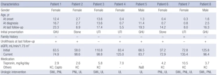
14.0 years). The clinical features of the patients are summarized in Table 1. The presenting symptoms were gross hematuria (n = 3) or urinary tract infection (n = 3) in association with renal stones, while renal stones were detected incidentally in two patients.
Family history of nephrolithiasis was absent in all patients, ex- cept for one (Patient 7), whose maternal uncle had a history of nephrolithiasis of unidentified composition. The initial renal func- tion was normal (estimated glomerular filtration rate [eGRF cal- culated using the Schwartz formula] ≥ 90 mL/min/1.73 m2) in 2 patients, and the remaining 6 patients had various degrees of renal dysfunction. Seven patients were treated with tiopronin (thiola®) with or without potassium citrate (n = 6) and captopril (n = 1). The remaining patient (Patient 5) had a single heterozy- gous SLC3A1 mutation and was treated with oral sodium bicar- bonate only. However, all patients suffered from recurrent at- tacks of symptomatic nephrolithiasis, which lead to urologic in-
Table 1. Clinical features of 8 patients with cystinuria
Characteristics Patient 1 Patient 2 Patient 3 Patient 4 Patient 5 Patient 6 Patient 7 Patient 8
Gender Female Female Female Female Male Female Female Male
Age, yr At onset At diagnosis At last follow-up
12.4 16.7 23.3
2.7 2.7 6.9
13.6 13.6 17.4
0.4 0.7 5.5
1.3 11.4 19.3
0.4 0.7 14.2
0.3 0.8 8.0
1.6 2.5 16.3
Initial presentation GHU Stone UTI UTI GHU Stone UTI GHU
Family history − − − − − − + −
Urolithiasis at last follow-up + + − + + + − +
eGFR, mL/min/1.73 m2 Initial
Current 63.5
74.9 58.0
98.6 110.8
98.8 83.4
125.0 66.5
83.7 37.2
72.9 72.8
135.4 125.8
96.4 Medication
Tiopronin, mg/kg/day
Others 2.9
KC, Capto 2.6
KC 5.8
KC 7.0
- -
NaB 4.2
KC 10.5
KC 3.7
KC
Urologic intervention SWL, PNL PNL, UL SWL, UL UL UL PNL, UL SWL, PNL, UL SWL, PNL
GHU = gross hematuria, eGFR = estimated glomerular filtration rate (calculated using the Schwartz formula), KC = potassium citrate, Capto = captopril, NaB = sodium bicar- bonate, SWL = shock wave lithotripsy, PNL = percutaneous nephrolithotomy, UL = ureteroscopic lithotripsy.
Table 2. Urinary excretion levels (μM/g of creatinine) of branched amino acids of the patients
Patients Cystine Ornithine Lysine Arginine NP test
Patient 1 7,036 2,332 11,813 7,740 n.d.
Patient 2 14,875 2,760 21,533 4,058 n.d.
Patient 3 2,778 1,436 9,884 1,101 n.d.
Patient 4 5,358 7,849 14,910 9,746 n.d.
Patient 5 276 1,074 8,532 3,342 n.d.
Patient 6 Mother Father
2,893 156 0
1,304 0 0
3,028 162 196
2,687 175 0
Positive Positive Negative
Patient 7 8,230 9,367 41,367 20,151 n.d.
Patient 8 Mother Father Sister
2,881 60 58 201
369 0 0 0
1,360 0 284 0
1,051 0 24 0
Positive Negative Negative Negative Reference values: Cystine 27–150 μM/g creatinine, Ornithine 0–44 μM/g creatinine, Lysine 62–513 μM/g creatinine, Arginine 0–44 μM/g creatinine.
NP test = cyanide nitroprusside test, n.d. = not done.
terventions. At the last follow-up, 3 patients had a mild-to-mod- erate degree of renal dysfunction.
Urinary excretion of cystine and dibasic amino acids were markedly increased in all patients, except for Patient 5 who ex- hibited only a mildly increased level of cystine. The cyanide ni- troprusside test was performed and positive in 2 patients (Table 2). Mutational analyses revealed biallelic SLC3A1 mutations (AA genotype) in 4 patients, a single heterozygous SLC3A1 mu- tation (A- genotype) in one patient, biallelic SLC7A9 mutations (BB genotype) in one patient, and a single heterozygous SLC7A9 mutation (B- genotype) in one patient. In one remaining patient, mutational analysis was not performed due to unavailable sam- ples (Table 3). The c.1820delT SLC3A1 mutation was detected in 3 of 5 patients with an AA genotype (4 of 10 alleles).
Urinary cystine excretion levels did not correlate with any clinical parameters or genotypes of the patients. We compared the clinical and laboratory findings of the 4 patients with the AA genotype to those of the others. The only difference observed was an older onset age in patients with the AA genotype. Uri- nary excretion levels of cystine did not correlate with any clini- cal parameters in the 4 patients with the AA genotype.
DISCUSSION
We report 8 cases of cystinuria, presenting with various symp- toms. Mutational analyses revealed the AA genotype in 4 pati- ents. Patients with the AA genotype had older onset ages than those with non-AA genotypes.
Several previous studies examining genotype-phenotype cor- relations in cystinuria did not show any correlation between pa- tients with type A genotype and patients with non-A genotypes (8,15,16). In a UK study (15), patients with at least one missense mutation in SLC3A1 had significantly lower levels of lysine, ar- ginine, and ornithine, but not cystine, than patients with all oth- er types of SLC3A1 mutations. Another UK study (16) revealed no difference between patients with type AA and patients with type BB in a variety of clinical parameters. In addition, patients with a single mutated allele also had variable disease severity and could not be differentiated from patients with 2 mutated alleles (16). In our study, the only difference between patients
with the AA genotype and those with non-AA genotypes was the later age of onset in the former group. Of the 4 patients in our study with the AA genotype, Patient 4 had 2 truncating mu- tations and the youngest onset age. However, urinary excretion levels of cystine were highest in Patient 2, who had 2 missense mutations. Other clinical parameters, including long-term prog- nosis, showed no correlation with the different genotypes.
The SLC3A1 p.T216M mutation was detected in 2 of our pa- tients. This mutation is common in South-eastern European (17, 18), Gypsy (19), and Greek populations (20) but has not been found in Chinese (21) and Japanese (22) patients. Conversely, the SLC7A9 p.P482L mutation is common in Japanese patients (22), but has not been found in European populations. In Pa- tient 5, we found a nucleotide variation, c.1976A>C (p.Q659P), in SLC3A1. Using Mutation Taster (http://www.mutationtaster.
org/), this variation was predicted to be a polymorphism, but was not found in ExAC (http://exac.broadinstitute.org/) or 1000G (http://www.1000genomes.org/). Mild but abnormal hyperex- cretion of cystine and early onset nephrolithiasis in Patient 5 suggested that the p.Q659P variation may be a hypomorphic mutation. Patient 6 had a BB genotype. However, a study of the patient’s family revealed type I cystinuria. A single heterozygous SLC7A9 mutation was detected in Patient 7. However, urinary excretion levels of branched amino acids in the patient were very high. Furthermore, her mother was heterozygous for the same mutation but was clinically silent, although urinary ami- no acid excretion levels were not measured in the mother. There- fore, it is quite possible that Patient 7 had another pathogenic mutation, not detected by Sanger sequencing, in SLC3A1 (AB genotype) or SLC7A9 (BB genotype). The genotype of Patient 8 was unavailable, but family study revealed type I cystinuria.
Progression to chronic kidney disease in patients with cystin- uria has been reported to be between 5 and 17% (23-25), and the prevalence of end stage renal disease has been reported to be up to 5% (26). In a recent UK study (16), the level of renal im- pairment observed was similar across all genotypes. Recently, a recent large retrospective study conducted by the French Cys- tinuria Group (27) showed that 5 (1.1%) of 442 patients with cys- tinuria progressed to end stage renal disease at a median age of 35.0 (11.8–70.7) years. Multivariate analyses revealed that pro- Table 3. Mutations detected in the patients
Patients Genes Mutation 1 Mutation 2 Genotypes
Patient 1 SLC3A1 c.647C>T, p.T216M c.1820delT, p.L607Hfs*4 AA
Patient 2 SLC3A1 c.647C>T, p.T216M c.2017T>C, p.C673R AA
Patient 3 SLC3A1 c.46A>T†, p.K16* c.1500+1G>A†, abnormal splicing AA
Patient 4 SLC3A1 c.1820delT, p.L607Hfs*4 c.1820delT, p.L607Hfs*4 AA
Patient 5 SLC3A1 c.1820delT, p.L607Hfs*4 c.1976A>C‡, p.Q659P A- (or AA)
Patient 6 SLC7A9 c.1445C>T, p.P482L c.1224+4166_1399+119del4972§ BB
Patient 7 SLC7A9 c.1224+4166_1399+119del4972§ ? B- (?)
Patient 8 Not done - - Unknown
†Novel mutations; ‡A nucleotide variation of uncertain pathogenicity; §This large deletion causes a total deletion of exon 12.
gression to chronic kidney disease was associated with age, hy- pertension, severe damage of renal parenchymal defined as a history of partial or total nephrectomy, and so on (27). In our study, 3 of 8 patients had a mild degree of renal dysfunction at the last follow-up.
In conclusion, we have analyzed the genotypes and pheno- types of cystinuria in 8 pediatric patients and identified two nov- el mutations. We did not observe an association between clini- cal course and genotype, except for earlier onset age in patients with non-AA genotypes. A big limitation of this study was the small number of subjects. Therefore, a large nationwide multi- center study is recommended.
DISCLOSURE
The authors have no potential conflicts of interest to disclose.
AUTHOR CONTRIBUTION
Conceptualization: Cheong HI. Data curation: Kim JH, Park E, Hyun HS, Lee BH, Kim GH, Park YS. Investigation: Kim JH, Park E, Hyun HS, Lee BH, Kim GH, Lee JH, Park YS, Kang HG, Ha IS, Cheong HI. Writing - original draft: Kim JH, Park E, Cheong HI.
Writing - review & editing: Kim JH, Park E, Kang HG, Ha IS, Cheong HI.
ORCID
Ji Hyun Kim http://orcid.org/0000-0002-3051-1691 Eujin Park http://orcid.org/0000-0002-4413-468X Hye Sun Hyun http://orcid.org/0000-0001-8525-1471 Beom Hee Lee http://orcid.org/0000-0001-9709-2631 Gu-Hwan Kim http://orcid.org/0000-0002-7056-7446 Joo Hoon Lee http://orcid.org/0000-0001-8010-3605 Young Seo Park http://orcid.org/0000-0001-7653-2036 Hee Gyung Kang http://orcid.org/0000-0001-8323-5320 Il-Soo Ha http://orcid.org/0000-0001-5428-6209 Hae Il Cheong http://orcid.org/0000-0001-7556-1265
REFERENCES
1. Eggermann T, Venghaus A, Zerres K. Cystinuria: an inborn cause of uroli- thiasis. Orphanet J Rare Dis 2012; 7: 19.
2. Palacín M, Borsani G, Sebastio G. The molecular bases of cystinuria and lysinuric protein intolerance. Curr Opin Genet Dev 2001; 11: 328-35.
3. Moe OW. Kidney stones: pathophysiology and medical management. Lan- cet 2006; 367: 333-44.
4. Knoll T, Zöllner A, Wendt-Nordahl G, Michel MS, Alken P. Cystinuria in childhood and adolescence: recommendations for diagnosis, treatment, and follow-up. Pediatr Nephrol 2005; 20: 19-24.
5. Claes DJ, Jackson E. Cystinuria: mechanisms and management. Pediatr Nephrol 2012; 27: 2031-8.
6. Shim M, Park HK. Multimodal treatments of cystine stones: an observa- tional, retrospective single-center analysis of 14 cases. Korean J Urol 2014;
55: 515-9.
7. Rosenberg LE, Downing S, Durant JL, Segal S. Cystinuria: biochemical evi- dence for three genetically distinct diseases. J Clin Invest 1966; 45: 365-71.
8. Dello Strologo L, Pras E, Pontesilli C, Beccia E, Ricci-Barbini V, de Sanctis L, Ponzone A, Gallucci M, Bisceglia L, Zelante L, et al. Comparison be- tween SLC3A1 and SLC7A9 cystinuria patients and carriers: a need for a new classification. J Am Soc Nephrol 2002; 13: 2547-53.
9. Choi JE, Yun BY, Park HW, Park JH, Ha IS, Jeong HI, Choi Y, Choi H, Kim IW. Cystinuria 3 cases. J Korean Pediatr Soc 1995; 38: 245-51.
10. Kang EG, Lee JH, Lee BH, Kim GH, Park YS. A case of cystinuria with mul- tiple renal stones in an 8-month-old girl. J Korean Soc Pediatr Nephrol 2013; 17: 122-6.
11. Lee EH, Kim YH, Hwang JS, Kim SH. Non-type I cystinuria associated with mental retardation and ataxia in a Korean boy with a new missence mu- tation (G173R) in the SLC7A9 gene. J Korean Med Sci 2010; 25: 172-5.
12. Lee ST, Cho H. Metabolic features and renal outcomes of urolithiasis in children. Ren Fail 2016; 38: 927-32.
13. Cho CH, Hahm KS, Park JK, Kim KH. A case of cystine stone in a child.
Korean J Urol 1986; 27: 933-8.
14. Hah ST, Yoon JH, Yoon JB. Cystine stone: report of two cases. Korean J Urol 1981; 22: 451-5.
15. Wong KA, Mein R, Wass M, Flinter F, Pardy C, Bultitude M, Thomas K. The genetic diversity of cystinuria in a UK population of patients. BJU Int 2015;
116: 109-16.
16. Rhodes HL, Yarram-Smith L, Rice SJ, Tabaksert A, Edwards N, Hartley A, Woodward MN, Smithson SL, Tomson C, Welsh GI, et al. Clinical and ge- netic analysis of patients with cystinuria in the United Kingdom. Clin J Am Soc Nephrol 2015; 10: 1235-45.
17. Saadi I, Chen XZ, Hediger M, Ong P, Pereira P, Goodyer P, Rozen R. Mo- lecular genetics of cystinuria: mutation analysis of SLC3A1 and evidence for another gene in type I (silent) phenotype. Kidney Int 1998; 54: 48-55.
18. Schmidt C, Vester U, Hesse A, Lahme S, Lang F, Zerres K, Eggermann T;
Arbeitsgemeinschaft Pädiatrische Nephrologie. The population-specific distribution and frequencies of genomic variants in the SLC3A1 and SL- C7A9 genes and their application in molecular genetic testing of cystin- uria. Urol Res 2004; 32: 75-8.
19. Popovska-Jankovic K, Tasic V, Bogdanovic R, Miljkovic P, Golubovic E, Soy- lu A, Saraga M, Pavicevic S, Baskin E, Akil I, et al. Molecular characteriza- tion of cystinuria in south-eastern European countries. Urolithiasis 2013;
41: 21-30.
20. Albers A, Lahme S, Wagner C, Kaiser P, Zerres K, Capasso G, Pica A, Pala- cin M, Lang F, Bichler KH, et al. Mutations in the SLC3A1 gene in cystin- uric patients: frequencies and identification of a novel mutation. Genet Test 1999; 3: 227-31.
21. Yuen YP, Lam CW, Lai CK, Tong SF, Li PS, Tam S, Kwan EY, Chan SY, Tsang WK, Chan KY, et al. Heterogeneous mutations in the SLC3A1 and SLC7A9 genes in Chinese patients with cystinuria. Kidney Int 2006; 69: 123-8.
22. Sakamoto S, Cheong HI, Naya Y, Shigeta Y, Fujimura M, Ueda T, Mikami K, Akakura K, Masai M, Ichikawa T. Genomic characteristics of Asian cys- tinuria patients. Eur Urol Suppl 2014; 13: e806.
23. Assimos DG, Leslie SW, Ng C, Streem SB, Hart LJ. The impact of cystinuria on renal function. J Urol 2002; 168: 27-30.
24. Worcester EM, Parks JH, Evan AP, Coe FL. Renal function in patients with
nephrolithiasis. J Urol 2006; 176: 600-3.
25. Worcester EM, Coe FL, Evan AP, Parks JH. Reduced renal function and benefits of treatment in cystinuria vs other forms of nephrolithiasis. BJU Int 2006; 97: 1285-90.
26. Gambaro G, Favaro S, D’Angelo A. Risk for renal failure in nephrolithiasis.
Am J Kidney Dis 2001; 37: 233-43.
27. Prot-Bertoye C, Lebbah S, Daudon M, Tostivint I, Bataille P, Bridoux F, Bri- gnon P, Choquenet C, Cochat P, Combe C, et al. CKD and its risk factors among patients with cystinuria. Clin J Am Soc Nephrol 2015; 10: 842-51.