Copyright © 2017 Korean Neurotraumatology Society 167
Introduction
Posterior reversible encephalopathy syndrome (PRES) is a clinical and radiological syndrome, first described by Hinchey et al.5) in 1996. Most common clinical manifesta- tions are headache, seizures, altered consciousness, tran- sient motor deficits and loss of vision. The main finding in neuroimaging is posterior white matter edema, which is pre- dominating in the occipital and parietal lobes and posteri- or fossa structures and which are potentially reversible, if prompt diagnosis and treatment would be performed.1) The disease has been more commonly described in adult population.11) The main causes of this condition include ec- lampsia, organ transplantation. Hypertensive crisis, immu- nosuppression, chemotherapeutic agents for lymphoma and
leukemia, severe hypercalcemia, thrombocytopenic syn- dromes, Henoch-Schönlein purpura, vasculitis, and renal failure.5,13) Although there have been reports of PRES in chil- dren following chemotherapy and tumor lysis syndrome, the prevalence of PRES among children is not well estab- lished. We would like to report PRES occurred after head trauma surgery in pediatric patient without any underly- ing disease.
Case Report
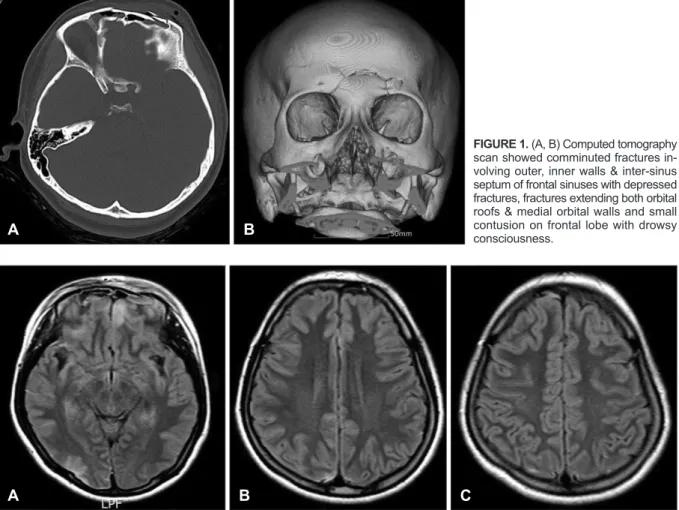
A 16-year-old girl without any medical history was re- ferred to emergency room for drowsy consciousness due to motorcycle accident. Computed tomography (CT) scan showed comminuted fractures involving outer walls, inner walls and inter-sinus septum of frontal sinuses with depressed fractures, extending both orbital roofs and medial orbital walls with small contusion on frontal lobe (Figure 1A and B). Ini- tial T2-weighted (T2W)/T2 fluid-attenuated inversion re- covery (FLAIR) image presented no other significant lesion except small contusion in posterior temporal area (Figure 2A-C). There was no scalp laceration and cerebrospinal fluid (CSF) leakage. Medical treatment including antibiot- ics was performed for 14 days. During hospitalization, neu- rologic state was improved and vital sign including blood
Posterior Reversible Encephalopathy Syndrome after Head Trauma Surgery in Pediatric Patient without Any Underlying Disease
Jae Eon Yoon, Cheol Young Lee, and Hyun Woo Kim
Department of Neurosurgery, Konyang University Hospital, Konyang University Collge of Medicine, Daejeon, Korea
Posterior reversible encephalopathy syndrome (PRES) is a neurological disorder characterized by signs of posterior cere- bral edema upon radiographic examination. A 16-year-old girl was involved in motorcycle accident and depressed frontal fracture was presented. She had generalized seizures 3 days after dural repair and fracture reduction. Signal changes was noted on both parietal lobes in the magnetic resonance images and it was completely resolved in 3 months follow-up. We would like to present the case that demonstrated PRES related hypertension after head trauma surgery for cerebrospinal fluid leakage in pediatric patient without any underlying disease.
(Korean J Neurotrauma 2017;13(2):167-170) KEY WORDS: Pediatric patient ㆍReversible posterior leukoencephalopathy syndrome ㆍSurgery.
Received: June 15, 2017 / Revised: September 11, 2017 Accepted: September 14, 2017
Address for correspondence: Cheol Young Lee
Department of Neurosurgery, Konyang University Hospital, Konyang University Collge of Medicine, 158 Gwanjeodong-ro, Seo-gu, Daejeon 35365, Korea
Tel: +82-42-600-9130, Fax: +82-42-600-6657 E-mail: [email protected]
cc This is an Open Access article distributed under the terms of Cre- ative Attributions Non-Commercial License (http://creativecommons.
org/licenses/by-nc/4.0/) which permits unrestricted noncommercial use, distribution, and reproduction in any medium, provided the original work is properly cited.
CASE REPORT
Korean J Neurotrauma 2017;13(2):167-170
pISSN 2234-8999 / eISSN 2288-2243 https://doi.org/10.13004/kjnt.2017.13.2.167
168 Korean J Neurotrauma 2017;13(2):167-170 Posterior Reversible Encephalopathy Syndrome
pressure (BP) was normal range. CSF leakage and menin- gitis sign were not observed. The patient was discharged without neurologic symptom and complication. After 14 days on discharge, the patient visited emergency room with rhi- norrhea. Despite of bed rest and lumbar drainage for three
days, CSF leakage was continued. Surgical treatment was planned. Dura repair, frontal sinus sealing, bony reconstruc- tion and galeal reposition on skull base were performed.
After surgery, CSF leakage was not observed and there were no surgical related complications. Three days after surgery,
FIGURE 1. (A, B) Computed tomography scan showed comminuted fractures in- volving outer, inner walls & inter-sinus septum of frontal sinuses with depressed fractures, fractures extending both orbital roofs & medial orbital walls and small contusion on frontal lobe with drowsy consciousness.
A B
FIGURE 2. Initial brain magnetic resonance imaging after head injury. (A-C) Initial T2-weighted/T2 fluid-attenuated inversion recov- ery image presented no other significant lesion except small contusion in posterior temporal area.
A B C
FIGURE 3. Brain magnetic resonance imaging (MRI) 3 days after the surgery with generalized seizures. (A-C) MRI showed high signal in T2-weighted/T2 fluid-attenuated inversion recovery image, suggesting cerebral edema, involving the subcortical white matter of bilateral frontal, temporal, parietal, and occipital lobes, with few areas of cortical involvement.
A B C
A B C
Jae Eon Yoon, et al.
http://www.kjnt.org 169 the patient presented several generalized tonic-clonic (GTC)
type seizure. The vital sign and blood examination showed hypertension and mild elevated creatinine (systolic BP [SBP], 140-160 mmHg; creatinine, 2.3 mg/dL). Followed by a retrospective review, BP was shown gradually increas- ing tendency after surgery. Magnetic resonance imaging (MRI) showed high signal in T2W/T2 FLAIR image, sug- gesting cerebral edema involving the subcortical white mat- ter of bilateral frontal, temporal, parietal, and occipital lobes, with few areas of cortical involvement (Figure 3C). Under pro- visional diagnosis of PRES based on radiologic findings and clinical symptoms, we started antiepileptic and antihy- pertensive agents. The patient improved clinically and dis- charged with no neurologic deficit. After 3 months on fol- low-up, MRI showed that previous edematous lesions were completely disappeared (Figure 4A-C). Antiepileptic and antihypertensive agents were stopped and BP has been well maintained in normal range.
Discussion
The most common clinical manifestations of PRES are headache, nausea and vomiting, altered mental status, de- creased alertness, seizures, cortical blindness, and transient motor deficits. In the patients with PRES, seizures are com- mon at the onset of neurologic symptoms but can also devel- op later. The seizures are usually GTC type and/or multiple.
Temporary restlessness and agitation may alternate with lethargy. Stupor and coma may develop. The patients are often confused and there may be some abnormalities of vi- sion such as hemianopia, blurred vision, and cortical blind- ness.12,14) Many predisposing factors have been proposed including hypertensive crisis, immunosuppressive drugs, eclampsia, and renal dysfunction. But, there were some dif-
ferences in the etiological factors between the children and the adults, as in the clinical features. Although significant elevation of the BP may not always be demonstrated, hy- pertension has often been emphasized as a common fea- ture of PRES-associated conditions.1) The pathophysiology underlying PRES is yet thoroughly demonstrated. Two the- ories are considered in the pathophysiology of PRES, the first being sudden increase in BP causing vasospasm and the other being failure of autoregulatory mechanism.13) With sud- den elevation in systolic BP, the autoregulatory capacity of brain vasculature is exceeded which results in a region of vasodilatation and vasoconstriction, especially in the arte- rial boundary zone. This causes breakdown of the blood- brain barrier with subsequent transudation of fluid along with hemorrhage.12) The preferential involvement of the posterior circulation has been postulated as being due to the sympathetic innervation protecting the brain from sudden increase in BP being relatively less in the arterioles supplied by the vertebrobasilar system than in the anterior circula- tion.7) Considering the rare frequency of arteriosclerosis and good plasticity of vessel walls in children, the vulnerabili- ty of vessel walls to hypertension might be decreased in childhood.8) But noteworthy, children sometimes may be regarded to be more vulnerable to cerebrovascular dysfunc- tion than adults under systemic hypertension because they have a narrower range of autoregulation in cerebral blood flow.3) Thus, it is possible that pediatric patients are more susceptible to PRES, and if so, this could be underrecog- nized in the literature.10) Postoperative hypertension is a common complication resulting from surgical intervention and occurs not only in those with pre-existing uncontrolled hypertension, but also in those who were normotensive or well controlled with medication. Given these characteris- tics, in our case, we thought PRES might be occurred due to FIGURE 4. Brain magnetic resonance imaging 3 months after the surgery. The previous edematous lesions were completely disap- peared (A-C).
A B C
170 Korean J Neurotrauma 2017;13(2):167-170 Posterior Reversible Encephalopathy Syndrome
post- operative transient hypertension. Some case reports show that CSF leak and intracranial hypotension can cause PRES.2,4) In this case, there is no evidence of CSF leak in the first stage of PRES, therefore it is reasonable that postop- erative hypertension cause PRES. The symptoms and le- sions of PRES may resolve completely if the diagnosis and treatment is prompt, as was seen in our patient. However, failure to diagnose may lead to irreversible infarction and death.6,9) Recurrence of PRES is rare and may be associated with infections and rapid rise in BP.13) The diagnosis may be overlooked, especially in children, unless a high index of suspicion and precise clinical history is maintained.
Conclusion
The PRES has been more commonly reported in adult population, especially with underlying disease. But because the pediatric patients may be more vulnerable to cerebrovas- cular dysfunction than adults under systemic medical prob- lems such as transient hypertension, if the children had no previous underlying disease, it is important that PRES should be kept as a possibility in children presenting with enceph- alopathy and seizures along with radiologic findings. Under surgery or treatment procedure in pediatric patients, more attention about vital sign and/or general function should be paid in intra and postoperative periods.
■ The authors have no financial conflicts of interest.
REFERENCES
1) Bartynski WS, Boardman JF. Distinct imaging patterns and lesion distribution in posterior reversible encephalopathy syndrome.
AJNR Am J Neuroradiol 28:1320-1327, 2007
2) Feil K, Forbrig R, Thaler FS, Conrad J, Heck S, Dorn F, et al. Re- versible cerebral vasoconstriction syndrome and posterior revers- ible encephalopathy syndrome associated with intracranial hypo- tension. Neurocrit Care 26:103-108, 2017
3) Girişgen I, Tosun A, Sönmez F, Ozsunar Y. Recurrent and atypical posterior reversible encephalopathy syndrome in a child with peri- toneal dialysis. Turk J Pediatr 52:416-419, 2010
4) Hammad T, DeDent A, Algahtani R, Alastal Y, Elmer L, Posteri- or reversible encephalopathy syndrome secondary to CSF leak and intracranial hypotension: a case report and literature review.
Case Rep Neurol Med 2015:538523, 2015
5) Hinchey J, Chaves C, Appignani B, Breen J, Pao L, Wang A, et al.
A reversible posterior leukoencephalopathy syndrome. N Engl J Med 334:494-500, 1996
6) Jones BV, Egelhoff JC, Patterson RJ. Hypertensive encephalopa- thy in children. AJNR Am J Neuroradiol 18:101-106, 1997 7) Koichihara R, Hamano S, Yamashita S, Tanaka M. Posterior revers-
ible encephalopathy syndrome associated with IVIG in a patient with Guillain-Barre syndrome. Pediatr Neurol 39:123-125, 2008 8) Kwon S, Koo J, Lee S. Clinical spectrum of reversible posterior leu- koencephalopathy syndrome. Pediatr Neurol 24:361-364, 2001 9) McCoy B, King M, Gill D, Twomey E. Childhood posterior revers-
ible encephalopathy syndrome. Eur J Paediatr Neurol 15:91-94, 10) McKinney AM, Short J, Truwit CL, McKinney ZJ, Kozak OS, 2011 SantaCruz KS, et al. Posterior reversible encephalopathy syn- drome: incidence of atypical regions of involvement and imaging findings. AJR Am J Roentgenol 189:904-912, 2007
11) Mirza A. Posterior reversible encephalopathy syndrome: a variant of hypertensive encephalopathy. J Clin Neurosci 13:590-595, 2006 12) Onder AM, Lopez R, Teomete U, Francoeur D, Bhatia R, Knowbi
O, et al. Posterior reversible encephalopathy syndrome in the pedi- atric renal population. Pediatr Nephrol 22:1921-1929, 2007 13) Ozcakar ZB, Ekim M, Fitoz S, Teber S, Hizel S, Acar B, et al. Hy-
pertension induced reversible posterior leukoencephalopathy syn- drome: a report of two cases. Eur J Pediatr 163:728-730, 2004 14) Prasad N, Gulati S, Gupta RK, Sharma K, Gulati K, Sharma RK,
et al. Spectrum of radiological changes in hypertensive children with reversible posterior leucoencephalopathy. Br J Radiol 80:
422-429, 2007