Is the Frozen Shoulder Classification a Reliable Assessment?
Ji-Yong Gwark1, Nitesh Gahlot2, Mincheol Kam1, Hyung Bin Park1,3
1Department of Orthopaedic Surgery, Gyeongsang National University Changwon Hospital, Changwon, Korea, 2Department of Orthopedic Surgery, All India Institute of Medical Science, Jodhpur, Rajasthan, India, 3Department of Orthopaedic Surgery, Gyeongsang National University School of Medicine, Changwon, Korea
Background: Although a common shoulder disease, there are no accepted classification criteria for frozen shoulder (FS). This study therefore aimed to evaluate the accuracy of the conventionally used FS classification system.
Methods: Primary FS patients (n=168) who visited our clinic from January 2010 to July 2015 were included in the study. After confirm- ing restrictions of the glenohumeral joint motion and absence of history of systemic disease, trauma, shoulder surgery, shoulder muscle weakness, or specific x-ray abnormalities, the Zuckerman and Rokito’s classification was employed for diagnosing primary FS. Following clinical diagnosis, each patient underwent a shoulder magnetic resonance imaging (MRI) and blood tests (lipid profile, glucose, hemo- globin A1c, and thyroid function). Based on the results of the blood tests and MRIs, the patients were reclassified, using the criteria pro- posed by Zuckerman and Rokito.
Results: New diagnoses were ascertained including blood test results (16 patients with diabetes, 43 with thyroid abnormalities, and 149 with dyslipidemia), and MRI revealed intra-articular lesions in 81 patients (48.2%). After re-categorization based on the above findings, only 5 patients (3.0%) were classified having primary FS. The remaining 163 patients (97.0%) had either undiagnosed systemic or intrin- sic abnormalities (89 patients), whereas 74 patients had both.
Conclusions: These findings demonstrate that most patients clinically diagnosed with primary FS had undiagnosed systemic abnormali- ties and/or intra-articular pathologies. Therefore, a modification of the Zuckerman and Rokito’s classification system for FS may be re- quired to include the frequent combinations, rather than having a separate representation of systemic abnormalities and intrinsic causes.
(Clin Shoulder Elbow 2018;21(2):82-86)
Key Words: Frozen shoulder; Classification; Blood tests; Magnetic resonance Clinics in Shoulder and Elbow Vol. 21, No. 2, June, 2018
https://doi.org/10.5397/cise.2018.21.2.82
Received December 7, 2017. Revised February 13, 2018. Accepted March 1, 2018.
Correspondence to: Hyung Bin Park
Department of Orthopaedic Surgery, Gyeongsang National University Changwon Hospital, Gyeongsang National University School of Medicine, 11 Samjeongja-ro, Seongsan-gu, Changwon 51472, Korea
Tel: +82-55-214-3744, Fax: +82-55-214-3259, E-mail: [email protected], ORCID: https://orcid.org/0000-0001-9468-6282 IRB approval: Gyeongsang National University Hospital (No. GNUH 2015-05-013).
Financial support: None. Conflict of interests: None.
Introduction
The definition of frozen shoulder (FS) remains largely un- changed since it was first expounded in 1934 by Codman.1) He described FS as a painful disease characterized by slow onset, restricted movements at the shoulder joint, and a grossly normal radiograph, and considered it to be a self-resolving disorder of unknown etiology.1) Codman’s description of FS as a disease of uncertain etiology has been supported by the American Acad- emy of Orthopaedic Surgery,2) which defines primary FS as a condition of uncertain etiology, characterized by restriction of both active and passive glenohumeral ranges of movement in
the absence of any underlying causes but having normal ra- diographs.2,3) However, investigators realized later that many cases of FS have underlying causes; Lundberg4) coined the term secondary FS to refer to such cases. Zuckerman and Rokito3) proposed a modification to the original Lundburg classification based on the etiology, and the secondary FS were sub-classified as intrinsic, extrinsic, and systemic subtypes.
However, debates still exist regarding diagnostic criteria and classification for FS. A uniform and accurate classification is man- datory for any disease to characterize the nature of the disorder and guide the treatment. Furthermore, the prognosis of the disease progression needs to be anticipated, so that consistent
reporting of the treatment and outcome is possible. This allows for accurate comparison of outcomes from different studies.5) A good classification system requires good inter- and intra-observer reliability, and enough validity to correctly describe the etiology.
However, the validity of previously reported classifications for FS have not been completely evaluated.5) The Zuckerman and Rokito’s FS classification system3) is based on clinical examina- tion, history and radiographs used to identify the etiology. Their diagnostic criteria, which have traditionally been used in clinical practices, are unable to fully diagnose the intrinsic or systemic causes, since simple radiographs and medical history are insuf- ficient to accurately diagnose these factors.
We hypothesized that most primary FS, as classified by Zuck- erman and Rokito’s FS criteria, are actually secondary FS, but cannot be identified with the traditional diagnostic methods of clinical examination. Therefore, the current study was under- taken to determine the accuracy of Zuckerman and Rokito’s FS classification using blood tests and shoulder magnetic resonance imaging (MRI).
Methods
This study was approved by the institutional review board of the Gyeongsang National University Hospital (GNUH 2015- 05-013). Medical records of patients diagnosed with primary FS from January 2010 to July 2015 were retrospectively reviewed.
Of the 465 patients reviewed, we excluded 70 patients who did not have the result of shoulder MRI, 62 patients who did not have laboratory results, 53 patients who did not have results of the physical examination, and 112 patients who had previous history of shoulder surgery, trauma, and systemic disease. The remaining 168 patients included in the study underwent blood tests (lipid profile, glucose level, glycosylated hemoglobin A1c [HbA1c], and thyroid function tests) and a shoulder MRI.
The initial diagnosis of primary FS was based on observations of the clinical examination which showed restriction in both the active and passive glenohumeral movements during flexion, ab- duction, and internal rotation (associated with >50% decrease in external rotation with arm at side), and on the basis of normal radiographic findings of the affected shoulders in true anteropos- terior, outlet, and axillary lateral views. Additionally, the diagnosis was based on a medical history of no underlying disease, sys- temic abnormality, shoulder surgery, or shoulder trauma. All the clinical assessments were carried out by the senior author (HBP).
The blood test results were analyzed in accordance with the standard criteria established for diagnosis of the respective dis- eases. Dyslipidemia was defined when any of following criteria of lipid profiles was positive: hypercholesterolemia (choles- terol≥200 mg/dl), hyper-low-density lipoproteinemia (≥100 mg/dl), hyper-triglyceridemia (≥150 mg/dl), hypo-high-density lipoproteinemia (HDL≤40 mg/dl in male, ≤50 mg/dl in female),
and hyper-non-HDLemia (non-HDL≥130 mg/dl).6) Diabetes was diagnosed when plasma levels of HbA1c were >6.4%, fast- ing plasma glucose was >125 mg/dl, or plasma glucose>199 mg/dl after two hours of a 75 g oral glucose load.7) Hyper- and hypo-thyroidism were based on the results of thyroid function tests, in which the serum free T4 levels>1.70 ng/dl indicated hyper-thyroidism, and <0.93 ng/dl indicated hypo-thyroidism.8)
Most patients (142/168, 84.5%) underwent MRIs at our insti- tute, with a 1.5 T scanner (Siemens Medical Systems, Erlangen, Germany); the remaining 26 patients performed MRIs outside our institute. All the MRI images included in this study, whether performed at our institute or elsewhere, were interpreted by a single experienced musculoskeletal radiologist who was blind to the clinical findings. All but 5 patients had their MRI examina- tions within two months of the outpatient visit. After compiling the results of the blood tests and MRI findings, we re-categorized the patients into appropriate sub-classifications, as proposed by Zuckerman and Rokito.3) We used the IBM SPSS Statistics ver.
21 Developer software (IBM Co., Armonk, NY, USA) to perform the frequency analysis of the data and to calculate the distribu- tion of patients in the various sub-classifications.
Results
A total of 168 patients, who were initially diagnosed with primary FS and who met the aforementioned inclusion criteria, were enrolled in this study. These included 66 males (39.3%) and 102 females (60.7%), with an average age of 53.5 ± 8.3 years. The right shoulders of 91 patients (54.2%) were affected, and the left shoulders of 77 patients (45.8%) (Table 1).
Table 1. Summary of Demographic Data (Gender and Systemic Disease after Blood Investigations)
Enrolled subject Percentage Mean age (yr)
Total enrolled subjects (n=168) 53.5 ± 8.3
Male 39.3 (66/168) 54.8 ± 5.9
Diabetes 7.6 (5/66) 55.3 ± 6.2
Abnormal thyroid function 18.2 (12/66) 54.2 ± 5.1
Dyslipidemia 83.3 (55/66) 54.3 ± 8.1
Female 60.7 (102/168) 52.4 ± 6.8
Diabetes 10.8 (11/102) 54.2 ± 7.1
Abnormal thyroid function 30.4 (31/102) 53.8 ± 6.2 Dyslipidemia 92.2 (94/102) 53.0 ± 5.3 Affected side
Right 54.2 (91/168) 54.2 ± 7.2
Left 45.8 (77/168) 53.0 ± 5.3
Values are presented as percent (number/total number) or mean ± standard deviation.
Based on the analyses of the blood tests, the newly diag- nosed afflictions were 16 cases of diabetes (9.5%), 43 cases of thyroid abnormalities (25.6%) (4 hyper-thyroidism and 39 hypo- thyroidism), and 149 cases of dyslipidemia (88.7%). A total of 156 patients (92.9%) were found to have one or more of the above systemic abnormalities, with dyslipidemia being the most common (149/168, 88.7%). Dyslipidemia was present in 68.8%
(11/16) of the diabetic patients, and in 88.4% (38/43) of patients with thyroid dysfunction (Table 2).
MRI examinations revealed 81 patients (48.2%) had intra- articular lesions of the shoulder joint, with the most commonly found lesion being a tear of the supraspinatus tendon present in 53 patients (31.5%). The spectrum of lesions detected on MRI is summarized in Table 3.
After compilation of the results and reclassification according to the Zuckerman and Rokito’s classification,3) only 5 patients (3.0%) were classified as primary FS. The remaining 163 patients (97.0%) had previously undiagnosed systemic and/or intrinsic abnormalities. Of the 163 newly diagnosed secondary FS pa- tients, 74 (45.4%) had both intrinsic lesions and systemic abnor- malities (Table 4).
Discussion
This study was undertaken to determine the accuracy of the Zuckerman and Rokito’s FS classification,3) after confirming blood tests and shoulder MRI outcomes. In accordance with our hypothesis, the results revealed that most of the primary FS were reclassified as secondary FS, having intrinsic lesions and/or sys- temic disease.
Zuckerman and Rokito3) differentiated secondary FS from primary FS using systemic, intrinsic, and extrinsic factors. The secondary FS group was later expanded by Kelley et al.9) and Nash and Hazleman10) to include diabetes, myocardial infarc- tion, and other neurological disorders that were associated with FS. Robinson et al.11) used a different classification system, wherein they divided the primary FS into idiopathic and sys- temic diseases. They separated diabetic FS from secondary FS, due to the former’s prognosis being worse than that of other af- flictions, and the incidence of FS being high in diabetes. Among the traditional definitions and diagnostic methods, Zuckerman and Rokito’s classification3) (an etiology-based classification) is simple and helpful to diagnose FS. However, this system is not universally accepted among shoulder surgeons; when polled to determine their opinions of the classification system, 34%
of the respondents expressed either disapproval or no opinion regarding appropriateness of secondary FS sub-classification. In our patient group, this classification led to a gross over-diagnosis of primary FS, and was not applicable to many patients in our study because of overlapping etiologies of combined systemic Table 2. Cases of Various Causes of Systemic Frozen Shoulder as Assessed after Blood Investigations
Variable Diabetes Abnormal thyroid function Dyslipidemia All three abnormalities
Diabetes 2 3 9
Abnormal thyroid function 2 36
Dyslipidemia 102
All three abnormalities 2
Table 3. Lesions Observed in the MRI Findings of the Affected Shoulders
MRI finding Specific lesions Percentage
Intraarticular lesion 48.2 (81/168)
Supraspinatus lesion Articular side partial tear 13.7 (23/168) Bursal side partial tear 4.8 (8/168) Interstitial partial tear 12.5 (21/168) Full thickness tear 0.6 (1/168) Subscapularis lesion Articular side partial tear 8.3 (14/168)
SLAP lesion 6.0 (10/168)
Biceps tendon lesion 1.8 (3/168)
Subscapularis partial
tear and SLAP lesion 0.6 (1/168)
Negative MRI finding 51.8 (87/168)
Values are presented as percent (number/total number).
MRI: magnetic resonance imaging, SLAP: superior labral tear from anterior to posterior.
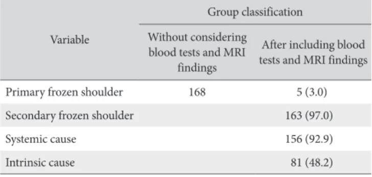
Table 4. Classification of Patients Based on Clinical and Radiographic Evalua
tions, with and without Considering Blood Tests and MRI Findings Variable
Group classification Without considering
blood tests and MRI findings
After including blood tests and MRI findings
Primary frozen shoulder 168 5 (3.0)
Secondary frozen shoulder 163 (97.0)
Systemic cause 156 (92.9)
Intrinsic cause 81 (48.2)
Values are presented as number only or number (%).
MRI: magnetic resonance imaging.
and intrinsic abnormalities. We found that physical examina- tion, simple radiological evaluation, and history evaluation was insufficient to identify 93% of our patients, who were eventually diagnosed to have systemic abnormalities. Therefore, we suggest some modifications to the currents FS classification system to ac- commodate the possibilities of simultaneous lesions and multiple etiologies.
Among the various systemic risk factors known to be associat- ed with FS, diabetes,12,13) hyper-thyroidism,14) hypo-thyroidism,15) and dyslipidemia16,17) have a relatively high prevalence, and can be easily diagnosed by blood tests. Early diagnosis of diabetes permits early intervention, which helps reduce the progressive stiffness,18,19) worsening of shoulder pain, and disability associ- ated with poor glycemic control.20,21) Similarly, thyroidectomy and normalization of thyroid hormone levels are reported to resolve shoulder stiffness.14,22) Patients enrolled in the current study were initially diagnosed with primary FS, but blood test diagnosed 16 patients (9.5%) with diabetes, 43 patients (25.6%) with thyroid abnormalities, and 149 (88.7%) with dyslipidemia.
This suggests that most patients clinically diagnosed with primary FS probably have undiagnosed systemic abnormalities and have lost the opportunity for early intervention. In view of this wealth of evidence, we believe blood tests to be essential for the initial diagnostic evaluation of FS to enable optimal early intervention.
In the current study, any intra-articular pathologies detected on MRI were considered to be intrinsic causes of FS.3) Of the few intra-articular lesions found, the most notable were the supraspinatus tear, followed by the subscapularis tear and the SLAP lesion. Although literature reports various MRI findings as factors associated with FS, there is no consensus as to whether MRI-detected intra-articular lesions are instrumental in causing FS.23,24) In a study using MR arthrography of primary FS patients, Yoo et al.25) reported findings similar to those of the current study: 61.7% of their patients had supraspinatus tendon pathol- ogies and 40% had rotator cuff tears. Since most of the lesions are commonly found in aged shoulders,26-28) it remains unclear whether they are causative factors of FS. With many authors advocating against routine use of MRI,29,30) no consensus has emerged regarding the inclusion of MRI for initial evaluations of FS. Longitudinal follow-up studies are required to determine whether MRI-detected intrinsic lesions are causative factors of FS. The current study found that 74 patients (44.0%) had both intrinsic lesions detected on MRI and systemic abnormalities found in blood tests, a circumstance not addressed by the Zuck- erman and Rokito’s classification.3)
This study has several limitations. First, the evaluation was confined to three systemic disease entities and did not include other systemic factors which are known to be associated with FS, for example, adrenocorticotropic hormone deficiency. Sec- ond, this is a cross-sectional observation study; hence, it was not possible to identify the causative relationships between various
factors and FS, particularly whether FS is merely age-related or whether MRI-detected intrinsic lesions trigger FS. Third, because we only included patients initially diagnosed with primary FS based on clinical findings, we were unable to evaluate any as- sociations between systemic causes and extrinsic causes. These limitations need to be addressed in future studies.
Conclusion
Findings of the current study demonstrate that most patients who were clinically diagnosed with primary FS had undiagnosed systemic abnormalities and/or intra-articular pathologies. We suggest a modification of the Zuckerman and Rokito’s classifica- tion system for FS to include frequent combinations, rather than a separate presentation of systemic abnormalities and intrinsic causes.
References
1. Codman EA. The shoulder: rupture of the supraspinatus ten- don and other lesions in or about the subacromial bursa. Bos- ton (MA): T. Todd Company; 1934. liii, 51, 29.
2. Zuckerman JD, Cuomo FC. Frozen shoulder. In: Matsen FA III, Fu FH, Hawkins RJ, eds. The shoulder: a balance of mobility and stability. Rosemont (IL): American Academy of Orthopae- dic Surgery; 1993. 253-67.
3. Zuckerman JD, Rokito A. Frozen shoulder: a consensus defini- tion. J Shoulder Elbow Surg. 2011;20(2):322-5.
4. Lundberg BJ. The frozen shoulder. Clinical and radiographical observations. The effect of manipulation under general anes- thesia. Structure and glycosaminoglycan content of the joint capsule. Local bone metabolism. Acta Orthop Scand Suppl.
1969;119:1-59.
5. Garbuz DS, Masri BA, Esdaile J, Duncan CP. Classifica- tion systems in orthopaedics. J Am Acad Orthop Surg.
2002;10(4):290-7.
6. Grundy SM, Cleeman JI, Merz CN, et al. Implications of recent clinical trials for the National Cholesterol Education Program Adult Treatment Panel III guidelines. Circulation.
2004;110(2):227-39.
7. American Diabetes Association. Diagnosis and classification of diabetes mellitus. Diabetes Care. 2010;33(Suppl 1):S62-9.
8. Baskin HJ, Cobin RH, Duick DS, et al. American Association of Clinical Endocrinologists medical guidelines for clinical prac- tice for the evaluation and treatment of hyperthyroidism and hypothyroidism. Endocr Pract. 2002;8(6):457-69.
9. Kelley MJ, McClure PW, Leggin BG. Frozen shoulder: evidence and a proposed model guiding rehabilitation. J Orthop Sports Phys Ther. 2009;39(2):135-48.
10. Nash P, Hazleman BL. Frozen shoulder. Baillieres Clin Rheu- matol. 1989;3(3):551-66.
11. Robinson CM, Seah KT, Chee YH, Hindle P, Murray IR. Frozen shoulder. J Bone Joint Surg Br. 2012;94(1):1-9.
12. Arkkila PE, Kantola IM, Viikari JS, Rönnemaa T. Shoulder capsulitis in type I and II diabetic patients: association with diabetic complications and related diseases. Ann Rheum Dis.
1996;55(12):907-14.
13. Thomas SJ, McDougall C, Brown ID, et al. Prevalence of symp- toms and signs of shoulder problems in people with diabetes mellitus. J Shoulder Elbow Surg. 2007;16(6):748-51.
14. Wohlgethan JR. Frozen shoulder in hyperthyroidism. Arthritis Rheum. 1987;30(8):936-9.
15. Bowman CA, Jeffcoate WJ, Pattrick M, Doherty M. Bilateral adhesive capsulitis, oligoarthritis and proximal myopathy as presentation of hypothyroidism. Br J Rheumatol. 1988;27(1):
62-4.
16. Bunker TD, Esler CN. Frozen shoulder and lipids. J Bone Joint Surg Br. 1995;77(5):684-6.
17. Hand GC, Athanasou NA, Matthews T, Carr AJ. The pathology of frozen shoulder. J Bone Joint Surg Br. 2007;89(7):928-32.
18. Sattar MA, Luqman WA. Periarthritis: another duration- related complication of diabetes mellitus. Diabetes Care.
1985;8(5):507-10.
19. Ogilvie-Harris DJ, Biggs DJ, Fitsialos DP, MacKay M. The resis- tant frozen shoulder. Manipulation versus arthroscopic release.
Clin Orthop Relat Res. 1995;(319):238-48.
20. Ramchurn N, Mashamba C, Leitch E, et al. Upper limb mus- culoskeletal abnormalities and poor metabolic control in dia- betes. Eur J Intern Med. 2009;20(7):718-21.
21. Laslett LL, Burnet SP, Redmond CL, McNeil JD. Predictors of shoulder pain and shoulder disability after one year in diabetic
outpatients. Rheumatology (Oxford). 2008;47(10):1583-6.
22. Oldham BE. Periarthritis of the shoulder associated with thyro- toxicosis. A report of five cases. N Z Med J. 1959;58:766-70.
23. Mengiardi B, Pfirrmann CW, Gerber C, Hodler J, Zanetti M.
Frozen shoulder: MR arthrographic findings. Radiology. 2004;
233(2):486-92.
24. Sofka CM, Ciavarra GA, Hannafin JA, Cordasco FA, Potter HG.
Magnetic resonance imaging of adhesive capsulitis: correlation with clinical staging. HSS J. 2008;4(2):164-9.
25. Yoo JC, Ahn JH, Lee YS, Koh KH. Magnetic resonance arthro- graphic findings of presumed stage-2 adhesive capsulitis: focus on combined rotator cuff pathology. Orthopedics. 2009;32(1):
22.
26. Pfahler M, Haraida S, Schulz C, et al. Age-related changes of the glenoid labrum in normal shoulders. J Shoulder Elbow Surg. 2003;12(1):40-52.
27. Sher JS, Uribe JW, Posada A, Murphy BJ, Zlatkin MB. Abnor- mal findings on magnetic resonance images of asymptomatic shoulders. J Bone Joint Surg Am. 1995;77(1):10-5.
28. Yamamoto A, Takagishi K, Osawa T, et al. Prevalence and risk factors of a rotator cuff tear in the general population. J Shoul- der Elbow Surg. 2010;19(1):116-20.
29. Manton GL, Schweitzer ME, Weishaupt D, Karasick D. Util- ity of MR arthrography in the diagnosis of adhesive capsulitis.
Skeletal Radiol. 2001;30(6):326-30.
30. Enders NK, ElHassan B, Higgins LD, Warner JJP. The stiff shoul- der. In: Rockwood CA Jr, Matsen FA III, Wirth MA, Lippitt SB, Fehringer EV, Sperling JW, eds. The shoulder. 4th ed. Philadel- phia: Saunders Elsevier; 2009. 1405-36.