INTRODUCTION
Hepatocellular carcinoma (HCC) is a common cause of mortality in cancer patients in Korea, with a death rate of approximately 21 per 100,000 (1). No treatment modality has been successful in increasing survival time in patients with advanced HCC, whereas effective treatments, such as liver transplantation, surgical resection, and local ablation therapies, are available for patients with small HCCs (2-6).
Although alpha-fetoprotein (AFP) measurement and ultra- sonography are useful surveillance tests for detecting HCCs at a stage at which they may be treated, both tests have limita- tions: AFP shows low sensitivity and specificity, while the results of ultrasonography are dependent on the skill of those performing the examination and on the condition of the patient.
Inactivation of tumor suppressor genes is important in the development of cancers, and leads to abnormal proliferation, transformation, invasion, and metastasis (7). Inactivation of tumor suppressor genes can occur due to silencing as a result of methylation of tumor suppressor gene promoters, as well as genetic mutation, loss of heterozygosity (LOH), or deletion of homozygosity (8-10).
The tumor suppressor gene p16INK4Ais located on chromo- some 9p21 and encodes the p16 protein, which binds selec- tively to CDK4 to inhibit activation of the CDK4/cyclin D complex in G1 phase (11). Recent studies have indicated the occurrence of structural changes in p16INK4Ain HCC (12, 13).
Inactivation of this gene, which normally inhibits progres- sion to the G1 phase of the cell cycle, is involved in the ini- tiation of tumors. The methylation of p16INK4Ais known to silence transcription of the gene (14).
The degree of p16INK4Amethylation shows a wide range of variation (from 0 to 94%) in tumor tissues of HCC patients (8, 15-22), and this change has been detected in the sera of such patients (20).
This study was performed to evaluate the incidence of methylated p16INK4Ain the sera of liver cirrhosis (LC) and HCC patients, and to examine its role as a tumor marker of HCC.
MATERIALS AND METHODS Subjects
This study included 23 patients with cirrhosis and 46 with HCC. Cirrhosis patients were selected on the basis of clini- cal, biochemical, and radiological findings, and follow-up was performed for at least 6 months to exclude undetected cancers. HCC patients (23) were selected consecutively from those diagnosed according to the criteria of the European association for the study of the liver (EASL) (Table 1).
Methods
Methylation-specific PCR (MSP) (24) was used to detect Hyung Jun Chu, Jeong Heo,
Soo Boon Seo, Gwang Ha Kim, Dae Hwan Kang, Geun Am Song, Mong Cho, Ung Suk Yang
Department of Internal Medicine, Pusan National University College of Medicine, Busan, Korea
Address for correspondence Mong Cho, M.D.
Department of Medicine, Pusan National University College of Medicine, 1-10 Ami-dong, Seo-gu, Busan 602-739, Korea
Tel : +82.51-240-7215, Fax : +82.51-244-8180 E-mail : [email protected]
83 J Korean Med Sci 2004; 19: 83-6
ISSN 1011-8934
Copyright � The Korean Academy of Medical Sciences
Detection of Aberrant p16
INK4AMethylation in Sera of Patients with Liver Cirrhosis and Hepatocellular Carcinoma
Hepatocellular carcinomas (HCCs) show genomic alterations, including DNA rear- rangements associated with HBV DNA integration, loss of heterozygosity, and chromosomal amplification. The genes most frequently involved are those encod- ing tumor suppressors. The p16INK4Atumor suppressor gene frequently displays genetic alteration in HCC tissues. The present study was performed to examine the incidence of methylated p16INK4Ain the sera of liver cirrhosis (LC) and HCC patients, and to evaluate its role as a tumor marker of HCC. The sera of 23 LC patients and 46 HCC patients were examined in this study. The methylation status of p16INK4Awas evaluated by methylation-specific PCR of serum samples. Methy- lated p16INK4Awas detected in 17.4% (4/23) of LC patients and in 47.8% (22/46) of HCC patients. No association was demonstrated between p16INK4Amethylation and serum AFP level. As the status of p16INK4Amethylation was not associated with serum AFP level, it may have a role as a tumor marker of HCC.
Key Words : p16INK4AMethylation; PCR, Methylation-Specific; Carcinoma, Hepatocellular; Liver Cirrhosis
Received : 12 May 2003 Accepted : 4 November 2003
84 H.J. Chu, J. Heo, S.B. Seo, et al.
abnormal methylation of the p16INK4Agene. Serum AFP was measured by radioimmunoassay (BioSource Europe S.A., Nivelles, Belgium).
Isolation and Quantification of DNA
To obtain serum, blood specimens from each patient were centrifuged at 3,000 RPM for 20 min. Sera were stored at -70
℃until DNA extraction. A total volume of 2 mL of serum was treated with an equal volume of 1% sodium dodecyl sulfate, 0.5 mg/mL proteinase K (1× SDS/PK) for over 16 hr at 58℃. The solution was extracted twice with an equal volume of PC-8 [250 mL of Aquaphenol, supplemented with pH 8.0 buffer (Qbiogene Inc., Carlsbad, CA, U.S.A.), 40 mL of distilled water, 2.5 mL of 0.5 M EDTA, and 200 mL of chloroform], and 7.5 M ammonium acetate was added to the supernatant. After addition of glycogen, the DNA was precipitated with ethanol. The DNA pellet was washed twice with 70% ethanol, dried, and dissolved in LoTE (30 mM Tris-HCl, 0.3 mM EDTA).
Modification Reaction
Bisulfite conversion of genomic DNA was performed using the reagents provided with a CpGenomeTMDNA modifica- tion kit, according to the manufacturer’s protocols (Intergen, Edinburgh, U.K.). The modified DNA was eluted into 20 L of TE (10 mM Tris-HCl, 1 mM EDTA), and was either used immediately as a template for MSP or stored at -20℃. Methylation-specific Polymerase Chain Reaction (MSP)
Bisulfite-modified DNA was amplified using the primers
provided with a CpG WIZTMp16INK4Aamplification kit (Inter- gen, Edinburgh, U.K.). The PCR mixture contained 1× uni- versal PCR buffer, dNTPs (each at 0.25 mM), methylated or unmethylated primer, 1 unit of AmpliTaq Gold polymerase (Perkin Elmer, Wellesley, MA), and modified DNA (100 ng) in a final volume of 12.5 L. Water was used as a negative control, while positive control DNA was supplied with the kit. Amplification was carried out in a thermal cycler (MWG- Biotech AG, Ebersberg, Germany) as follows: 94℃for 5 min;
35 cycles of 94℃for 45 sec, 56℃for 45 sec, and 72℃for 1 min; with a final extension of 10 min at 72℃. PCR prod- ucts (12.5 L) were separated on non-denaturing 10% poly- acrylamide gels, stained with ethidium bromide, and visu- alized under UV illumination.
Statistical Analysis
Statistical analysis was performed using the SPSSTM‚ version 11.0 for Windows software (SPSS Inc., Chicago, IL, U.S.A.).
The significance of variations between groups was examined by chi-square test, Fisher’s exact test, or Student’s t-test.
RESULTS Clinical Features of Patients
The average age of patients was 57±11 yr (mean standard deviation), and ranged from 30 to 86. The patient population consisted of 45 men and 24 women. Forty patients (58.0%)
*Four techniques considered. US, spiral CT, MRI, and angiography.
Cytohistological criteria
Non-invasive criteria (restricted to cirrhotic patients) 1. Radiological criteria: two coincident imaging techniques*
Focal lesion >2 cm with arterial hypervascularization 2. Combined criteria: one imaging technique associated with AFP
Focal lesion >2 cm with arterial hypervascularization AFP levels >400 ng/mL
Table 1.Diagnostic criteria for hepatocellular carcinoma
*Fisher’s exact test. �Student’s t-test.
Liver cirrhosis HCC p value
Number of patients 23 46
Sex (M/F) 13/10 32/14 0.290*
Age (yr) 58±14 56±9 0.624�
Etiology (%) 0.010*
Chronic hepatitis B 43.5 (10/23) 65.2 (30/46) Chronic hepatitis C 13.0 (3/23) 21.7 (10/46)
Alcohol 34.8 (8/23) 4.3 (2/46)
Unknown 8.7 (2/23) 10.9 (5/46)
Alpha-fetoprotein (ng/mL) 82.9±197.7 11,327.9±23,485.9 0.003� Table 2.Clinical and laboratory features of cirrhosis and HCC patients
Fig. 1.Detection of aberrant p16INK4Amethylation (sample numbers 14, 38, 39 and 40, as indicated with arrows) in the sera of patients with liver cirrhosis (LC) and hepatocellular carcinoma (HCC). U, unmethylated; M, methylated; bp, base pairs.
LC 10 U M
LC 11 U M
LC 12 U M
LC 13 U M
LC 14 U M
HCC 32 U M
HCC 36 U M
HCC 37 U M
HCC 38 U M
HCC 39 U M
HCC 40 U M
HCC 41 U M
(+) U M
control
154 bp 145 bp
(-) U M
p16INK4AMethylation in Liver Cirrhosis and Hepatocellular Carcinoma 85
were positive for HBsAg and 13 (18.8%) were positive for anti-HCV antibody. The clinical and laboratory features of each group are shown in Table 2.
Detection of Aberrant p16INK4AMethylation in Serum Using MSP
Methylated p16INK4Awas detected in 47.8% (22/46) of HCC patients, a significantly higher rate than the 17.4% (4/23) seen in LC patients (p=0.014) (Fig. 1).
Sensitivity, Specificity, and Positive Predictive Value of the Detection of p16INK4AMethylation in the Diagnosis of Hepatocellular Carcinoma
Aberrant serum p16INK4Amethylation showed 47.8% (22/46) sensitivity, 82.6% (19/23) specificity, and 84.6% (22/26) positive predictive value in the diagnosis of HCC.
Association of p16INK4AMethylation Status and Serum AFP Level or Tumor Size in Patients with Hepatocellular Carcinoma
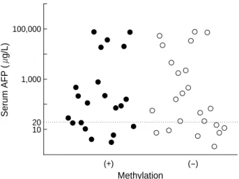
There were no associations between the status of p16INK4A methylation and serum AFP level in HCC patients (Table 3, Fig. 2) or tumor size (Table 4).
DISCUSSION
The tumor suppressor gene p16INK4Aexhibits variations, including methylation, that are involved in the process of carcinogenesis. A causal relationship between genetic varia- tion and methylation has yet to be determined. Kondo et al.
(8) studied LOH, microsatellite instability, and DNA methy- lation in HCC and in non-cancerous surrounding tissue by microdissection. Their results suggested that methylation often occurs in both tissue types, that it precedes LOH, and that methylation is involved in the early genesis of HCC as the cause of LOH.
The reported rate of incidence of p16INK4Amethylation is quite variable, ranging from 0% to 94% in HCC (8, 15, 22), and from 29.4% to 83% in cirrhosis (8, 21, 22). This may be due to the lack of a standardized method of detection and to the diversity in the clinical courses of patient groups. There have been few serum studies of p16INK4Amethylation in HCC patients. Wong et al. (20) reported abnormal p16INK4Amethy- lation in 60% of sera and in 73% of tissue in HCC patients.
In the present study, abnormal p16INK4Amethylation was detected in 48% of the sera of HCC patients. Further stud- ies on plasma or sera are required; large variations in inci- dence may be expected due to differences in sample materi- als, detection methods, and/or subjects selected.
Wong et al. (20) reported that p16INK4Amethylation was not detected in the plasma of patients with either liver cirrhosis or hepatitis, whereas in the present study it was detected in the sera of 17.4% of cirrhosis patients. We infer that abnor- mal p16INK4Amethylation can also be detected in the sera of patients with cirrhosis because it is frequently found in non- tumorous tissues (8). AFP and albumin mRNA were also de- tected in the sera of hepatitis and cirrhosis patients, as well as in those of HCC patients (25). More meticulous methods may be required to detect p16INK4Amethylation in cirrhosis patients, as the amount of circulating DNA is lower in the sera of cir- rhosis patients than in that of HCC patients. The incidence of methylated p16INK4A DNA did not differ between small (≤3 cm) and large HCCs, and was not correlated with AFP levels in the sera of HCC patients. Wong et al. (26) reported a significant correlation between the methylation of circulat- ing DNA and serum AFP levels in HCC patients. There is a danger in generalizing from this result, however, as that study included only six cases of HCC without methylation.
Our study suggested that methylated p16INK4ADNA may
*Two patients were excluded due to long interval between the AFP test date and serum sampling for methylation. �chi-square test.
Methylation (%)
AFP ( g/L) p value
0.690� Positive Negative
≥20 (n=28) 67.0 61.0
< 20 (n=16) 33.0 39.0
Table 3.Association between p16INK4Amethylation and serum AFP levels in patients with HCC*
*Chi-square test.
Methylation (%)
Size (cm) p value
0.226� Positive Negative
>3 (n=25) 63.6 45.8
≤3 (n=21) 36.4 54.2
Table 4.Association between p16INK4Amethylation and tumor size
Serum AFP (g/L)
100,000
1,000
20 10
(+) (-) Methylation
Fig. 2.Serum AFP levels of 44 HCC patients according to status of p16INK4Amethylation.
86 H.J. Chu, J. Heo, S.B. Seo, et al.
play an important role as a tumor marker in detection of HCC. The state of serum p16INK4Amethylation discriminated HCC from cirrhosis with a sensitivity of 47.8% and a speci- ficity of 82.6%. As there is no correlation between serum methylation and AFP, it may be a useful complementary tool to the AFP test.
REFERENCES
1. Korea National Statistical Office. Korea Statistical Yearbook. Dae- jeon 2002.
2. Shiina S, Tagawa K, Unuma T, Terano A. Percutaneous ethanol injec- tion therapy for treatment of hepatocellular carcinoma. Am J Reon- tgenol 1990; 154: 947-51.
3. Okuda S. Local ablation therapy for hepatocellular carcinoma.
Semin Liver Dis 1999; 19: 323-8.
4. Yuen MF, Cheng CC, Lauder IJ, Lam SK, Ooi CG, Lai CL. Early detection of hepatocellular carcinoma increases the chance of treat- ment: Hong Kong experience. Hepatology 2000; 31: 330-5.
5. Nagasue N, Uchida M, Makino Y, Takemoto Y, Yamanoi A, Hayashi T, Chang YC, Kohno H, Nakamura T, Yukaya H. Incidence and factors associated with intrahepatic recurrence following resection of hepatocellular carcinoma. Gastroenterology 1993; 105; 488-94.
6. Shimada M, Takenaka K, Gion T, Fujiwara Y, Kajiyama K, Maeda T, Shirabe K, Nishizaki T, Yanaga K, Sugimachi K. Prognosis of recurrent hepatocellular carcinoma: a 10-year surgical experience in Japan. Gastroenterology 1996; 111: 720-6.
7. Greenblatt MS, Bennett WP, Hollstein M, Harris CC. Mutation in the p53 tumor suppressor gene: clues to cancer etiology and molec- ular pathogenesis. Cancer Res 1994; 54: 4855-78.
8. Kondo Y, Kanai Y, Sakamoto M, Mizokami M, Ueda R, Hirohashi S. Genetic instability and aberrant DNA methylation in chronic hepatitis and cirrhosis: A comprehensive study of loss of heterozy- gosity and microsatellite instability at 39 loci and DNA hypermethy- lation on 8 CpG islands in microdissected specimens from patients with hepatocellular carcinoma. Hepatology 2000; 32: 970-9.
9. Chaubert P, Gayer R, Zimmermann A, Fontolliet C, Stamm B, Bosman F, Shaw P. Germ-line mutations of the p16INK4 (MTS1) gene occur in a subset of patients with hepatocellular carcinoma. Hepatology 1997; 25: 1376-81.
10. Merlo A, Herman JG, Mao L, Lee DJ, Gabrielson E, Burger PC, Baylin SB, Sidransky D. 5′CpG island methylation is associated with transcriptional silencing of the tumour suppressor p16/CDKN2/MTS1 in human cancers. Nat Med 1995; 1: 686-92.
11. Serrano M, Hannon GJ, Beach D. A new regulatory motif in cell- cycle control causing specific inhibition of cyclin D/CDK4. Nature 1993; 366: 704-7.
12. Thorgeirsson SS, Grisham JW. Molecular pathogenesis of human hepatocellular carcinoma. Nat Genet 2002; 31: 339-46.
13. Tannapfel A, Wittekind C. Genes involved in hepatocellular carci-
noma: deregulation in cell cycling and apoptosis. Virchows Arch 2002; 440: 345-52.
14. Razin A. CpG methylation, chromatin structure and gene silencing:
a three-way connection. EMBO J 1998; 17: 4905-7.
15. Kim JR, Kim SY, Kim MJ, Kim JH. Alterations of CDKN2 (MTS1/
p16INK4A) gene in paraffin-embedded tumor tissues of human stom- ach, lung, cervix and liver cancers. Exp Mol Med 1998; 30: 109-14.
16. Kita R, Nishida N, Fukuda Y, Azechi H, Matsuoka Y, Komeda T, Sando T, Nakao K, Ishizaki K. Infrequent alterations of the p16INK4A gene in liver cancers. Int J Cancer 1996; 67: 176-80.
17. Lin YW, Chen CH, Huang GT, Lee PH, Wang JT, Chen DS, Lu FJ, Sheu JC. Infrequent mutations and no methylation of CDKN2A (p16/MTS1) and CDKN2B(p15/MTS2) in hepatocellular carcinoma in Taiwan. Eur J Cancer 1998; 34: 1789-95.
18. Chaubert P, Gayer R, Zimmermann A, Fontolliet C, Stamm B, Bos- man F, Shaw P. Germ-line mutations of the p16INK4 (MTS1) gene occur in a subset of patients with hepatocellular carcinoma. Hepa- tology 1997; 25: 1376-81.
19. Liew CT, Li HM, Lo KW, Leow CK, Chan JY, Hin LY, Lau WY, Lai PB, Lim BK, Huang J, Leung WT, Wu S, Lee JC. High fre- quency of p16INKA4 gene alterations in hepatocellular carcinoma.
Oncogene 1999; 18: 789-95.
20. Wong IH, Lo YM, Zhang J, Liew CT, Ng MH, Wong N, Lai PB, Lau WY, Hjelm NM, Johnson PJ. Detection of aberrant p16 methy- lation in the plasma and serum of liver cancers patients. Cancer Res 1999; 59: 71-3.
21. Roncalli M, Bianchi P, Bruni B, Laghi L, Destro A, Di Gioia S, Gennari L, Tommasini M, Malesci A, Coggi G. Methylation frame- work of cell cycle gene inhibitors in cirrhosis and associated hepa- tocellular carcinoma. Hepatology 2002; 36: 427-32.
22. Kaneto H, Sasaki S, Yamamoto H, Itoh F, Toyota M, Suzuki H, Ozeki I, Iwata N, Ohmura T, Satoh T, Karino Y, Satoh T, Toyota J, Satoh M, Endo T, Omata M, Imai K. Detection of hypermethylation of the p16(INK4A) gene promoter in chronic hepatitis and cirrhosis associated with hepatitis B or C virus. Gut 2001; 48: 372-7.
23. Bruix J, Sherman M, Llovet JM, Beaugrand M, Lencioni R, Bur- roughs AK, Christensen E, Pagliaro L, Colombo M, Rodes J, EASL Panel of Experts on HCC. Clinical management of hepatocellular carcinoma. Conclusions of the Barcelona-2000 EASL conference.
European Association for the Study of the Liver. J Hepatol 2001;
35: 421-30.
24. Herman JG, Gaff JR, Myohanen S, Nelkin BD, Baylin SB. Methy- lation-specific PCR: a novel PCR assay for methylation status of CpG islands. Proc Natl Acad Sci USA 1996; 93: 9821-6.
25. Wong IH, Lau WY, Leung T, Johnson PJ. Quantitative comparison of alpha-fetoprotein and albumin mRNA levels in hepatocellular carcinoma/adenoma, non-tumor liver and blood: implications in cancer detection and monitoring. Cancer Lett 2000; 156: 141-9.
26. Wong IH, Lo YM, Lai PB, Johnson PJ. Relationship of p16 methy- lation status and serum alpha-fetoprotein concentration in hepato- cellular carcinoma patients. Clin Chem 2000; 46: 1420-2.