CONTENTS
Ⅰ. INTRODUCTION
Ⅱ. MATERIALS AND METHODS
Ⅲ. RESULTS
Ⅳ. DISCUSSION
Ⅴ. CONCLUSIONS REFERENCES KOREAN ABSTRACT EXPLANATION OF FIGURES
Ⅰ. INTRODUCTION
The oral cavity is an important organ playing a role of many functions, in such as respiratory, digestive, phonetic and protective system, which has very complex, detail structures and functions.
The oral mucosa separate and protect deeper tissues and organs in the oral region from environment of the oral cavity. This shows a number of adaptations of both the epithelium and the connective tissue to withstand mechanical, thermal insults produced during eating food. In addition, many of microorganisms in oral cavity produce
substances that have a toxic effect on tissues. The epithelium of the oral mucosa acts as the major barrier to these threats.
1)The oral mucosa is particularly well adapted for the functions it has to perform.
Saliva has a variety of functions. It keeps the mouth moist, facilitates speech, lubricates food, and helps with taste by acting as a solvent for the food molecules. It also contains a digestive enzyme and antibodies as well as acts like a buffer and therefore plays an important role in maintaining the pH of the oral cavity. In addition, it cleanses the mouth.
1)A lack of saliva results in, e.g., a painful tongue and mucosa, problems with taste, swallowing, chewing, and phonation, dental caries and tooth loss, and increased risk for infection.
2)3)4)5)This lack of saliva can be caused by several circumstances, including aging, disease, radiotherapy of the head-neck region, certain medications
6)and psychological stress.
7)It is becoming increasingly apparent that changes in the orofacial tissues may be a result of underlying systemic disease, such as gastrointestinal tract disease, hematological disorder, nutritional defi- ciency, psychological or immunological disorder.
.8)Ultrastructural Changes of Submandibular Gland in Rat under Moderate Restraint and Cold Stresses
Sun-Q Kim
1, D.M.D.,M.S.D, Jung-Won Kim
1, D.M.D., Yang-Hyun Chun
2, D.M.D.,M.S.D.,Ph.D.,
Jung-Pyo Hong
1, D.M.D.,M.S.D.,Ph.D.
Department of Oral Diagnosis & Oral Medicine, College of Dentistry, Kyung Hee University1, Institute of Oral Biology, Kyung Hee University2
* This research was supported by grant No. 1U0101039 (2001) from Institute of Oral Biology, Kyung Hee University.
That is, oral cavity is closely associated with one's health state.
Stress can have an effect on human body which is apt to maintain the homeostasis and modulate three main systems such as autonomic nervous, hormonal and immune response systems.
9)So the term 'psychoneuroimmunology' has developed in order to elucidate the relationship between psycho- social stress and diseases.
10)For a better understanding of the effects of psychological stress on the human body, we studied ultrastructural changes of the submandibular gland in rats, which produce saliva for protecting oral tissues, under restraint stress.
Ⅱ. MATERIALS AND METHODS 1. Experimental animals
Sprague-Dawley rats (8-week-old, 323-367 g/bw) were purchased from Dae-Han Experimental Animal Research Center, Seoul, Korea. They were maintained at 20-23℃ and fed ad libitum on a normal laboratory diet. The rats were divided into 3 groups:
1) Normal control group; 2) Restraint stress group;
3) Cold stress group : the rats of restraint stress group were placed in the stress cage throughout the period of experiment and the rats of cold stress group were immersed in cold water (4℃) for 6 min once a day throughout the experiment. All the animals were then sacrificed at day 0, 1, 3, 5, and 7 day of the experiment and the submandibular glands were excised immediately and stored in the glutaradehyde in phosphate buffer (PB) after rinsed in PB.
2. Electron Microscopy
The tissues were rinsed in 0.1M cacodylate buffer 3 times for 10 min each, postfixed in 1% osmium tetroxide for 90 min, and rinsed in 0.1M malate buffer 3 times for 5 min each. They were prestained with 1% uranyl acetate for 90 min and washed in 0.1 M malate buffer 3 times 5 min each, and dehydrated through an ascending series of ethanol concentration
(50% to 100%, 15 min each). And they were placed in 100% ethanol and prophylene oxide (1:1) for 45 min, prophylene oxide for 45 min, and prophylene oxide and epon (1:1) for 1 hr. After then, they were placed in epon in a vacuum oven overnight, embedded with fresh epon which was polymerized at 60℃ for 3 days. The embedded tissues were cut with a diamond knife in 50nm thick and stained with uranyl acetate and lead citrate. The tissues were observed under the transmission electron microscopy (JEOL Ltd , Japan).
Ⅲ. RESULTS
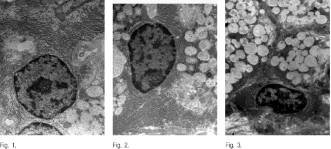
In the normal control group, many lamellated rough endoplasmic reticulum(rER), mitochondria, Golgi complex and some electron-lucent vacuoles were observed in the salivary gland acinar cells.
In the moderate restraint stress group, in lapse of time, according to the restraint duration, ultrastructural changes appeared gradually.
1. Under the moderate restraint stress,
1. Submandibular gland acinar cells were showed apoptotic changes in some areas.
2. The number of the secretory granules were decreased.
3. Mitochondrias were partially condensed but almost intact.
4. rERs were dilated and decreased.
5. Electron-lucent vacuoles were increased and their membranes were distructed irregularly.
6. Double layer of nuclear envelope had the irregular structures at day 7
And under the cold stress,
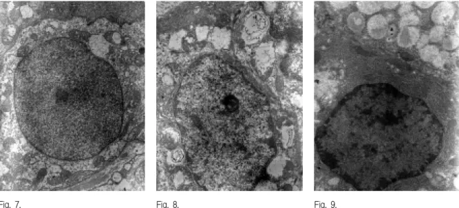
1. Mitochondria, secretory granules, Golgi complex and nuclear structures had no changes.
2. The lamellated structures of rERs were changed
into loose pattern at day 5, but were recovered at
day 7.
Ⅳ. DISCUSSION
The oral cavity has many functions. It is the site for ingestion food, for starting the process of digestion, for tasting and for articulating speech. It also has protective properties against various noxious agents such as microorganisms and physical trauma. The structure of the oral mucosa and its interaction with saliva contribute to all these tasks.
11)The oral mucosa is in intimate contact and interrelates with many general systems of the body.
A lots of oral lesions are associated with general health and/or disease such as immuopathologic disorders (lichen planus, benign mucous membrane pemphigoid, pemphigus vulgaris, erythema multi- forme, recurrent aphthous stomatitis, lupus erythe- matoses, progressive systemic sclerosis), oral mucosal disorders of the keratinizing process (genokeratoses, erythroplakia, leukoplakia, verrucous hyperplasia, verrucous carcinoma, squamous cell carcinoma), and viral (HIV, HBV, HCMC, EBV, HSV, HPV, VZV, enterovirus) and fungal infections (Candida).
Its blood supply is excellent, making healing of injuries almost invariably uneventful. Its association with the immunologic system and contents of immunologically competent cells contributes to the body's defense against exogenous "non-self", bioactive or other chemical agents. The mucosa also contains neuologic structures with many different functions, from propagating the sense of taste to contributing to proprioception.
In all these respects, different parts of the oral mucosa have different properties and, accordingly, they show quite different macro and microchara- cteristics. The differences may even justify categorizing the mucosa into several classes of tissues. The very thin non-keratinized lining mucosa of the floor of the mouth has little in common with the tough keratinized mucosa of the hard palate and the gingiva propria or the complicated mucosa of the dorsal tongue surface with its numerous papillae.
5)Saliva is an extremely complex fluid containing mucin, enzymes, immunoglobulins, growth factors,
etc. and has several functions. In addition to moistening the oral tissues as an aid for speech, and functioning as an aqueous solvent necessary for taste, it acts as a masticatory wetting agent which assists swallowing. Certain glands initiate the digestive process by secreting an enzyme. Also, saliva has an antibacterial action which inhibits or prevents the onset of dental caries and other infections
It is well documented that saliva plays an important role in the development and maintenance of a healthy oral cavity.
12)The salivary glands are controlled by the autonomic nervous system. Parasympathetic stimulation increases salivary flow, while anticholinergic drugs inhibit secretions. However, the sympathetic influence on the salivary gland function is more complex. Generally, sympathetic activity inhibits salivation, but the opposite case is also observed. Emotional stress induces a variety of responses in the body. In most instances alarm-like reactions and depression and anxiety inhibit, while pleasurable, relaxed sensations promote increase salivation.
12)13)So stress can be an important factor to affect on apoptosis of salivary gland
14)15)and may cause hyposalivation.
A lack of saliva results in, e.g., a painful tongue and mucosa, problems with taste, swallowing, chewing, and phonation, tooth decay and tooth loss, and increased risk for infection
2)3)4)5)Stress is the nonspecific response of the body to any demand
16)and can also be defined as an interactional process between the individual and the environment.
9)The theory of stress in oriental medicine is introduced from the concepts of wholism between
‘nature and human,’ ‘parts and whole’ and ‘mind and body.’ Stressor contains all pathological factors such as ‘endogenous,’ ‘exogenous’ and ‘non endo- exogenous pathological factors’
17)Stress can have an effect on human body which
is apt to maintain homeostasis and modulate three
main systems such as autonomic nervous system,
the hormonal system and immune response system.
9)A large body of evidence substates the contention that the central nervous system(CNS) and the endocrine system are involved in reciprocal interactions with the immune system.
18)19)20)Various immune factors alter neural and endocrine functions:
neural and endocrine signals modify both the humoral and the cellular arms of the immune response.
21)The neural influence on the immune system is best illustrated by the effect of stressors on immune functions.
22)It has been suggested that the activation of the hypothalamic-pituitary- adrenal(HPA) axis plays an important role in this process.
23)The events occurring in the brain in response to stress, specifically at the hypothalamic-pituitary- adrenal(HPA) axis, are similar to those occurring in response to an activated immune system. In both systems, the HPA axis is stimulated by corticotrophin-releasing factor(CRF), resulting in a down-regulation of immune function.
24)Therefore, stress-induced alterations in endocrine function and the potential pathophysiologic effects of such endocrine changes lead to a depletion of the subject's ability to resist stress, which ultimately leads to the disease process.
25)The stress could either suppress or enhance immune responses depending on the type and duration of the stressor.
26)27)28)29)Numerous studies have revealed that severe environmental and/or psychological stress have strong supressive effects on the immune system.
30)Such suppression of the immune system has significant implications for disease susceptibility and progression. Investigations in both humans and animals have revealed that stress could promote tumor development,
31)32)autoimmunity,
33)and infectious diseases by influen- cing the onset, course, and outcome of the pathological processes.
34)35)The relationship of stress to the orofacial area can be considered in four aspects
9): normal physiological and psychological functions of the mouth, stress-relieving orofacial activities, stress and dental treatment, and stress-related orofacial disorders and diseases.
Recently a few studies have suggested that stress is strongly associated with orofacial diseases. Chun and Hong
36)indicated that stress causes various forms of diseases in the region including orofacial psychosomatic diseases in which emotional stress appears to play a major role (lichen planus, aphthous stomatitis), orofacial diseases in which psychologic factors appear to play a role (erythema multiforme, benign mucous membrane pemphigoid, geographic tongue), orofacial infections where emotional stress is a significant predisposing factor (recurrent herpes labialis, acute necrotizing ulcerative gingivitis), orofacial lesions induced by neurotic habits inflicting trauma (biting of oral tissues, physical trauma with foreign objects, leukoplakia due to smoking, bruxism and clenching), neurotic orofacial symptoms (xerostomia, halitosis, burning mouth syndrome, altered or loss of taste perception, pain or discomfort with no tissue change), and orofacial pain induced by emotional stress (temporomandibular disorders, muscle tension headache, atypical odontalgia).
In multicellular organisms, homeostasis is main- tained through a balance between cell proliferation and cell death. Although much is known about the control of cell proliferation, less is known about the control of cell death. Physiologic cell death occurs primarily through an evolutionarily conserved form of cell suicide termed apoptosis.
37)Over the recent years cell death (apoptosis) has
been extensively studied. Apoptosis is found to play
a key role in proliferation, differentiation, embryo-
genesis, morphogenesis and homeostatic processes.
38)Stress can induce apoptosis which may be
important in body defense mechanism. It is reported
that psychophysical stress and its related hormones
modulate the apoptosis of the immune cells,
suggesting that psychoneural systems may exert an
effect on host defense.
39)40)41)42)43)44)The alterations in
cell survival contribute to the pathogenesis of a
number of human diseases, including cancer, viral
infections, autoimmune diseases, neurodegenerative
disorders, and AIDS (acquired immunodeficiency
syndrome). Treatments designed to specifically alter
the apoptotic threshold may have the potential to
change the natural progression of some of these diseases.
37)As stated above, stress is important in the pathogenesis including apoptosis and causes stress-related symptom such as xerostomia, burning mouth syndrome(BMS), halitosis and many other oral ulcers. Our study is based on the hypothesis that these symptoms may be associated with dysfunction of salivary glands resulting from apoptosis. Hong et al.
14)investigated apoptosis in submandibular gland of rats under restraint stress. They have reported that as an early response to stress, clusterin is expressed in the salivary glands to protect the glandular cells from the stress. But if stress is strong or prolonged that it can exceed the stress adaptability of the cells, then the cells may undergo apoptosis instead of producing clusterin. So I can suggest that the clusterin is an antiapoptotic and cytoprotective protein to environmental changes or readily detectable marker in cells undergoing apoptosis. And they
15)also suggested that caspase-3, which is known as a key enzyme in apoptosis and can be expressed prior to morphological changes in apoptosis, occurred as a result of cell response to psychological stress for apoptosis but then gradually disappeared. It is likely that if stress far exceeds the acceptable range for adaptation, apoptosis may occur in the submandibular gland and cause pathologic change of the gland.
In this study, we would observe what happen to intracellular structure of submandibular gland under restraint stress. We chose the submandibular gland among the salivary glands because there are many researches that subamndibular glands have more steady function and highly sensitivity to cold stress than other salivary glands.
The salivary glands, the major salivary glands including the parotid, the submandibular and the sublingual glands, and the several minor glands, produce and secrete saliva. Saliva is formed by units of secretory cells of which there are 3 types, serous, mucous and seromucous, and is modified by cells of the ductal system as it passes toward the oral cavity.
13)The composition of the final saliva, as well as the primary saliva, is quite possibly unique for each salivary gland and is determined by the nature of the secretory endpiece cells and ductal elements of that gland.
12)General ultrastructures of salivary gland are as followings :
The morphological criteria for seromucous cells advocated by Bernard T. et al.
45)include 1) relatively large, discrete secretory granules and 2) a moderately electron-dense secretory granule matrix, often with one or more dense inclusions. Seen in the transmission electron microscope, serous cells in salivary glands have all the structural hallmarks of cells geared for synthesis of proteins for export.
46)As with the exocrine pancreas cell, serous cells of salivary glands have an extensive infranuclear rough endoplasmic reticulum (RER) consisting of numerous, usually parallel cisternae,
47)but, in unstimulated mouse parotid
48)and stimulated rat parotid glands
49)neighboring cisternae may unite to form complex filigreed patterns. The Golgi apparatus in most salivary serous cells is an extensive organelle that may wend a sinuous course through the supranuclear cytoplasm.
45)The nuclei of serous cells tend to be centrally placed. In the human parotid gland, serous cells sometimes contains two nuclei.
50)Serous cell nuclei are largely euchromatic in appearance and occasionally have a fairly prominent nucleolus. Serous cells of the human parotid gland may have intranuclear inclusions. Most serous cells contain a few stray elements of smooth endoplasmic reticulum(SER). And, mitochondria usually are fairly abundant between adjacent RER cisternae and in the cytoplasmic interstices between secretory granules.
Regardless of size, all mitochondria in these cells
contain an array of randomly arranged dense ribbons
in their matrix compartment : the nature of these
ribbons is unknown.
45)In keeping with their
secretory function serous cells exhibit a great many
surface specializations, including a greatly
augmented basal surface in the form of slender basal
folds, lateral gutters in the form of intercellular
canaliculi, and varying numbers of luminal microvilli.
51)52)
Mucus, a major component of whole saliva, is thick, ropy, viscous, and above all slippery.
53)It is this latter property that allows salivary mucus to act as a lubricant for both chewing and swallowing, especially when the food in question has a dry consistency and, in human beings, for the free movement of the tongue, lips, and cheeks during speech. In addition, mucus provides a protective coating on both the hard and the soft tissues of the oral cavity.
54)At the electron microscopic level, the most prominent feature of fully charged mucous cells is the large number of mucous droplets that fill the supra-and peri-nuclear cytoplasm. Typical mucous droplets tend to be 50% larger in diameter than serous granules. Usually, they have a lucent matrix.
55)Ultrastructurally the mucous cell differs from the seromucous cell in that it contains more prominent Golgi complexes (shich reflect the cell's increased carbohydrate metabolism). In resting cells the RER and other cytoplasmic organelles(e.g., mitochondria) are less conspicuous than in seromucous cells and are mainly confined to the base and lateral aspects of the cell. The interdigitations between adjacent mucous cells tend to be fewer than between seromucous cells.
1)In typical mucous cells, the morphology of the organelles devoted to secretion depends in large measure on the stage of the secretory cycle. In cells that are replete with mucous droplets, the RER is reduced to a few cisternae in the cell base and in the interstices of the secretory droplets. The Golgi apparatus similarly is masked by the mucous droplets. In contrast, a mucous cell just embarking on a new round of mucus secretion usually has an extensive infranuclear RER
56)that in some organs consists of dilated cisternae with a moderately dense flocculent content.
57)The Golgi apparatus, not yet hidden by a collection of mucous droplets, is prominent.
55)The cellular changes in apoptosis are numerous, but it is still not clear which of them are directly associated with death, and which are of the greatest
physiological importance.
Apoptosis characteristically affects scattered single cells, not groups of adjoining cells, as is the case with necrosis. The earliest recognized morphologic changes are compaction and segregation of the nuclear chromatin, with the formation of sharply delineated, uniformly finely granular masses that become marginated against the nuclear envelope, and condensation of the cytoplasm.
Progression of the condensation is accompanied by convolution of the nuclear and cell outlines, and this is followed by breaking up of the nucleus into discrete fragments that are surrounded by a double-layered envelope and by budding of the cell as a whole to produce membrane-bounded apoptotic bodies. The size and composition of the latter vary considerably; many contain several nuclear fragments whereas others lack a nuclear component.
The cytoplasmic organelles of newly formed apoptotic bodies remain well preserved. Apoptotic bodies arising in tissues are quickly ingested by nearby cells and degraded within their lysosome.
There is no inflammatory process.
58)According to standard morphological descriptions, mitochondria were long thought to remain unchanged during apoptosis
59)but to swell during necrosis. However, a review of the past literature on cell death, before apoptosis had even been described, reveals abnormal mitochondria in types of cell death that, retrospectively, can be classified as apoptosis.
The most frequent abnormalities are a reduction in mitochondria size and a hyperdensity of their matrix, features often referred to as ‘mitochondrial pyknosis’.
60)61)62)
Namely, Following Bcl-2-related proapoptotic protein Bax insertion into the outer mitochondrial membrane, mitochondria first condense(‘pyknotic mitochondria’), possibly fragment, and then cluster around the nucleus. The condensed mitochondria have lost their cytochrome C.
63)Hiroyuki H. et al.
studied apoptosis in rat submandibular gland
development. Electron microscopic analysis showed
that certain morphological features of apoptosis,
including increase of electron-lucent vacuole, fewer
and smaller secretory granule, decrease of RER,
presence of apoptotic bodies in the cytoplasm and phagocytosis. But, nuclear changes, such as condensation and aggregation of the chromatin near the periphery of the nucleus, were only rarely observed. Nuclear fragmentation and cytoplasmic blebbing were not observed.
64)65)In this present study, during the moderate restraint stress, it was observed that secretory granules were decreased and rERs were dilated and decreased remarkably, electron-lucent vacuoles were increased and their size or morphology had been changed irregularly in lapse of time. Mitochondria were just a little condensed but intact in almost.
While the cold stress was applied, mitochondria, secretory granules, Golgi complex and nuclear structures had no changes. The rERs were transformed into loose lamellated pattern at day 5, but were recovered at day 7.
The overall results reported that restraint stress(psychological stress) induced apoptosis in rat submandibular gland and ultrastructural changes of apoptotic cell and that rat submandibular gland cells were adapted to cold stress(physical stress). And this conclusion contribute to clarifying the mechanism of stress-related symptom in orofacial region such as xerostomia, BMS, halitosis and many other oral ulcers.
Apoptotic signals are generally believed to be mediated through a hierarchy of caspase activation controlled by one of two distinct pathways that are associated with either caspase-8(i.e. death receptors) or caspase-9(i.e. mitochondria). Some studies demonstrated that caspase-8 can be activated in caspase-9 dependent manner. However, neither study was able to determine whether caspase-9- mediated activation of caspase-8 is physiologically relevant of merely a bystander effect.
66)In present study, we observed almost intact mitochondria under restraint stress. Therefore we think that apoptosis may be originated from the membrane of cell, but further study will be necessary using the caspase-8 and caspase-9, because this results is insufficient to discover exactly the origin of apoptosis.
Ⅴ. CONCLUSIONS
The belief that stress leads to illness has a long history. A number of the orofacial diseases are also closely associated with stress. Despite research in the relationship of stress and the orofacial diseases leading to statistically, significant correlations and the accurate pathology remains vague.
In the previous studies, we suggested that stress can induce apoptosis and decrease of function in salivary gland. And, in this study, we will try to observe ultarstructural changes under restraint stress.
Eighteen Sprague-Dawley rats (8-week-old, 32 3-367 g/bw) were used for the experiment and the rats were divided into 3 groups: 1) Normal control group; 2) Restraint stress group; 3) Cold stress group : the rats of restraint stress group were placed in the stress cage throughout the period of experiment and the rats of cold stress group were immersed in cold water (4℃) for 6 min once a day throughout the experiment. All the animals were then sacrificed at day 0, 1, 3, 5, and 7 day of the experiment and the submandibular glands were excised immediately and fixed in the glutaraldehyde in phosphate buffer. The submandibular gland samples were subjected to transmission electron microscopy.
The results were as follows:
In the normal control group, many rough endoplasmic reticulum(rER) with lamellated structure, mitochondria, Golgi complex and less electron-lucent vacuole were observed in the salivary gland acinar cell.
In lapse of the time according to the restraint stress, ultrastructural changes appeared gradually.
Under the moderate restraint stress,
1. Submandibular gland acinar cells were showed apoptotic changes in some areas.
2. The number of the secretory granules were decreased.
3. Mitochondrias were partially condensed but almost
intact.
4. rERs were dilated and decreased.
5. Electron-lucent vacuoles were increased and their membranes were distructed irregularly.
6. Double layer of nuclear envelope had the irregular structures at day 7
And under the cold stress,
7. Mitochondria, secretory granules, Golgi complex and nuclear structures had no changes.
8. The lamellated structures of rERs were changed into loose pattern at day 5, but were recovered at day 7.
It is likely that stress may cause apoptotic cellular changes. It also appears that stress-induced damage of submandibular cells may lead to functional disturbances of salivary gland and develope many oral diseases.
In present study, we observed almost intact mitochondria under mild restraint stress. Therefore we suggest that apoptosis may be originated from the cell membrane in this situation, but further study will be necessary using the caspase-8 and caspase-9, to discover the exact origin of apoptosis.
REFERENCES
1. Ten Cate A.R. : Oral histology : development, struc- ture, and function. 4th ed, Mosby-Year Book, Inc, St.
Louis, 1994.
2. Vissink A. : Xerostomia. development, properties and application of mucin, containing saliva substitute(dissertation). University of Groningen, Groningen, the Netherlands, 1985.
3. Fox PC, Van der Ven PF, Sonies BC, Weiffenbach JM and Baum BJ : Xerostomia ; Evaluation of a symptom with increasing significance. J. Am. Dent.
Assoc., 110 : 519-525, 1985.
4. Mandel ID : The role of saliva in maintaing oral homeostasis. J. Am. Dent. Assoc., 119 : 298-304, 1989.
5. Axell T. : The oral mucosa as a mirror of general health of disease. Scand. J. Dent. Res. 100 : 9-16, 1992.
6. Valdez IH and Fox PC : Diagnosis and management of salivary dysfunction. Crit. Rev. Oral Med., 4 : 271-277, 1993
7. Bergdahl M and Bergdahl J. : Low unstimulated salivary flow and subjective oral dryness: association with medication, anxiety, depression, and stress. J.
Dent. Res. Sep ; 79(9) : 1652-1658, 2000.
8. Michael A O Lewis and Philip-John L. : Clinical oral medicine. Wright. Butterworth-Heinemann Ltd., London, England, 1993.
9. Selye H : Selye's guide to stress research. Vol. I. Van Nostrand Reinhold Ltd., Canada, 1980.
10. Shklar G and McCarthy PL : The oral manifestations of systemic disease. Butterworths, Boston and London, 1976.
11. Blackwell : Oral mucosa in health and disease.
DOLBY AE., London, 1975.
12. Dobrosielski-Vergona K. : Biology of the salivary glands. CRC Press, Inc, Boca Raton, 1993.
13. Lynch M.A., Brightman V.J. and Greenber M.S : Burket's oral medicine. J.B. 9th Ed, Lippincott Co., philadelphia, 1994.
14. H.K. Park, Y.H. Chun, J.Y. Lee, H.K. Cho and J.P.
Hong : Exprossion of clusterin in the salivary gland under restraint stress. The Korean J. Stress Res.
6(2):33-44, 1998.
15. Chung WB, Jung SH, Chun, YH, Lee JY and Hong JP
; Caspase-3 expression in the submandibular gland of rat under restraint stress, J of Korean Academy of Oral Medicine 25(3):265-278, 2000.
16. Kutash I.L. and Schlesinger L.B. : Handbook of stress and anxiety. Jossey-Bass Inc, California, 1980.
17. Kim J.W. : The view on stress in oriental medicine J Kyung Hee med, 13(3) ; 252-258, 1997.
18. Blalock, F.E. : A molecular basis for bidirectional communication between the immune and neuroendocrine systems. Physiol. Rev., 69 : 1-32, 1989.
19. Ader, R., Relten, D. and Cohen, N. : Interaction between the brain and the immune system. Ann. Rev.
Pharmacol. Toxicol., 30 : 561-602, 1990.
20. Reichlin, S. : Neuroendocrine-immune interactions. N.
Engl. J. Med., 329:1246-1253, 1993.
21. Nora T., Haim O., David W. W. and Joseph W. : Restraint stress-induced thymic involution and cell apoptosis and dependent on endogenous glucocorticoids. J. Neuroimmunology, 82 : 40-46, 1998.
22. Weiss, J.M. and Sundar, S. : Effects of stress on cellular immune responses in animals. Rev.
Psychiatry, 11 : 25-48, 1992.
23. Besedovsky, H.O. and del Rey, A. : Immune-neuro-
endocrine interactions ; Facts and hypotheses.
Endocr. Rev., 17 : 64-102, 1996.
24. Black PH : Psychoneuroimmunology : Brain and immunity. Sci. Am. Sci. Med., 2 : 16-25, 1995.
25. Ronald C. Auvenshine, Psychoneuroimmuology and its relationship to the defferential diagnosis of temporomandibular disorders Dental Clin. of Nor.
Am. 41(2) April : 279-296, 1997
26. Nieman. DC. : Exercise immunology ; practical applications, Int. J. Sports Med., 18(Suppl.) 1 ; S91-S100, 1997.
27. Nieman. DC., and B.K. Pedersen. : Exercise and immune function. Recent developments. Sports Med., 27:73-80, 1999.
28. Dhabhar. F.S., and B.s. McEwen. : Acute stress enhances while chronic stress suppresses cell- mediated immunity in vivo ; a potential role for leukocyte trafficking. Brain Behav. Immun., 11 : 286-306, 1997.
29. Dhabhar. F.S., and B.S. McEwen. : Enhancing versus suppressive effects of stress hormones on skin immune function. Proc. Natl. Acad. Sci. USA., 96 : 1059-1064, 1999.
30. Ader, R., and N. Cohen. : Psychoneuroimmunology ; conditioning and stress. Annu. Rev. Psychol., 44 : 53-85, 1993.
31. Sklar, S.S., and H. Anisman. : Stress and coping factors influence tumor growth. Science., 205 : 513-515, 1979.
32. Temoshok, L. : Personality, eoping style, emotion and cancer ; towards an integrative model. Cancer Surv., 6 : 545-567., 1987.
33. Shanks, N., and A.W. Kusnecov. : Differential immune reactivity to stress in BALB/cByJ and C57BL/6J mice ; in vivo dependence on macrophages. Physiol.
Behav., 65 : 95-103, 1998.
34. Olson, G.A., R.D. Olson, A.L. Vaccarino, and A.J.
Kastin. : Endogenous opiates ; 1997. Peptides., 19 : 1791-1843., 1998.
35. Sheridan, J.F., C. Dobbs, J.Jung, X. Chu, A.
Konstatinos, D. Padgett, and R. Glaser. : Stres-induced neuroendocrine modulation of viral pathogenesis and immunity. Ann. NY Acad. Sci., 840 : 803-808, 1998.
36. Chun Y.H. and Hong J.P. : Stress and orofacial diseases. Kor. Stress Res., 3(1) : 57-72, 1995.
37. Craig B. Thompson : Apoptosis in the pathogenesis and treatment of disease. Science, 267(10) : 1456-1462, 1995.
38. Khanson K.P. : Programmed cell death (apoptosis):
molecular mechanisms and the role in biology and medicine. Vopr Med Khim, 43(5) : 402-16, 1997.
39. Sendo F., Kato T. and Yazawa H. : Modulation of neutrophil apoptosis by psychological stress and glucocorticoid. Int. J. Immunopharmacol., 19(9-10) : 511-516, 1997.
40. Tomei L.D., Kiecolt-Glaser J.K., Kennedy S. and Glaser R : Psychological stress and phorbol ester inhibition of radiation-induced apoptosis in human peripheral blood leukocytes. Psychiatry Res., 33(1) : 59-71, 1990.
41. Wang J.H., Redmond H.P., Watson R.W. and Bouchier-Hayes D. : Induction of human endothelial cell apoptosis requires both heat shock and oxidative stress responses. Am J Physiol, 272(5 Pt 1) : C1543-1551, 1997.
42. Viard I., Wehrli P., Jornot L., Bullani R., Vechietti J.L., Schifferli J.A., and Tschopp J. : Clusterin gene expression mediates resistance to apoptotic cell death induced by heat shock and oxidative stress. J Invest Dermatol, 112(3) : 290-296, 1999.
43. Concordet J.P. and Ferry A. : Physiological programmed cell death in thymocytes is induced by physical stress (exercise). Am. J. Physiol., 265(3 Pt 1) : C626-629, 1993.
44. Fuchs E. and Flugge G. : Stress, glucocorticoids and structural plasticity of the hippocampus. Neurosci Biobehav Rev, 23(2) : 295-300, 1998.
45. Tandler, B. and Carleton J. Phillips : Structure of serous cells in salivary glands. Microscopy Res. and Tech., 26 : 32-48, 1993
46. Palade, G.E., : Intracellular aspects of the process of protein systhesis. Science, 189 : 347-358, 1975.
47. Tandler, B. and Erlandson, R.A. : Ultrastructure of the human submaxillary gland. Ⅳ. Serous granules. Am.
J. Anat., 135 : 419-434, 1972.
48. Parks, H.F. : Unusual formations of ergastoplasm in parotid acinous cells of mice. J. Cell Biol., 14 : 221-234, 1962.
49. Lillie, J.H. and Han, S.S. : Secretory protein synthesis in the stimulated rat parotid gland. Temporal dissociation of the maximal response from secretion.
J. Cell Biol., 59 : 708-721, 1973.
50. Hand,A.R. : Salivary glands. In ; Orban's Oral Histology and Embryology, 9th Ed. S.N. Bhaskar, ed.
C.V. Mosby, St. Louis, pp.336-370, 1980.
51. Pinkstaff,C.A. : The cytology of salivary glands. Int.
Rev. Cytol., 63 : 141-261, 1980.
52. Riva, A. and Riva-Testa, F. : Fine structure of acinar cells of human parotid gland. Anat. Rec., 176 : 149-166, 1973.
53. Schwarz, W.H. : The rheology of saliva. J. Dent. Res., 66 : 660-664, 1987.
54. Tabak, L.A. : Structure and function of human salivary mucins. Oral Biol. Med., 1 : 229-234, 1990.
55. Tandler B. : Structure of mucous cells in salivary glands, Microscopy Res. and Tech. 26 : 49-56, 1993.
56. Tandler, B., Denning, C.R., Mandel, I.D., and Kutscher, A.H. : Ultrastructure of human labial salivary glands. I. Acinar secretory cells. J. Morphol., 127 : 383-408s, 1969.
57. Tandler, B., and Poulsen, J.H. : Ultrastructure of the cat sublingual gland. Anat. Rec., 187 : 153-172, 1977.
58. John F.R. Kerr, Clay M. Winterford and Brian V.
Harmon : Apoptosis ; Its significance in cancer and cancer therapy. Cancer, 73(8) : 2013-2026, 1994.
59. Kerr, F.F.R. et al. : Apoptosis ; a basic biological phenomenon with wide-ranging implications in tissue kinetics. Br. J. Cancer, 26 : 239-257, 1972.
60. Mannweiler, K. et al. : Recherches ultrastructurales sur une tumeur renale experimentale du hamster. J.
Ultrastruct. Res., 1 : 158-169, 1957.
61. Rouiller, C. : Contribution de la microscopie electronique a l'etude du foie normal et pathologique.
Ann. Anat. Pathol., 2 : 548-562, 1957.
62. Hackenbrock, C.R. : Chemical and physical fixation of isolated mitochondria in low-energy and high-energy states. Proc. Natl. Acad. Sci. U.S.A., 61 : 598-605, 1968.
63. Desagher S and Martinou JC : Mitochondria as the central control point of apoptosis. Trends Cell Biol., 10(9) : 369-377, 2000.
64. Hiroyuki H., Atoru O., Kazuko W., Ikuko N., and Minoru O. : Morphological aspects of the postnatal development of submandibular glands in male rats ; involvement of apoptosis. J. Histochem. and Cytochem., 48(5) : 695-698, 2000.
65. Rachelle H., Michael C., Luigi M., William D. Bal, and Arthur R. Hand : Cell death during development of intercalated ducts in the rat submandibular gland.
Anat. Rec., 258 : 349-358, 2000.
66. Damu T., Jill M.L. and Vincent J.K. : Caspase-8 activation and bid cleavage contribute to MCF7 cellular execution in a caspase-3-dependent manner during staurosporine-mediated apoptosis. J. Biol.
Chem., 275(13) : 9303-9307, 2000.
Corresponding Author : Jung-Pyo Hong, Professor, Department of Oral Diagnosis & Oral Medicine, School of Dentistry, Kyung Hee University, 1 Hoegi-Dong, Dongdaemun-Ku, Seoul 130-701, Korea
국문초록
한냉 및 중등도의 구속 스트레스 시 백서 악하선 조직의 변화에 대한 전자현미경적 연구
경희대학교 치과대학 구강내과학 교실
1, 경희대학교 구강생물학 연구소
2김선규
1․김정원
1․전양현
2․홍정표
1현대에는 날로 복잡해지는 생활 양식의 변화에 따라 다양한 스트레스에 노출되고 그로 인한 면역계, 신경계, 내분비계 및 면역계의 변화를 수반한 항상성의 파괴로 수많은 정신적, 육체적 질병을 야기시키고, 특히 구강안면 영역에서는 다양한 구강점막질환과 구강건조증, 구강작열감 증후군 등을 발생시킨다.
특히 스트레스에 의한 구강건조증이나 구강작열감 증후군을 호소하는 환자들의 많은 수가 임상검사나 타액선 기능 검사에서 악하선의 기능이 현저하게 저하되어 있는 소견을 관찰할 수 있었으며, 실험적으로도 백서에 완전 구속 스트레스를 가하였을 때, 악하선 세포가 완전히 자사되는 것이 관찰되었던 바 있다.
이에 본인은 중등도의 구속 스트레스와 한냉 스트레스를 부여하였을 때 백서 악하선 세포의 미세구조 변화를 전자 현미경으로 관찰하여 다양한 정도의 스트레스 시 악하선 세포가 변화되는 것을 증명하고자 한다.
본 실험에서는 생후 8주된 Sprague-Dawley계 웅성 백서 (322-367 g/bw)를 대조군으로 3마리, 실험군으로 30 마리를 배정하였으며, 실험군 중 중등도의 구속 스트레스군은 실험 전 기간에 걸쳐 다소의 굴신이 허용되는 구속 장치에 구속시켰고, 한냉 스트레스군은 하루에 한번 6분씩 4℃의 물에 잠수시켜 스트레스를 부여하였다. 그 후 모든 실험동물의 악하선을 적출하여, 전자현미경으로 조직변화를 관찰한 결과 다음과 같은 결론을 얻었다.
전자현미경에 의한 점차적인 미세구조적 변화는 다음과 같다.
중등도의 구속 스트레스에 의하여
1. 백서 악하선 세포는 부분적으로 자사되었다.
2. 전자현미경상에서 백서 악하선 세포의 분비과립은 점차 감소되었다.
3. 전자현미경상에서 백서 악하선 세포의 사립체는 부분적으로 기질이 농축되었으나, 대부분 건전한 형태로 핵 주위에 다수 출현하였다.
4. 전자현미경상에서 백서 악하선 세포의 과립내형질망은 그 수가 점차 수가 감소되고 심하 게 이완되었다.
5. 전자현미경상에서 백서 악하선 세포의 전자밀도가 낮은 소포는 크기나 모양이 점차 불규 칙하게 관찰되었다.
6. 실험 7일군에서, 핵 이중막의 불규칙한 구조가 관찰되었다.
또한 한냉 스트레스 하에서는,
7. 전자현미경상에서 사립체, 분비과립, 골지체, 핵의 변화는 관찰되지 않았다.
8. 전자현미경상에서 치밀한 층판구조의 과립내형질망은 실험 5일군에서 느슨한 구조로 관찰되었으나, 실험 7일 군에서 회복되었다.
중등도의 구속 스트레스와 한냉 스트레스 부여 후 악하선 세포의 미세구조를 관찰한 결과, 세포 내 미세구조
및 세포간극의 변화가 있었던 것은 스트레스에 의해 악하선 세포가 변성될 수 있다는 것을 의미하는 것으로, 이는
스트레스와 관련된 구강안면동통 등의 질병 기전을 밝히는데 도움이 되리라 사료되며, 세포 내 미세구조 중 특히
사립체의 변화가 거의 없었던 것으로 보아 세포 자사의 기원이 세포막일 가능성도 보여지나 이것에 대하여는 향
후 추가적인 조직학적, 분자생물학적 연구가 필요하리라 생각된다.
EXPLANATION OF FIGURES
Electromicroscopic features of the rat submandibular gland
Fig. 1. The submandibular gland of the normal control rat. (X10,000)
Fig. 2. The submandibular gland of the rat under moderate restraint stress at day 0 of the experiment.
(X10,000)
Fig. 3. The submandibular gland of the rat under moderate restraint stress at day 1 of the experiment.
(X10,000)
Fig. 4. The submandibular gland of the rat under moderate restraint stress at day 3 of the experiment.
(X10,000)
Fig. 5. The submandibular gland of the rat under moderate restraint stress at day 5 of the experiment.
(X10,000)
Fig. 6. The submandibular gland of the rat under moderate restraint stress at day 7 of the experiment.
(X10,000)
Fig. 7. The submandibular gland of the rat under cold stress at day 0 of the experiment. (X10,000) Fig. 8. The submandibular gland of the rat under cold stress at day 1 of the experiment. (X10,000) Fig. 9. The submandibular gland of the rat under cold stress at day 3 of the experiment. (X10,000) Fig. 10. The submandibular gland of the rat under cold stress at day 5 of the experiment. (X10,000) Fig. 11. The submandibular gland of the rat under cold stress at day 7 of the experiment. (X10,000)
Fig. 1. Fig. 2. Fig. 3.
Fig. 4. Fig. 5. Fig. 6.
Fig. 7. Fig. 8. Fig. 9.
Fig. 10. Fig. 11.