Introduction
These days, the increase in global mixing from one country to another country, the spread of infectious diseases become faster and thus it became very serious issue [1]. In order to cope with these issues of global mixing and spread of infec- tious diseases, the fast and accurate detection of the target disease are important for improving the curative efficiency and
preventing the spread of disease. Traditional methods, such as immunological or biochemical identification, are time-consum- ing and laborious, which are not applicable on-site detection of diseases. In addition, antibody detection methods have limita- tions; for example, they cannot discriminate previously infected patients from currently infected patients. Hence, the use of DNA-based molecular diagnostic methods in various areas including human health, animal health, and food safety has Int J Oral Biol 44:8-13, 2019
pISSN: 1226-7155 • eISSN: 2287-6618 https://doi.org/10.11620/IJOB.2019.44.1.8
Optimization of ultra-fast convection polymerase chain reaction conditions for pathogen detection with nucleic acid lateral flow immunoassay
Tae-Hoon Kim
1, Hyun Jin Hwang
2, and Jeong Hee Kim
1,3*
1Department of Life and Nanopharmaceutical Sciences, Graduate School, Kyung Hee University, Seoul 02447, Republic of Korea
2R&D Center, Ahram Biosystems Inc., Seoul 04794, Republic of Korea
3Department of Oral Biochemistry and Molecular Biology, School of Dentistry, Kyung Hee University, Seoul 02447, Republic of Korea
Recently, the importance of on-site detection of pathogens has drawn attention in the field of molecular diagnostics.
Unlike in a laboratory environment, on-site detection of pathogens is performed under limited resources. In this study, we tried to optimize the experimental conditions for on-site detection of pathogens using a combination of ultra-fast convection polymerase chain reaction (cPCR), which does not require regular electricity, and nucleic acid lateral flow (NALF) immunoassay. Salmonella species was used as the model pathogen. DNA was amplified within 21 minutes (equivalent to 30 cycles of polymerase chain reaction) using ultra-fast cPCR, and the amplified DNA was detected within approximately 5 minutes using NALF immunoassay with nucleic acid detection (NAD) cassettes. In order to avoid false-positive results with NAD cassettes, we reduced the primer concentration or ultra-fast cPCR run time.
For singleplex ultra-fast cPCR, the primer concentration needed to be lowered to 3 µM or the run time needed to be reduced to 14 minutes. For duplex ultra-fast cPCR, 2 µM of each primer set needed to be used or the run time needed to be reduced to 14 minutes. Under the conditions optimized in this study, the combination of ultra-fast cPCR and NALF immunoassay can be applied to on-site detection of pathogens. The combination can be easily applied to the detection of oral pathogens.
Keywords: Molecular diagnostics, Ultra-fast convection polymerase chain reaction, Nucleic acid lateral flow
immunoassay
Received January 13, 2019; Revised February 11, 2019; Accepted February 15, 2019
*Correspondence to: Jeong Hee Kim, E-mail: [email protected] https://orcid.org/0000-0002-3884-4503 Copyright © The Korean Academy of Oral Biology
Original Article IJOB
become more popular [2-5]. Molecular diagnostic approaches including rapid detection of pathogens using polymerase chain reaction (PCR) draw the most attention because of the speed and higher accuracy than the traditional serology-based micro- bial serotyping [6-9].
Recently, studies on on-site usable technology development are reported [10-12]. In order to be used for on-site detec- tion of diseases, where resources are limited, it will be good if the equipment used are regular electricity-independent and the assay can be completed in a short time. And yet, the on- site detection assay should retain the same specificity and sensitivity which are obtained in the laboratory. In this study, we used battery-operated convection PCR (cPCR) device and nucleic acid lateral flow (NALF) immunoassay for detection of a model pathogen, Salmonella species. cPCR is recently developed method which can perform DNA amplification in ultra-fast speed and has been used for on-site detection of pathogens and meat identification [4,5]. The NALF assay has been recently developed for the detection of foodborne patho- gens, and pathogens and gene mutations that underlie human diseases [8,13-16]. We optimized the assay conditions for on- site cPCR-based molecular detection of pathogens in combi- nation of NALF immunoassay which is critical for the on-site usable assay development.
Materials and Methods
1. Bacteria strains and preparation of genomic DNA
The bacterial strains used in the current study were Sal- monella Enteritidis, Salmonella Typhimurium (NCCP 14771 and NCCP 12219) from the National Culture Collection for Pathogens (NCCP) at the Korea Center for Disease Control &
Prevention (Cheongju, Korea). Bacteria were grown overnight in LB broth (Duchefa Biochemic, Haarlem, The Netherlands) at 37°C with shaking.
Genomic DNA (gDNA) was extracted using DNeasy Blood and Tissue kit (Qiagen, Hilden, Germany) according to the manufacturer’s instructions. DNA concentration was deter- mined using a NanoDrop spectrophotometer (Thermo Scientif- ic, Waltham, MA, USA). gDNA copy numbers were calculated for 1 ng of DNA, based on the molecular weight of double- stranded DNA and chromosomal DNA size (http://sciencep- rimer.com/copy-number-calculator-for-realtime-pcr), as 1.9
× 105 copies/ng of chromosomal DNA for Salmonella.
2. Convection polymerase chain reaction
The primers used were; for detection of Salmonella spp., Biotin-CACGTCGGGCAATTCGT and FAM-GCTTTCCCTTTC- CAGTACGC (SPP primers); for detection of S. Enteritidis, Biotin-GTCAGTGCCATACTTTTAATGACTGC and FAM-GTAC- TATGTCGATACGGTGGGT (SE primers); for detection of S.
Typhimurium, Biotin-GCTGTATTTGTTCACTTTTTACCCCT and Digoxigenin-ACCCTGACAGCCGTTAGATATTC (ST primers).
The cPCR mixture (20 µL) contained 1 × PalmTaq HS buffer (Ahram Biosystems, Inc., Seoul, Korea) (supplemented with 1.5 mM MgCl2), 0.2 mM deoxyribonucleotide triphosphates, 0.4 U PalmTaq high-speed DNA polymerase (Ahram Biosystems), and primers for single or multiple PCR detection. For Salmo- nella detection using single-line NALF (NAD1), SPP primers were used (singleplex detection). To differentiate between S.
Enteritidis and S. Typhimurium using double-line NALF (NAD2), ST primers and/or SE primers were used (duplex detection).
The concentration of primers used for each experiment was 10 µM each, otherwise stated in the text. Generally, 1.6 ng (3
× 105 copies) of Salmonella gDNA was used as a template.
PCR was performed using a battery-operated convection thermal cycler Palm PCR device (G2-12; Ahram Biosystems).
The speed was set to T1 (30 cycles in 21 minutes), and the annealing temperature was set to 56°C. All experiments were performed at least in triplicate.
3. Detection of polymerase chain reaction amplification products by electrophoresis and nucleic acid lateral flow analysis
Upon completion of the cPCR, an aliquot of the PCR mixture was analyzed by 1.5% agarose gel electrophoresis (25 minutes at 135 V). PCR products were visualized with ethidium bromide staining and an imaging system (Ultra-Lum, Carson, CA, USA).
The NAD1 and NAD2 cassettes (Ahram Biosystems) were designed to indirectly detect amplified PCR products using antibodies against primer tags, such as FAM, DIG, and biotin.
When the NAD cassettes were used to detect DNA amplifica- tion, 2 µL of PCR product was diluted with 100 µL of the NAD buffer. The diluted solution was added to the reservoir of the NAD cassette, and waited for until the lateral flow was com- pleted. Typically, the reaction development took ca. 5 minutes.
The assay outcomes were documented by photographing.
Results and Discussion
1. Optimization of convection polymerase chain reaction conditions for single line nucleic acid detection
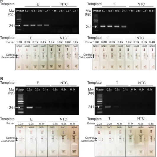
The purpose of this study is to optimize the experimental conditions of ultra-fast cPCR and NALF detection using NAD cassette which can be used for on-site detection of patho- gens for various diseases. Since the color development of NAD cassette is not dependent of the specific amplification of the target DNA but the presence of the conjugation of the antibody with its antigen, there is possibility of having non-specific color development with non-specifically amplified DNA. Many of these non-specific backgrounds come from the redundant primers in the PCR reaction. Therefore, we tried to optimized primer concentrations of Salmonella spp., S. Enteritidis and S. Thyphimurium, in the cPCR (Fig. 1A and 1B). cPCR was performed at speed level T1 for 21 minutes and the anneal-
ing temperature was 56℃. When 10 µM of the primers were used with 1.6 ng of gDNA of S. Enteritidis, non-specific band in no template control (NTC) appeared on both agarose gel and NAD1 detection. However, when the primer concentration was reduced to 3 µM (0.3×), no non-specific band was ob- served. Similarly, the primer concentration of S. Typhimurium was reduced to 3 µM (0.3×) and non-specific DNA band was observed neither in agarose nor NAD1 detection (Fig. 1B).
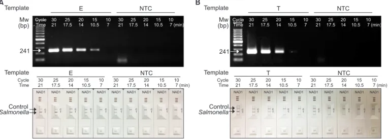
One of the way to obtain DNA amplification product before the amplified DNA products reach the saturation phase is to reduce the number of PCR cycles or the PCR running time.
We reduced the number of PCR cycles from 30 to 10 cycles (equivalent to 21 minutes to 7 minutes of cPCR running time) with an interval of 5 cycles. The other PCR conditions were kept as the same as the experiment described above. As shown in Fig. 2, when cPCR was performed with S. Enteritidis (Fig. 2A) and S. Typhimurium (Fig. 2B) with SPP primers, the intensity of DNA amplification product was gradually reduced as the PCR running time was reduced from 21 minutes to
A
E NTC
Primer 1.0 0.8 0.6 0.4 1.0 0.8 0.6 0.4
1.0 0.8 0.6 0.4 1.0 0.8 0.6 0.4
E NTC
NAD1 NAD1 NAD1 NAD1 NAD1 NAD1 NAD1 NAD1
C T
C T
C T
C T
C T
C T
C T
C T
Template Mw (bp)
241
Template Primer
Control Salmonella
BTemplate E NTC
Mw (bp)
241
Primer 0.3x 0.2x 0.1x 0.3x 0.2x 0.1x
Template Primer
E NTC
NAD1 NAD1 NAD1 NAD1 NAD1 NAD1
C T
C T
C T
C T
C T
C T
Control Salmonella
0.3x 0.2x 0.1x 0.3x 0.2x 0.1x
Template T NTC
Primer 1.0 0.8 0.6 0.4 1.0 0.8 0.6 0.4
Template
Primer 1.0 0.8 0.6 0.4 1.0 0.8 0.6 0.4
T NTC
NAD1 NAD1 NAD1 NAD1 NAD1 NAD1 NAD1 NAD1
C T
C T
C T
C T
C T
C T
C T
C T
Template T NTC
Template Primer
NAD1 NAD1 NAD1 NAD1 NAD1
C T
C T
C T
C T
C T
C T
Primer 0.3x 0.2x 0.1x 0.3x 0.2x 0.1x
0.3x 0.2x 0.1x 0.3x 0.2x 0.1x
T NTC
NAD1
Control Salmonella
Control Salmonella Mw (bp)
241
Mw (bp)
241
Fig. 1. Optimization of primer concnetra- tion of convection polymerase chain reaction (cPCR) for nucleic acid lateral flow (NALF) immunoassay using single-line NALF (NAD1).
Genomic DNA (1.6 ng each) isolated from Salmonella Enteritidis (E) and Salmonealla Typhimurium (T) were used as templates in the ultra-fast cPCR and NALF immunoassay with a single test line. Primer concentrations were varied from 10 µM (1.0×) to 4 µM (0.4×) (A) or from 3 µM (0.3×) to 1 µM (0.1×) (B).
DNA amplicons were analyzed by agarose gel electrophoresis or with NAD1 cassettes.
Speed mode was set to T1 and the annealing temperature was 56°C. The PCR operation time was 21 minutes (equivalent to 30 cycles).
Mw, molecular weight marker; NTC, no tem-
7 minutes. Non-specific dimeric DNA production was also reduced in the NTC lanes as the cPCR running time was re- duced. The same results were obtained with the agarose gel electrophoresis (Fig. 2A and 2B, upper panels) and with NAD cassettes (Fig. 2A and 2B, lower panels). The results obtained
with 14 minutes of cPCR running gave the clear appearance of the DNA amplification band with Salmonella DNA template and no detectable band in NTC experiments. The 14 minutes cPCR running was corresponding to the 20 cycles of cPCR.
ATemplate E NTC
Mw (bp)
241 Cycle
Time
Template E NTC
NAD1 NAD1 NAD1 NAD1 NAD1 NAD1 NAD1 NAD1
C T
C T
C T
C T
C T
C T
C T
C T
Control Salmonella
30 21
25 17.5
20 14
15 10.5
10 7
30 21
25 17.5
20 14
15 10.5
10 7 (min)
NAD1 NAD1
C T
C T
Cycle Time
30 21
25 17.5
20 14
15 10.5
10 7
30 21
25 17.5
20 14
15 10.5
10 7 (min)
B Template T NTC
Cycle Time
Template T NTC
NAD1 NAD1 NAD1 NAD1 NAD1 NAD1 NAD1 NAD1
C T
C T
C T
C T
C T
C T
C T
C T
30 21
25 17.5
20 14
15 10.5
10 7
30 21
25 17.5
20 14
15 10.5
10 7 (min)
NAD1 NAD1
C T
C T
Cycle Time
30 21
25 17.5
20 14
15 10.5
10 7
30 21
25 17.5
20 14
15 10.5
10 7 (min)
Control Salmonella Mw (bp)
241
Fig. 2. Optimization of polymerase chain reaction running time of convection polymerase chain reaction (cPCR) for nucleic acid lateral flow (NALF) immunoassay using single-line NALF (NAD1). Genomic DNA (1.6 ng each) of Salmonella Enteritidis (E) (A) and Salmonealla Typhimurium (T) (B) were used as templates. Primer concentration was 10 µM. cPCR running time was varied from 21 minutes to 7 minutes (equivalent to 30 cycles to 10 cycles). DNA amplicons were analyzed by agarose gel electrophoresis or with NAD1 cassettes. Speed mode was set to T1 and the annealing temperature was 56°C. Mw, molecular weight marker; NTC, no template control.
A B
E Primer
0.3 0.2 0.1 0.4
NAD2
Template E+T NTC
T Primer
E+T NTC
E Primer Template
NAD2 NAD2 NAD2 NAD2
C T2 T1
C T2 T1
C T2 T1
C T2 T1
T Primer 0.4 0.3 0.2 0.1
NAD2 NAD2 NAD2
0.3 0.2 0.1 0.4
0.3 0.2 0.1
0.4 0.4 0.3 0.2 0.1
0.3 0.2 0.1 0.4
0.3 0.2 0.1
0.4 0.4 0.3 0.2 0.1
C T2 T1
C T2 T1
C T2 T1
C T2 T1
E Primer 0.4
Mw (bp)
T Primer 1.0 0.8 0.6 0.4
NAD2
C T2 T1
Control ST SE
Template E+T NTC
0.4
321 409
0.4 T Primer 1.0 0.8 0.6 0.4
E+T NTC
0.4 E Primer
Template
NAD2 NAD2 NAD2 NAD2
C T2 T1
C T2 T1
C T2 T1
C T2 T1
Control ST SE Mw (bp)
321 409
Fig. 3. Optimization of primer concnetration of convection polymerase chain reaction (cPCR) for nucleic acid lateral flow (NALF) immunoassay using double- line NALF (NAD2). cPCR was performed with genomic DNA (gDNA) from Salmonella Enteritidis (E) and Salmonealla Typhimurium (T), and a 1 : 1 mixture of the two gDNA types (E + T). S. Enteritidis (SE) and S. Typhimurium (ST)-specific primer sets were mixed at different ratio (A) or the same ratio (B), and used in multiplex cPCR. The resulting amplicons were analyzed by agarose gel electrophoresis and with the NAD2 cassettes. Speed mode was set to T1 and the annealing temperature was 56°C. The polymerase chain reaction operation time was 21 minutes (equivalent to 30 cycles). 1.0× indicates 10 µM of primer concentration. Mw, molecular weight marker; NTC, no template control.
2. Optimization of conditions for double line nucleic acid detection
Next, we tried to detect both of S. Enteritidis and S. Ty- phimurium with a NAD2 cassettes. For balanced detection of S. Enteritidis and S. Typhimurium, the ratio of the primers was optimized. As shown in Fig. 3, the concentration of SE primer which detects S. Enteritidis was fixed to 4 µM, and the con- centration of ST primer was varied from 10 to 4 µM. cPCR was run for 21 minutes (equivalent to 30 cycles).
The PCR amplification of targets of S. Enteritidis and S. Ty- phimurim were well balanced on agarose gel analysis where the ratio of two primer sets were 1 : 1 (0.4 µM : 0.4 µM) on agarose gel analysis. However, due to the generation of non- specific dimer bands, the red lines were appeared on NAD2 cassettes as false-positive. Thus, we reduced the concentra- tion of primer sets while maintaining the ratio of SE and ST primers as 1 : 1. When the primer concentration was 0.2 µM the two DNA amplicons were appeared with almost equal in-
tensity and at the same time no detectable non-specific band was not observed on the agarose gel (Fig. 3B, upper panel).
The similar pattern was observed with NAD2 cassettes: two red lines were appeared with Salmonella DNA templates and no band with NTC when both primer set were used at concen- tration of 0.2 µM (Fig. 3B, lower panel).
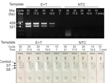
Next, we tried to reduce the PCR running time for the du- plex cPCR. As shown in Fig. 4, the cPCR running time was reduced from 21 minutes to 7 minutes (corresponding to 30 cycles to 10 cycles of cPCR). The concentration of the primer was adjusted to have balanced red-line appearance with NAD2 cassettes. The concentration gave the best results were 8 µM of SE primers and 10 µM of ST primers (data not shown).
Duplex cPCR amplification was successfully completed with various cPCR running time. When cPCR was performed for 14 minutes (20 cycles), amplified duplex DNA bands appeared on the agarose gel analysis and two red lines were appeared on T section of NAD2 cassette (Fig. 4). At the same conditions, NTC control showed no DNA amplification on the agarose gel analysis and no red line in NAD2 cassette.
Our data suggested that the combination of cPCR [4,5] and lateral flow immunoassay [8,13-16] using NAD cassettes can be applied for a pathogen detection in field. The cPCR conditions to be used with NAD cassettes were found to be optimized by reduction of the primer concentration to 3 µM for singleplex or 2 µM for duplex amplification, or reduction of cPCR running time to 14 minutes which is equivalent to 20 cy- cles. The assay developed here can be applied to the detection of oral pathogens including Aggregatibacter actinomycetem- comitans, Porphyromonas gingivalis, Prevotella intermedia, Treponema denticola, and Fusobacterium nucleatum both in clinics and laboratory.
Acknowledgements
This work was supported by Grant 10080151 from Korea Evaluation Institute of Industrial Technology, funded by the Ministry of Trade, Industry and Energy, Korea.
Conflicts of Interest
No potential conflict of interest relevant to this article was reported.
Template
Control ST SE
Cycle Time
E+T NTC
30 21
25 17.5
20 14
15 10.5
10 7
30 21
25 17.5
20 14
15 10.5
10 7 (min)
NAD2
C T2 T1
Template E+T NTC
Mw (bp)
409 321
Cycle Time
30 21
25 17.5
20 14
15 10.5
10 7
30 21
25 17.5
20 14
15 10.5
10 7 (min)
NAD2 NAD2 NAD2 NAD2 NAD2 NAD2 NAD2 NAD2 NAD2
C T2 T1
C T2 T1
C T2 T1
C T2 T1
C T2 T1
C T2 T1
C T2 T1
C T2 T1
C T2 T1
Fig. 4. Optimization of polymerase chain reaction running time of convec- tion polymerase chain reaction (cPCR) for nucleic acid lateral flow (NALF) immunoassay using double-line NALF (NAD2). cPCR was performed with genomic DNA (gDNA) from Salmonella Enteritidis (E) and Salmonealla Typhimurium (T), and a 1 : 1 mixture of the two gDNA types (E + T). S. En- teritidis (SE) and S. Typhimurium (ST)-specific primer sets were mixed at a ratio of 0.8 : 1.0 and used in multiplex cPCR. The resulting amplicons were analyzed by agarose gel electrophoresis and with the NAD2 cassettes.
Speed mode was set to T1 and the annealing temperature was 56°C. The PCR operation time was varied from 21 minutes to 7 minutes (from 30 cy- cles to 10 cycles). Mw, molecular weight marker; NTC, no template control.
References
1. Pardee K, Green AA, Takahashi MK, Braff D, Lambert G, Lee JW, Ferrante T, Ma D, Donghia N, Fan M, Daringer NM, Bosch I, Dudley DM, O’Connor DH, Gehrke L, Collins JJ. Rapid, low-cost detection of Zika virus using programmable biomo- lecular components. Cell 2016;165:1255-66. doi: 10.1016/
j.cell.2016.04.059.
2. Brugman VA, Kristan M, Gibbins MP, Angrisano F, Sala KA, Dessens JT, Blagborough AM, Walker T. Detection of malaria sporozoites expelled during mosquito sugar feeding. Sci Rep 2018;8:7545. doi: 10.1038/s41598-018-26010-6.
3. Kawamori F, Shimazu Y, Sato H, Monma N, Ikegaya A, Ya- mamoto S, Fujita H, Morita H, Tamaki Y, Takamoto N, Su H, Shimada M, Shimamura Y, Masuda S, Ando S, Ohashi N.
Evaluation of diagnostic assay for Rickettsioses using duplex real-time PCR in multiple laboratories in Japan. Jpn J Infect Dis 2018;71:267-73. doi: 10.7883/yoken.JJID.2017.447.
4. Song KY, Hwang HJ, Kim JH. Ultra-fast DNA-based multiplex convection PCR method for meat species identification with possible on-site applications. Food Chem 2017;229:341-6.
doi: 10.1016/j.foodchem.2017.02.085.
5. Kim TH, Hwang HJ, Kim JH. Development of a novel, rapid multiplex polymerase chain reaction assay for the detection and differentiation of Salmonella enterica serovars Enteritidis and Typhimurium using ultra-fast convection polymerase chain reaction. Foodborne Pathog Dis 2017;14:580-6. doi:
10.1089/fpd.2017.2290.
6. Liu B, Zhou X, Zhang L, Liu W, Dan X, Shi C, Shi X. Develop- ment of a novel multiplex PCR assay for the identification of Salmonella enterica Typhimurium and Enteritidis. Food Control 2012;27:87-93. doi: 10.1016/j.foodcont.2012.01.062.
7. Suo B, He Y, Tu SI, Shi X. A multiplex real-time polymerase chain reaction for simultaneous detection of Salmonella spp., Escherichia coli O157, and Listeria monocytogenes in meat products. Foodborne Pathog Dis 2010;7:619-28. doi:
10.1089/fpd.2009.0430.
8. Kamphee H, Chaiprasert A, Prammananan T, Wiriyachaiporn N, Kanchanatavee A, Dharakul T. Rapid molecular detection
of multidrug-resistant tuberculosis by PCR-nucleic acid lat- eral flow immunoassay. PLoS One 2015;10:e0137791. doi:
10.1371/journal.pone.0137791.
9. He X, Xu X, Li K, Liu B, Yue T. Identification of Salmonella enterica Typhimurium and variants using a novel multiplex PCR assay. Food Control 2016;65:152-9. doi: 10.1016/
j.foodcont.2016.01.015.
10. Pardee K, Slomovic S, Nguyen PQ, Lee JW, Donghia N, Burrill D, Ferrante T, McSorley FR, Furuta Y, Vernet A, Lewandowski M, Boddy CN, Joshi NS, Collins JJ. Portable, on-demand biomolecular manufacturing. Cell 2016;167:248-59. doi:
10.1016/j.cell.2016.09.013.
11. Meagher RJ, Negrete OA, Van Rompay KK. Engineer- ing paper-based sensors for Zika virus. Trends Mol Med 2016;22;529-30. doi: 10.1016/j.molmed.2016.05.009.
12. Takahashi MK, Tan X, Dy AJ, Braff D, Akana RT, Furuta Y, Donghia N, Ananthakrishnan A, Collins JJ. A low-cost paper- based synthetic biology platform for analyzing gut micro- biota and host biomarkers. Nat Commun 2018;9:3347. doi:
10.1038/s41467-018-05864-4.
13. Shan S, Lai W, Xiong Y, Wei H, Xu H. Novel strategies to enhance lateral flow immunoassay sensitivity for detecting foodborne pathogens. J Agric Food Chem 2015;63:745-53.
doi: 10.1021/jf5046415.
14. Singh J, Sharma S, Nara S. Evaluation of gold nanoparticle based lateral flow assays for diagnosis of enterobacteriaceae members in food and water. Food Chem 2015;170:470-83.
doi: 10.1016/j.foodchem.2014.08.092.
15. Wang C, Chen X, Wu Y, Li H, Wang Y, Pan X, Tang T, Liu Z, Li X. Lateral flow strip for visual detection of K-ras mutations based on allele-specific PCR. Biotechnol Lett 2016;38:1709- 14. doi: 10.1007/s10529-016-2161-9.
16. Zhang H, Ma L, Ma L, Hua MZ, Wang S, Lu X. Rapid de- tection of methicillin-resistant Staphylococcus aureus in pork using a nucleic acid-based lateral flow immunoas- say. Int J Food Microbiol 2017;243:64-9. doi: 10.1016/
j.ijfoodmicro.2016.12.003.