• 검색 결과가 없습니다.

A multiplex quantitative real-time polymerase chain reaction panel for detecting neurologic pathogens in dogs with meningoencephalitis JVS

N/A
N/A
Protected

Academic year: 2022

Share "A multiplex quantitative real-time polymerase chain reaction panel for detecting neurologic pathogens in dogs with meningoencephalitis JVS"

Copied!
7
0
0

로드 중.... (전체 텍스트 보기)

전체 글

(1)

J Vet Sci 2015, 16(3), 341-347ㆍhttp://dx.doi.org/10.4142/jvs.2015.16.3.341

JVS

Received 13 Oct. 2014, Revised 6 Mar. 2015, Accepted 4 Apr. 2015

*Corresponding author: Tel: +82-43-261-3151; Fax: +82-43-261-3224; E-mail: [email protected]

Present address: Laboratory of Wildlife Disease, College of Veterinary Medicine, Chonbuk National University, Iksan 570-752, Korea

Journal of Veterinary Scienceㆍⓒ 2015 The Korean Society of Veterinary Science. All Rights Reserved.

This is an Open Access article distributed under the terms of the Creative Commons Attribution Non-Commercial License (http://creativecommons.org/licenses/

by-nc/4.0) which permits unrestricted non-commercial use, distribution, and reproduction in any medium, provided the original work is properly cited.

pISSN 1229-845X eISSN 1976-555X

A multiplex quantitative real-time polymerase chain

reaction panel for detecting neurologic pathogens in dogs with meningoencephalitis

Jae-Ik Han

1,†

, Dong-Woo Chang

2

, Ki-Jeong Na

1,

*

Laboratories of 1Veterinary Laboratory Medicine and 2Veterinary Radiology, College of Veterinary Medicine, Chungbuk National University, Cheongju 361-763, Korea

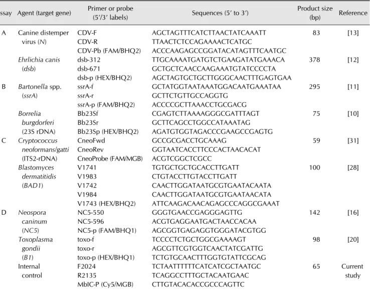
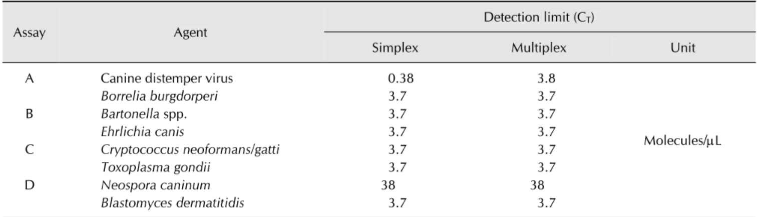
Meningoencephalitis (ME) is a common inflammatory disorder of the central nervous system in dogs. Clinically, ME has both infectious and non-infectious causes. In the present study, a multiplex quantitative real-time polymerase chain reaction (mqPCR) panel was optimized for the detection of eight canine neurologic pathogens (Blastomyces dermatitidis, Cryptococcus spp., Neospora caninum, Borrelia burgdorferi, Bartonella spp., Toxoplasma gondii, Ehrlichia canis, and canine distemper virus [CDV]). The mqPCR panel was subsequently applied to 53 cerebrospinal fluid (CSF) samples collected from dogs with ME. The analytic sensitivity (i.e., limit of detection, expressed as molecules per 1 L of recombinant vector) was 3.8 for CDV, 3.7 for Ehrlichia canis, 3.7 for Bartonella spp., 3.8 for Borrelia burgdorferi, 3.7 for Blastomyces dermatitidis, 3.7 for Cryptococcus spp., 38 for Neospora caninum, and 3.7 for Toxoplasma gondii. Among the tested CSF samples, seven (15%) were positive for the following pathogens in decreasing order of frequency: Cryptococcus spp. (3/7), Blastomyces dermatitidis (2/7), and Borrelia burgdorferi (2/7). In summary, use of an mqPCR panel with high analytic sensitivity as an initial screen for infectious agents in dogs with ME could facilitate the selection of early treatment strategies and improve outcomes.

Keywords: canine, meningoencephalitis, multiplex polymerase chain reaction, neurologic pathogen

Introduction

Meningoencephalitis (ME) results from concurrent inflammation of the meninges (meningitis) and brain (encephalitis) [22]. ME can be classified as granulomatous meningoencephalomyelitis, necrotizing meningoencephalitis, or necrotizing leukoencephalitis based on histopathologic features [28]. However, in veterinary practice, this classification is challenging because it requires biopsy and histopathologic examination. Thus, a simple and rapid strategy for identifying the causative agent of ME would be useful for selecting an appropriate therapeutic regimen.

Multiple infectious agents can cause ME in dogs. The major infectious agents include viruses (canine distemper virus [CDV]), fungi (Blastomyces [B.] dermatitidis, Cryptococcus [C.]

neoformans, Histoplasma capsulatum, and Coccidioides spp.), bacteria (Borrelia [B.] burgdorferi, Bartonella spp., and

Ehrlichia [E.] canis), and parasites (Neospora [N.] caninum and Toxoplasma [T.] gondii) [4,15,17,19,23,25,31]. The pathogenesis of ME can also involve immune-mediated or idiopathic etiologies that provoke aseptic inflammatory responses in the central nervous system (CNS).

Rapid and reliable detection of the causative pathogens of

ME is essential for implementation of timely and effective

therapeutic strategies. Routine techniques in veterinary diagnostic

laboratories include bacterial and fungal cultures,

immunohistochemistry, antigen-capturing enzyme-linked

immunosorbent assay (ELISA), and polymerase chain reaction

(PCR) [4,15,17,19,23,25,31]. These methods are laborious,

expensive, and time-consuming, and their sensitivity depends

on specimen quality, timing of sampling, and prior antimicrobial

therapy [7]. Furthermore, different assays for detecting the

various pathogens have variable sensitivities and specificities,

leading to biased outcomes [3]. Recent studies have used

(2)

multiplex quantitative real-time PCR (mqPCR) as a diagnostic tool for multifactorial diseases because this approach can simultaneously detect multiple pathogens with good diagnostic performance and is faster than conventional methods [8,29].

The present study was conducted to develop an mqPCR-based diagnostic approach for ME (hereafter, ME mqPCR) that simultaneously detects eight infectious agents (B. dermatitidis, C. neoformans, N. caninum, B. burgdorferi, Bartonella spp., T.

gondii, E. canis, and CDV) and to assess the prevalence of these pathogens among cases of canine ME in South Korea.

Materials and Methods

Biological materials and vectors

Type strains of C. neoformans (ATCC 32045), Staphylococcus aureus (ATCC 29213, 33592, BAA-44, BAA-41, and BAA-1683), Escherichia coli (ATCC 25922), and Malassezia pachydermatis (ATCC 14522) were purchased from the American Type Culture Collection (ATCC; USA). Field isolates of B. burgdorferi (n = 2), T. gondii (n = 2), E. canis (n = 1), E. ewingii (n = 1), Anaplasma phagocytophilum (n = 2), candidatus Mycoplasma haematoparvum (n = 1), S. pseudintermedius (n = 5), Candida spp. (n = 3), Malassezia spp. (n = 5), Microsporum canis (n = 3), Microsporum gypseum (n = 2), Aspergillus spp. (n = 3), canine parvovirus (n = 3), canine coronavirus (n = 3), and CDV (n = 6) were identified at Chungbuk National University Veterinary Diagnostic Laboratory. Recombinant plasmid vectors for B.

dermatitidis, C. neoformans, N. caninum, B. burgdorferi, Bartonella spp., T. gondii, E. canis, and CDV were used to assess the analytical specificity and as positive controls.

Nucleic acids (DNA and RNA) extracted from whole blood in two dogs were used as negative controls. All field isolates were identified by culture, conventional PCR, or antigen-capturing ELISA and verified by DNA sequencing. The vectors were constructed by amplifying synthetic oligonucleotides based on sequences obtained from the GenBank database (National Center for Biotechnology Information, USA) using the primers listed in Table 1 and cloned into a T-Blunt PCR Cloning Kit (SolGent, Korea) according to the manufacturer’s instructions.

Correct insertion into the vector was verified by sequencing.

Each recombinant vector was prepared in diethylpyrocarbonate- treated distilled, deionized water as a series of 10-fold serial dilutions and then used to assess the analytic sensitivity of the test.

Nucleic-acid extraction

Total nucleic acid was extracted from the samples using the MagMAX Total Nucleic Acid Isolation Kit (Applied Biosystems, USA) according to the manufacturer’s instructions. Briefly, 200 L of each sample were added to a bead tube containing zirconia beads, followed by 235 L of lysis/binding solution.

The bead tube was then placed on an Ambion Vortex Adapter

and beaten with a Vortex-Genie 2 (Scientific Industries, USA) at maximum speed for 15 min. After beating, the tubes were centrifuged at 16,000 × g for 3 min, and the supernatant was then carefully transferred into clean microcentrifuge tubes.

Following another centrifugation step at 16,000 × g for 6 min, 115 L of the supernatant was transferred to a 96-well, deep-well microplate containing 20 L of paramagnetic beads and 65 L of 100% isopropanol. The plate was shaken for 1 min on an orbital multi-well-plate shaker at maximum speed without spilling the sample, after which it was placed on a magnetic stand for 5 min and the supernatant was carefully discarded. The bead pellet was then washed twice each with Wash solution I and II (150 L). Next, the plate was vigorously shaken for 2 min, after which the resultant pellet was resuspended in 50 L of elution buffer. Finally, the extracted total nucleic acid in elution buffer was stored at −80

o

C until use in PCR.

Primers and probes

Primer and probe sequences used for ME mqPCR were based on published reports [10,11,12,13,16,20,27,30] and are listed in Table 1. All primers and probes were synthesized and purchased from Bioneer (Korea), except probes for Minor Groove Binder (Applied Biosystems). A 65 bp oligonucleotide was used as an internal control (IC) to monitor false negatives resulting from failure of PCR due to the presence of inhibitory substances. The IC contained a non-specific 16S ribosomal RNA gene sequence flanked by the uvrC gene sequence of Mycobacterium (M.) bovis to minimize cross-reactivity with M. bovis. The IC (0.001

M) was spiked into each reaction, which resulted in a positive signal with threshold cycle (C

T

) values between 36 and 38.

mqPCR panel

Primers and probes were categorized as mqPCR A, B, C, and

D (Table 1). Each mqPCR reaction included two primer/probe

sets for the target pathogen, one primer/probe set for the IC, and

a reference dye. To minimize cross-interference of the dyes for

each probe, a combination of FAM and HEX was used for the

target probes. Cy5 was used for the IC, and ROX was used as the

reference dye. PCR amplification was conducted using an Eco

Real-Time PCR system (Illumina, USA). The Path-ID Multiplex

One-Step RT-PCR kit (Applied Biosystems) was used for PCR

A, and the QuantiTect Multiplex PCR (Qiagen, USA) kit was

used for PCR B, C, and D according to the manufacturer’s

recommended protocols. Each 20 L reaction contained 0.4 M

of each primer, 0.2 M of each probe, and 5 L (for PCR A) or

2 L (for PCR B, C, and D) of the template. Cycling conditions

were as follows: reverse transcription for 10 min at 45

o

C

(omitted for PCR B, C, and D), 10 min (for PCR A) or 15 min

(for PCR B, C, and D) initial activation at 95

o

C, and then 35

cycles of 15 sec at 95

o

C and 60 sec (for PCR A) or 90 sec (for

PCR B, C, and D) at 60

o

C. A C

T

of 35 or lower was considered

positive for a given pathogen.

(3)

Table 1. Primer and probe sequences used for multiplex quantitative real-time polymerase chain reaction (mqPCR) in this study Assay Agent (target gene) Primer or probe

(5’/3’ labels) Sequences (5’ to 3’) Product size

(bp) Reference A Canine distemper

virus (N)

CDV-F CDV-R

CDV-Pb (FAM/BHQ2)

AGCTAGTTTCATCTTAACTATCAAATT TTAACTCTCCAGAAAACTCATGC

ACCCAAGAGCCGGATACATAGTTTCAATGC

83 [13]

Ehrlichia canis (dsb)

dsb-312 dsb-671

dsb-p (HEX/BHQ2)

TTGCAAAATGATGTCTGAAGATATGAAACA GCTGCTCAACCAAGAAATGTATCCCCTA AGCTAGTGCTGCTTGGGCAACTTTGAGTGAA

378 [12]

B Bartonella spp.

(ssrA)

ssrA-f ssrA-r

ssrA-p (FAM/BHQ2)

GCTATGGTAATAAATGGACAATGAAATAA GCTTCTGTTGCCAGGTG

ACCCCGCTTAAACCTGCGACG

295 [11]

Borrelia burgdorferi (23S rDNA)

Bb23Sf Bb23Sr

Bb23Sp (HEX/BHQ2)

CGAGTCTTAAAAGGGCGATTTAGT GCTTCAGCCTGGCCATAAATAG AGATGTGGTAGACCCGAAGCCGAGTG

75 [10]

C Cryptococcus neoformans/gatti (ITS2-rDNA)

CneoFwd CneoRev

CneoProbe (FAM/MGB)

GCCGCGACCTGCAAAG

GGTAATCACCTTCCCACTAACACAT ACGTCGGCTCGCC

59 [31]

Blastomyces dermatitidis (BAD1)

V1741 V1983 V1742 V1984

V1743 (HEX/BHQ2)

TGTGCTGCTGCACCTTGATT CTGTACCTTGTACCTTGATT

CAACTTGGATAATGCGTGAATACAATA CAACTTGGATAATGCGTGAATAACATA ATTCAAGACAACAGAGCCCAGGCGAAAT

100 [28]

D Neospora caninum (NC5)

NC5-550 NC5-596

NC5-p (FAM/BHQ1)

GGGTGAACCGAGGGAGTTG ACGTGAGGAATGACTAACCACAA AGCGGTGAGAGGTGGGATACGTGG

142 [16]

Toxoplasma gondii (B1)

toxo-f toxo-r

toxo-p (HEX/BHQ1)

TCCCCTCTGCTGGCGAAAAGT AGCGTTCGTGGTCAACTATCGATTG TCTGTGCAACTTTGGTGTATTCGCAG

98 [20]

Internal control

F2024 R2135

MbIC-P (Cy5/MGB)

TCTAATTTTTTCATCATCGCTAATGC TCAGGCCTTTGCTACAATGAAC CTTGTACACACCGCCCAGTTC

65 Current study

Limit of detection measurement

To compare and optimize the PCR conditions, analytical sensitivity was assessed for each pathogen in triplicate as a singleplex assay and the multiplex panel by using the corresponding recombinant vector. The singleplex PCR assay was performed using the same regents and PCR conditions as the multiplex panel. The reaction for E. canis was performed using two mqPCR kits (Path-ID Multiplex One-Step real time-PCR kit; Applied Biosystems; QuantiTect Multiplex PCR; Qiagen) with the recombinant vector as a template to determine whether the two kits had comparable sensitivity.

Cerebrospinal fluid (CSF) samples

A total of 47 CSF samples collected from dogs with ME or submitted to Chungbuk National University between January 2010 and August 2013 and six CSF samples collected from healthy dogs were included in this study. All dogs with ME presented with non-specific neurological symptoms, such as

seizure, incoordination, or altered consciousness, and had no

laboratory abnormalities consistent with a metabolic disorder

or radiographic abnormalities (i.e., complete blood count,

serum biochemistry profile, urinalysis, X-ray and abdominal

ultrasonography) that would suggest non-CNS disease as the

cause of symptoms. The criteria for case selection included: (1)

evidence of focal or multifocal brain lesions during neurological

examination without signs of spinal cord lesions or lesions in

the peripheral nervous system; (2) abnormal result of CSF

examination indicating inflammation and tissue necrosis

(reference interval: < 5 white blood cells/L, total protein <

0.3 g/L); and (3) evidence of focal or multifocal intracranial

lesions upon computed tomography (CT) or magnetic resonance

imaging (MRI) [5,6,14,21,29]. After diagnosis, the residual

CSF samples were used for the mqPCR assay.

(4)

Fig. 1. Detection of viral and bacterial meningoencephalitis pathogens in canine cerebrospinal fluid samples by mqPCR. Specific primer and probe sets were used to amplify sequences from (A) canine distemper virus, Ehrlichia (E.) canis, Bartonella spp., Borrelia (B.) burgdorferi, (B) Blastomyces (B.) dermatitidis, Cryptococcus (C.) neoformans, Neospora (N.) caninum, and Toxoplasma (T.) gondii.

Known quantities (i.e., copy number per L) of recombinant vector encoding the target gene from each pathogen were prepared as serial 10-fold dilutions and used as controls. Cycle threshold (C

T

) values are shown on the Y-axis. Each regression line was obtained from triplicate measurements.

Table 2. Detection limits and corresponding threshold cycle (Ct) values from simplex and multiplex PCR assays

Assay Agent Detection limit (C

T

)

Simplex Multiplex Unit

A Canine distemper virus 0.38 3.8

Molecules/L

Borrelia burgdorperi 3.7 3.7

B Bartonella spp. 3.7 3.7

Ehrlichia canis 3.7 3.7

C Cryptococcus neoformans/gatti 3.7 3.7

Toxoplasma gondii 3.7 3.7

D Neospora caninum 38 38

Blastomyces dermatitidis 3.7 3.7

Results

Analytical specificity and sensitivity

All mqPCR assays detected only the target pathogen and did not produce any non-specific amplification products or products of other pathogens in repeated experiments. In addition, there were no false positives due to cross-talk between dye signals within each reaction. Standard curves for all multiplex assays generated from 10-fold serial dilutions of each template showed correlation coefficients (R

2

) ranging from 0.970 to 0.995 and slopes of 3.41–3.59 (Fig. 1), indicating good linearity of PCR amplification.

Next, the performance of the mqPCR panel and singleplex

PCR for each of the eight pathogens were compared directly to identify any negative effects of multiplexing on PCR detection.

The analytical sensitivity (i.e., limit of detection) of the PCR

was estimated using serially diluted recombinant vectors with

known copy numbers (molecules per L). The estimated

mqPCR detection limits of the mqPCR panel were 3.8 for CDV,

3.7 for E. canis, 3.7 for Bartonella spp., 3.8 for B. burgdorferi,

3.7 for B. dermatitidis, 3.7 for C. neoformans, 38 for N. caninum,

and 3.7 for T. gondii (Table 2). The results of singleplex and

multiplex PCR were identical except for CDV, and C

T

differences

between PCR reactions were not significant, demonstrating that

multiplexing did not have a significant negative effect on

sensitivity of the PCR.

(5)

The C

T

value obtained for E. canis using the Path-ID Multiplex One-Step RT-PCR kit (Applied Biosystems) was slightly lower than that obtained with the QuantiTect Multiplex PCR kit (Qiagen); however, the difference in C

T

was less than 1, suggesting that the kits had comparable performance.

CSF sample analysis

Of the 47 CSF samples obtained from dogs diagnosed with ME, seven (15%) were positive for the target pathogen. The most common pathogens were C. neoformans (3/7), B.

dermatitidis (2/7), and B. burgdorferi (2/7). In contrast, conventional CSF analysis and culture was negative for all pathogens. None of the samples were positive for more than one pathogen. For the seven samples with discrepant results between the mqPCR panel and routine test methods, agarose gel electrophoresis, sequencing, and comparison with sequences available in the GenBank database confirmed the presence of the identified pathogen. Among the six CSF samples from healthy dogs, all were negative for the eight pathogens of the mqPCR panel, suggesting that mqPCR did not falsely detect normal CSF constituents as target materials.

Discussion

In the present study, a panel of four mqPCR assays (one qRT-PCR and three qPCRs) that can simultaneously detect eight pathogens (B. dermatitidis, C. neoformans, N. caninum, B. burgdorferi, Bartonella spp., T. gondii, E. canis, and CDV) was optimized and applied to CSF samples. The analytic sensitivity of the panel was as good as each singleplex assay, and there was no interference among primer/probe sets, suggesting that the panel could be substituted for the singleplex assay. The panel optimized in this study was also designed for use with one-step extraction and PCR procedures in a 48-well format. Accordingly, simultaneous testing of a large number of samples can be achieved at a lower cost, with less time and effort than routine diagnostic techniques, such as individual PCR, cultures, microscopic examination, and ELISA.

ME is a common inflammatory CNS disease in dogs [22].

Histopathology can be used to classify ME based on the tissue manifestations of the disease. However, histopathological diagnosis has little relevance for clinical outcome because treatments are not specific for the ME subtype. A previous study employed PCR to examine the presence of viral agents that cause human ME in a small number of canine ME cases and found that none were positive for human ME viruses [26]. In contrast, the present study focused on eight known neurologic pathogens that have caused outbreaks in Korea and confirmed their relatively high prevalence among dogs with ME (7/47).

Therefore, it is possible that the prevalence of neurologic infections causing ME in dogs has been underestimated.

Although the cases of ME that tested positive for pathogens

were not confirmed by histopathology, one case of infection with Cryptococcus spp. exhibited dramatic clinical improvement after a two-month treatment with oral fluconazole. Moreover, after observation for one year, there was no relapse of neurological signs, suggesting the importance of early pathogen detection for the selection of an effective treatment strategy. In the present study, five of the seven cases of infection were with a fungal agent that could not be eradicated by conventional antibiotics or anti-inflammatory agents, demonstrating the importance of proper diagnosis.

Although uncommon, fungal infections in dogs pose a diagnostic and therapeutic challenge [19]. Symptoms of fungal infections vary depending on the location and distribution of the lesion, making it difficult to distinguish them from non- infectious inflammatory or neoplastic disorders based on clinical manifestations [19]. For example, Cryptococcus spp., the most common fungal pathogen in this study, tends to infect the cerebellum, resulting in non-specific neurological symptoms, such as seizures and obtundation [2,19]. B. dermatitidis, which is endemic to North America, is seldom associated with CNS infection [1] and usually presents with lymphadenopathy or pulmonary infiltration [17]. B. dermatitidis infection is rare among humans in Korea [24]; however, it was found to be the second most common pathogen in the present study. Interestingly, these cases did not exhibit typical clinical signs, indicating that appropriate laboratory tests are critical for achieving a definitive diagnosis. This is the first report to implicate B.

dermatitidis in canine ME in Korea.

Lyme disease is a zoonotic tick-borne disease caused by B.

burgdorferi that affects various organs in dogs, including the skin, lymph nodes, aorta, and kidney [4,9]. Although the bacterium also infects the meninges and choroid plexus, CNS involvement is not pathognomonic of Lyme disease [18]. In the present study, two cases positive for B. burgdorferi had a history of ixodid tick infestation. In both cases, the locations of CNS lesions included the choroid plexus, which can be associated with neurologic signs in dogs afflicted by neuroborreliosis.

However, it was not possible to confirm whether the lesions were caused by B. burgdorferi because the owners declined additional tests and therapeutic trials.

In conclusion, an mqPCR-based test panel for eight canine

neurologic pathogens was developed and applied to 47 CSF

samples in this study. The panel with high analytic sensitivity

confirmed that seven (15%) samples were positive to the target

pathogen, suggesting the necessity for screening of infectious

agents in dogs with ME. Because treatment strategies for

different histopathologic subtypes of ME are not specific, initial

screening for infectious agents using the developed panel

followed by CT or MRI would be useful for selecting an

appropriate treatment in a timely manner.

(6)

Acknowledgments

The authors thank the staff of Nowon N and Helix Animal Medical Centers for assistance collecting samples and interpreting the MRI results. This study was supported by the Wildlife Center of Chungbuk and aniDAP Laboratories in Korea.

Conflict of Interest

There is no conflict of interest.

References

1. Arceneaux KA, Taboada J, Hosgood G. Blastomycosis in dogs: 115 cases (1980-1995). J Am Vet Med Assoc 1998, 213, 658-664.

2. Berthelin CF, Bailey CS, Kass PH, Kegendre AM, Wolf AM.

Cryptococcosis of the nervous system in dogs. Part 1:

Epidemiologic, clinical, and neuropathologic features. Prog Vet Neurol 1994, 5, 88-97.

3. Bhat N, O’Brien KL, Karron RA, Driscoll AJ, Murdoch DR;

Pneumonia Methods Working Group. Use and evaluation of molecular diagnostics for pneumonia etiology studies. Clin Infect Dis 2012, 54 (Suppl 2), S153-158.

4. Chang YF, Novosel Y, Chang CF, Summers BA, Ma DP, Chiang YW, Acree WM, Chu HJ, Shin S, Lein DH.

Experimental induction of chronic borreliosis in adult dogs exposed to Borrelia burgdorferi-infected ticks and treated with dexamethasone. Am J Vet Res 2001, 62, 1104-1112.

5. Cherubini GB, Platt SR, Anderson TJ, Rusbridge C, Lorenzo V, Mantis P, Cappello R. Characteristics of magnetic resonance imags of granulomatous meningoencephalomyelitis in 11 dogs. Vet Rec 2006, 159, 110-115.

6. Cherubini GB, Platt SR, Howson S, Baine E, Brodbelt DC, Dennis R. Comparison of magnetic resonance imaging sequences in dogs with multi-focal intracranial disease. J Small Anim Pract 2008, 49, 634-640.

7. Cho YI, Han JI, Wang C, Copper V, Schwartz K, Engelken T, Yoon KJ. Case-control study of microbiological etiology associated with calf diarrhea. Vet Microbiol 2013, 166, 375-385.

8. Cho YI, Kim WI, Liu S, Kinyon JM, Yoon KJ. Development of a panel of multiplex real-time polymerase chain reaction assays for simultaneous detection of major agents causing calf diarrhea in feces. J Vet Diagn Invest 2010, 22, 509-517.

9. Chou J, Wünschmann A, Hodzic E, Borjesson DL. Detection of Borrelia burgdorferi DNA in tissues from dogs with presumptive Lyme borreliosis. J Am Vet Med Assoc 2006, 229, 1260-1265.

10. Courtney JW, Kostelnik LM, Zeidner NS, Massung RF.

Multiplex real-time PCR for detection of Anaplasma phagocytophilum and Borrelia burgdorferi. J Clin Microbiol 2004, 42, 3164-3168.

11. Diaz MH, Bai Y, Malania L, Winchell JM, Kosoy MY.

Development of a novel genus-specific real-time PCR assay for detection and differentiation of Bartonella species and genotypes. J Clin Microbiol 2012, 50, 1645-1649.

12. Doyle CK, Labruna MB, Breitschwerdt EB, Tang YW, Corstvet RE, Hegarty BC, Bloch KC, Li P, Walker DH, McBride JW. Detection of medically important Ehrlichia by quantitative multicolor TaqMan real-time polymerase chain reaction of the dsb gene. J Mol Diagn 2005, 7, 504-510.

13. Elia G, Decaro N, Martella V, Cirone F, Lucente MS, Lorusso E, Di Trani L, Buonavoglia C. Detection of canine distemper virus in dogs by real-time RT-PCR. J Virol Methods 2006, 136, 171-176.

14. Flegel T, Henke D, Boettcher IC, Aupperle H, Oechtering G, Matiasek K. Magnetic resonance imaging findings in histologically confirmed Pug dog encephalitis. Vet Radiol Ultrasound 2008, 49, 419-424.

15. Gerber JE, Johnson JE, Scott MA, Madhusudhan KT. Fatal meningitis and encephalitis due to Bartonella henselae bacteria. J Forensic Sci 2002, 47, 640-644.

16. Ghalmi F, China B, Kaidi R, Daube G, Losson B. Detection of Neospora caninum in dog organs using real time PCR systems. Vet Parasitol 2008, 155, 161-167.

17. Kerl ME. Update on canine and feline fungal diseases. Vet Clin North Am Small Anim Pract 2003, 33, 721-747.

18. Krimer PM, Miller AD, Li Q, Grosenbaugh DA, Susta L, Schatzberg SJ. Molecular and pathological investigations of the central nervous system in Borrelia burgdorferi-infected dogs. J Vet Diagn Invest 2011, 23, 757-763.

19. Lavely J, Lipsitz D. Fungal infections of the central nervous system in the dog and cat. Clin Tech Small Anim Pract 2005, 20, 212-219.

20. Lin MH, Chen TC, Kuo TT, Tseng CC, Tseng CP. Real-time PCR for quantitative detection of Toxoplasma gondii. J Clin Microbiol 2000, 38, 4121-4125.

21. Lowrie M, Smith PM, Garosi L. Meningoencephalitis of unknown origin: investigation of prognostic factors and outcome using a standard treatment protocol. Vet Rec 2013, 172, 527.

22. Menaut P, Landart J, Behr S, Lanore D, Trumel C. Treatment of 11 dogs with meningoencephalomyelitis of unknown origin with a combination of prednisolone and cytosine arabinoside. Vet Rec 2008, 162, 241-245.

23. Panciera RJ, Ewing SA, Confer AW. Ocular histopathology of ehrlichial infections in the dog. Vet Pathol 2001, 38, 43-46.

24. Park KS, Ki CS, Lee NY. Laboratory experience in phenotypic and molecular identification of Blastomyces dermatitidis first isolated in Korea. Korean J Clin Microbiol 2012, 15, 114-116.

25. Rudd PA, Bastien-Hamel LE, von Messling V. Acute canine distemper encephalitis is associated with rapid neuronal loss and local immune activation. J Gen Virol 2010, 91, 980-989.

26. Schatzberg SJ, Haley NJ, Barr SC, de Lahunta A, Sharp NJH. Polymerase chain reaction screening for DNA viruses in paraffin-embedded brains from dogs with necrotizing meningoencephalitis, necrotizing leukoencephalitis, and granulomatous meningoencephalitis. J Vet Intern Med 2005, 19, 553-559.

27. Sidamonidze K, Peck MK, Perez M, Baumgardner D, Smith

G, Chaturvedi V, Chaturvedi S. Real-time PCR assay for

identification of Blastomyces dermatitidis in culture and in

tissue. J Clin Microbiol 2012, 50, 1783-1786.

(7)

28. Talarico LR, Schatzberg SJ. Idiopathic granulomatous and necrotising inflammatory disorders of the canine central nervous system: a review and future perspectives. J Small Anim Pract 2010, 51, 138-149.

29. Thonur L, Maley M, Gilray J, Crook T, Laming E, Turnbull D, Nath M, Willoughby K. One-step multiplex real time RT-PCR for the detection of bovine respiratory syncytial virus, bovine herpesvirus 1 and bovine parainfluenza virus 3. BMC Vet Res 2012, 8, 37.

30. Veron V, Simon S, Blanchet D, Aznar C. Real-time polymerase chain reaction detection of Cryptococcus neoformans and Cryptococcus gattii in human samples. Diagn Microbiol Infect Dis 2009, 65, 69-72.

31. Williams JH, Köster LS, Naidoo V, Odendaal L, van

Veenhuysen A, de Wit M, van Wilpe E. Review of idiopathic

eosinophilic meningitis in dogs and cats, with a detailed

description of two recent cases in dogs. J S Afr Vet Assoc

2008, 79, 194-204.

수치

Table 1. Primer and probe sequences used for multiplex quantitative real-time polymerase chain reaction (mqPCR) in this study Assay Agent (target gene) Primer or probe
Table 2. Detection limits and corresponding threshold cycle (Ct) values from simplex and multiplex PCR assays

참조

관련 문서

 Multiplex real time RT-PCR 호흡기 바이러스 & 세균..

This solution includes Smart Monitoring, which checks the status of miners and the mining in real-time, and provides the information necessary for optimal decision

This paper proposes the improved real time object detection and distance measurement system using IR-UWB radar.. Firstly, the IR-UWB signal is applied for

Wald-Type Tests for Detecting Breaks in The Trend Function of a Dynamic

The object of this study is to analyse the bacteria of periapical bascess and fascial space abscess using multiplex real-time polymerase chain reaction (MRT-PCR) before

Although there was a trend for a greater decline in quantitative HBsAg titers in the group that achieved HBeAg seroclearance, the difference in the kinetics of qHBsAg

- Increase the rise time significantly if the extra pole is within a factor of 4 of the real part of the complex poles. of 4 of the real part of

- For gas-phase reactions, the volumetric flow rate most often changes during the course of the reaction due to a often changes during the course of the reaction due to a change