Effectiveness of Arch Support Taping is Subjects With Excessive Foot Pronation: A Meta-analysis
So-yeon Park
1, PhD, PT
1Dept. of Physical Therapy, College of Health Science, Sangji University
Abstract
1)Background: An excessive pronated foot is defined as a flattening or complete loss of the medial longitudinal arch. Excessive foot pronation is considered to have high risk factors of overuse injuries in the lower limb. Various treatments have been investigated in attempts to control excessive pronation.
Objects: This meta-analysis identifies the effects of an anti-pronation taping technique using different materials.
Methods: The electronic databases used include MEDLINE, the Physiotherapy Evidence Database (PEDro), Science Direct, the Korean Studies Information Service System (KISS), the Research Information Sharing Service (RISS), the Korea National Library, and the Korean Medical Database (studies published up to July 31, 2019). The database search used the following keywords: "foot drop" OR "foot arch" OR
"foot pronation" OR "flat foot (pes planus)" AND "taping" OR "support." Eight eligible studies were analyzed to determine the effectiveness of anti-pronation taping in study and control groups.
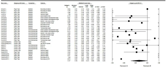
Results: The overall random effect size (Hedges’ g) of the anti-pronation taping technique was 0.147 (95% confidence interval [CI]: -.214 to .509). When the effect (Hedges’ g) was compared by the type of tape material, rigid tape (RT; Lowdye taping) was .213 (95% CI: -.278 to .704) and kinesiotape (KT; arch support taping) was -.014 (95% CI: -.270 to .242). Based on this meta-analysis, it was not possible to identify the extent to which anti-pronation taping was effective in preventing navicular drop, improving balance, or changing foot pressure. Only three of the eight eligible studies applied KT on excessive pronated feet, and the outcome measure areas were different to those of the RT studies. The KT studies used EMG data, overall foot posture index (FPI) scores, and rear foot FPI scores. In contrast, the RT studies measured navicular heights, various foot angles, and foot pressure.
Conclusion: This review could not find any conclusive evidence about the effectiveness of any taping method for patients with pronated feet. Future studies are needed to develop the anti-pronation taping technique based on the clinical scientific evidence.
Key Words: Kinesio taping; Meta-analysis; Rigid taping; Pronated foot.
Introduction
Pes planus has been defined as a loss or flat- tening of the medial longitudinal arch; it is also called excessive pronated foot (Kodithuwakku Arachchige et al, 2019). Foot pronation action is nor- mally needed for shock absorption during the gait’s intial stance phases (Lafortune et al, 1994). However abnormal foot pronation tends have other structural
causes, such as tibial internal rotation or foot adduc- tion, altering normal biomechanical mechanism. The flat foot population endures decreased postural stabil- ity, which is associated with higher incidence of lower extremity overuse injuries (Dahle et al, 1991;
Levinger et al, 2010). Such problems typically include medial tibial stress syndrome, patellofemoral pain syndrome, metatarsal stress fracture, plantar fasciitis, Achilles tendinitis, and hallux ridius (Cheung and Corresponding author: So-yeon Park [email protected]
This research was supported by Sangji University Research Fund, 2018.
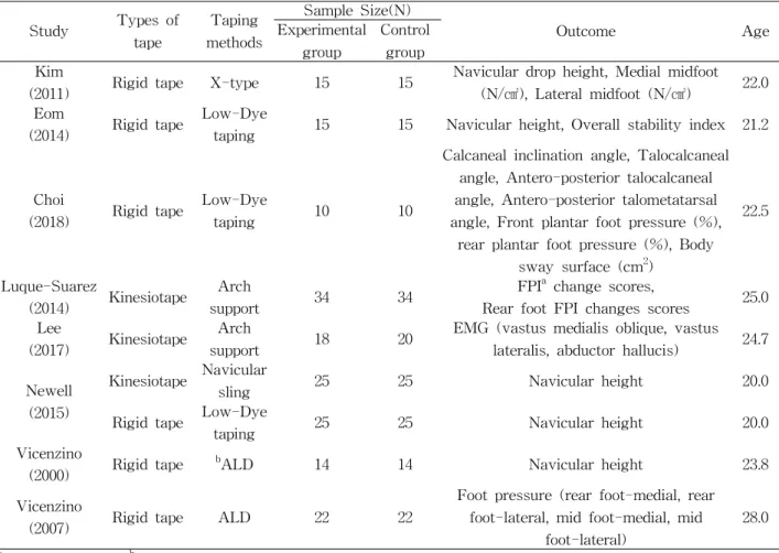
Study Types of tape
Taping methods
Sample Size(N)
Outcome Age
Experimental group
Control group Kim
(2011) Rigid tape X-type 15 15 Navicular drop height, Medial midfoot (N/㎠), Lateral midfoot (N/㎠) 22.0 Eom
(2014) Rigid tape Low-Dye
taping 15 15 Navicular height, Overall stability index 21.2
Choi
(2018) Rigid tape Low-Dye
taping 10 10
Calcaneal inclination angle, Talocalcaneal angle, Antero-posterior talocalcaneal angle, Antero-posterior talometatarsal angle, Front plantar foot pressure (%),
rear plantar foot pressure (%), Body sway surface (cm
2)
22.5
Luque-Suarez
(2014) Kinesiotape Arch
support 34 34 FPI
achange scores,
Rear foot FPI changes scores 25.0 Lee
(2017) Kinesiotape Arch
support 18 20 EMG (vastus medialis oblique, vastus lateralis, abductor hallucis) 24.7 Newell
(2015)
Kinesiotape Navicular
sling 25 25 Navicular height 20.0
Rigid tape Low-Dye
taping 25 25 Navicular height 20.0
Vicenzino
(2000) Rigid tape
bALD 14 14 Navicular height 23.8
Vicenzino
(2007) Rigid tape ALD 22 22
Foot pressure (rear foot-medial, rear foot-lateral, mid foot-medial, mid
foot-lateral)
28.0
a