ISSN 1225-6552, eISSN 2287-7630 https://doi.org/10.7853/kjvs.2020.43.4.237
< Original Article >
Veterinary Service
Available online at http://kjves.org
*Corresponding author: Dongseob Tark, E-mail. [email protected] ORCID https://orcid.org/0000-0001-7499-4253
These first two authors contributed equally to this work.
Development of sandwich enzyme-linked immunosorbent assay for a large-scale detection of porcine transmissible gastroenteritis virus in feces
Yeonsu Oh
1, Sang-Joon Lee
1, Ho-Seong Cho
2, Dongseob Tark
3*
1
Department of Veterinary Pathology, Collage of Veterinary Medicine and Institute of Veterinary Science, Kangwon National University, Chuncheon 24341, Korea
2
College of Veterinary Medicine and Bio-Safety Research Institute, Jeonbuk National University, Iksan 54596, Korea
3
Korea Zoonosis Research Institute, Jeonbuk National University, Iksan 54531, Korea (Received 24 November 2020; revised 15 December 2020; accepted 17 December 2020)
Abstract
Porcine transmissible gastroenteritis (TGE) has been a significant cause of economic losses in pig farm- ing industry since 1950s. Although transmissible gastroenteritis virus (TGEV) has declined in recent years, it should not be excluded because of its characteristics; the frequency of gene mutation, the mortality in piglets, and the possibility for sudden incidence. Therefore, the herd-level monitoring of the virus is important to prevent further circulation of TGE. The aim of this study is to develop a large-scale sand- wich enzyme-linked immunosorbent assay (ELISA) with high specificity to rapidly detect TGEV in fe- ces by using monoclonal antibodies (Mabs). The TGEV specific Mabs were produced in hybridoma cells.
Among the Mabs belonged to the IgG class developed by this study, the final selected 8H6, 1B7, 4G3, and 1F8 were identified to have the neutralization ability against TGEV. The sandwich ELISA was es- tablished using 8H6 as a reporter antibody and 1B7 and the reported 5C8 as a capture antibody. The developed sandwich ELISA was able to distinguish TGEV from other pathogenic diarrheal agents (porcine rotavirus, porcine reovirus, porcine epidemic diarrhea virus (PEDV), E. coli, and C. perfringens) in tis- sue culture as well as fecal samples. And the detection rate of TGEV in feces was 80% compared with RT-PCR. The results suggested that the developed sandwich ELISA may be useful in the herd-level monitoring for effective preventive measures due to the early diagnosis of TGEV using a large amount of samples.
Key words : Porcine transmissible gastroenteritis virus, Sandwich ELISA, Herd-level monitoring
INTRODUCTION
Diarrhea in swine is acting as one of the biggest fac- tors which reduce productivity in the industry. Several types of gastrointestinal diseases with diarrhea occur throughout the year and cause significant economic loss- es to pig farms. Pathogens such as bacteria, viruses, and protozoa can cause the various types of diarrhea, but the most problematic are viral diarrhea; transmissible gastro- enteritis (TGE) and porcine epidemic diarrhea (PED).
In recent years, the porcine epidemic diarrhea virus (PEDV) has been recognized as one of the most noto- rious viruses for porcine diarrhea in terms of the num- ber of positive cases and the economic losses. And the transmissible gastroenteritis virus (TGEV) has declined with its innocent relative, non-enteropathogenic porcine respiratory coronavirus (PRCV). However, there are reasons to monitor transmissible gastroenteritis virus periodically.
First, the TGEV belongs to the genus Alphacoronavirus,
family Coronaviridae like PEDV. Therefore, despite the
presence of a vaccine, TGEV also continues to occur
due to frequent gene mutations. Second, the mortality
rate of TGEV can approach up to 100% in piglets under
1 week of age and either infected or recovered pigs may be acting as a carrier with excreting viruses through fe- ces for a long period of time. Finally, according to KAHIS (Korea Animal Health Integrated System), the number of TGE positive cases is 1,729 since 2012 and 1,682 for only 2014. This indicate that an explosive in- crease may occur without any notice and suggest that periodic monitoring may be necessary. Therefore, the herd-level monitoring of the virus is important to pre- vent further circulation of the disease.
The aim of this study is to develop a large-scale di- agnostic method for detection of swine TGEV in feces.
In order to establish an effective preventive measure, it is necessary to develop a diagnostic method capable of discriminating the causative agents of swine diarrhea such as PEDV, rotavirus infection and coliform diarrhea, and detecting TGEV quickly and accurately in a large quantity. The diagnostic methods to detect TGEV were developed in various ways (Bohac et al, 1975; Saif et al, 1977; Chu et al, 1982; Asagi et al, 1986; Van Nieuwstadt et al, 1988; Oh and Tark, 2019) including fluorescence antibody test (Pensaert et al, 1970; Black, 1971). Dulac et al. (1977) attempted to isolate TGEV from field specimens using cell culture and piglets.
However, isolation and identification of TGEV by cell culture may take a long time and may not detect the causative agent. Although electron microscopy can de- tect the causative agents in the feces (Saif et al, 1977;
Van Nieuwstadt et al, 1988), both methods have the dis- advantage of requiring expertise and facilities in the in- spection process. In general, the fluorescence antibody test has the advantage of being able to rapidly test the small intestine by frozen section or mucous membrane smearing. However, there are many subjective factors in the test result, and freshness materials are needed. The aim of this study was to develop a sandwich ELISA with high specificity to rapidly detect TGEV in feces by using monoclonal antibodies that specifically reacts with TGEV.
MATERIALS AND METHODS
Viruses and bacteria used in the study
Total six TGEV strains, four Korean isolates strains (NVRI 48 strain, NVRI 41 strain, WP strain, and Pyeong- taek strain) and two standard viruses (Purdue strain and Miller strain), were used in the study. The proliferation and potency of each virus was measured respectively in the swine testicular (ST) cell line (ATCC, MD, USA).
Porcine endemic diarrhea virus (Wey strain and Japanese vaccine strain), porcine rotavirus (Korean isolate, OSU type), porcine reovirus (Korean field isolate), and por- cine pathogenic bacteria, Escherichia coli (K88ac) and Clostridium perfringens, were used. All of the viruses and bacteria were distributed from the Animal and Plant Quarantine Agency (APQA), and Ministry for Agriculture, Food and Rural Affairs (MAFRA) in Republic of Korea.
Fecal sample collection after TGEV challenge
3-day-old SPF pigs were orally inoculated with 10
7.0TCID
50/mL of TGEV (NVRI 48 strain-10 passages), and fecal samples were collected daily. At the peak of viral infection pigs were necropsied and intestinal contents were collected to be stored at −80°C until use. Pigs were fed with milk replacer ad libitum throughout ex- perimental periods in accordance with the institutional animal ethical standards (IACUC no. JBNU 2020-0127).
Preparation of the monoclonal antibody to TGEV
Monoclonal antibodies against TGEV were produced according to the literature (Coyle et al, 1992; Oh and Tark, 2019). Briefly, when the Sf9 cell infected with pF9AH-bac was showed cytopathic effect completely, the cell was harvested, sonicated, and mixed with in- complete Freund’s adjuvant (Sigma-Aldrich, MO, USA) for immunization. The prepared antigen was inoculated on the footpad of BALB/c mice under mild anesthesia.
The popliteal lymph nodes were collected 10 days after
inoculation, made into single cells, and fused with mur-
ine myeloma cell line P3X63 (ATCC, MD, USA). Pro-
duction of TGEV specific monoclonal antibody from the
hybridoma cells was confirmed as follows: the ST cell infected with TGEV and intact ST cell were prepared and fixed with acetone. The fixed cells were first re- acted with the hybridoma cell culture supernatant, fol- lowed by the rabbit anti-mouse immunoglobulins (IgG, IgA, and IgM) FITC conjugate (Sigma-Aldrich, MO, USA). The strong positive hybridoma cells for fluores- cent antibodies were cultured in feeder cell plates de- rived from ICR mouse peritoneal cavity. Screening and cloning were performed twice in a single well contain- ing hybridoma cells. Fully cloned hybridoma cells were stored in a liquid nitrogen tank until use.
The virus neutralization test for the selected mono- clonal antibody was performed by serial two-fold dilu- tion of antibodies with 200 TCID
50/50 µL of TGEV (Pyeongtaek strain) in a 96-well microplate. After sensi- tization, ST cells suspended in α-MEM medium supple- mented with 5% FCS were injected into all wells and cultured in a CO
2incubator for 4 ∼5 days. The result was expressed as the reciprocal of the highest serum di- lution factor neutralizing the 100 TCID
50/100 µL TGEV.
Antibody isotyping was performed using a mouse-hy- bridoma subtyping kit (Roche Diagnostics, Mannheim, Germany) according to the manufacturer’s procedure.
Purification of the monoclonal antibody and conjugation with horseradish peroxidase
The monoclonal antibody was purified using fast pro- tein liquid chromatography (FPLC) (Pharmacia, NJ, USA). The protein was quantified by the Micro BCA protein assay reagent kit (Pierce, MA, USA). The horse- radish peroxidase (HRP) was conjugated according to the periodate coupling method (Nakane and Kawaoi, 1974).
In brief, after dissolving 5 mg of HRP (peroxidase, Type VI-A) (Sigma-Aldrich, MO, USA) in 1.2 mL of distilled water, 0.3 mL of sodium periodate (0.1 M)/sodium phosphate (10 mM; pH 7.0) was added, and allowed to stand at room temperature for 20 minutes, followed by dialysis overnight in sodium acetate (1 mM; pH 4.0).
The dialyzed HRP was mixed with 6.6 mg of the puri- fied monoclonal antibody suspended in 0.5 mL of car- bonate (20 mM; pH 9.5), and incubated at room temper- ature for 2 hours. Then, 100 µL of sodium borohydride
(4 mg/mL) was added and incubated at 4 °C for 2 hours, followed by dialysis in PBS. The dialyzed conjugate was dispensed in 0.1 mL aliquots and stored at −20°C until use.
Sandwich ELISA
Sandwich ELISA to detect TGEV antigen in feces or cell culture medium was developed with modification as described previously (Bernard et al, 1986; Van Nieuwstadt et al, 1988). 5C8 and 1B7 which specifically binds to TGEV spike protein, as virus capture antibodies were diluted to 4.2 µg/mL and 6.6 µg/mL, respectively in 100 µL of carbonate-bicarbonate buffer (0.05 M; pH 9.6) per well of an ELISA plate (Maxi-sorp, Nunc, Denmark) and incubated at 37 °C overnight. Then, 150 µL of blocking solution (Tris, 0.01 M, pH 7.5; NaCl, 0.15 M;
gelatin, 1%; horse serum, 10%) was added per well and
incubated at 37 °C for 1 hour. The fecal samples were
diluted to 1/10 with diluting solution (Tris, 0.01 M, pH
7.5; NaCl, 0.15 M; gelatin, 1%; horse serum, 10%; Tween
20, 0.05%) and incubated at 37 °C for 2 hours in the
sensitized solid phase. Then, HRP-conjugated TGEV-spe-
cific monoclonal antibody was distributed. After 1 hour
of incubation at 37 °C, the 3,3’, 5,5’-tetramethyl-benzi-
dine (KPL, MD, USA) was added and reacted for 30
minutes. Finally, the reaction was stopped and the ab-
sorbance was measured at 450 nm using a microplate
reader (Tecan, Mannedorf, Switzerland). Between all
steps, washings were carried out four times with washes
(0.01 M Tris pH 7.5, 0.5 M NaCl, 0.05% Tween 20),
except for the blocking step, all reaction solutions were
100 µL per well. The results were obtained by adding
samples to the wells (S) containing the monoclonal anti-
body, and the wells (B) containing no antibody. The re-
sults were divided by the absorbance of S and that of
B. When the value was 2 or more, positive, incon-
clusive when the value was between 1.5 and 2, and
negative when the value was less than 1.5. The setting
of this range is based on the detection of TGEV in tis-
sue culture with a titer greater than or equal to 10
5.0TCID
50/mL.
Fig. 1. Immunofluorescence pat- terns of the TGEV infected swine testicle cells that reacted with Mabs (A-8H6, B-1B7, C-1F8, D- 4G3, E-tissue culture fluid) pro- duced by recombinant transmiss- ible gastroenteritis virus spike pro- tein.
Reverse transcription polymerase chain reaction (RT-PCR) to detect the TGEV antigen
The RT-PCR was performed as a control for the TGEV detection effect by the developed sandwich ELISA.
Viral RNA in feces was extracted using TRIzol
ⓇReagent (GibcoBRL, NY, USA). For the synthesis of cDNA, the extracted viral RNA was mixed with a re- verse primer (5’-TTCTAATGTAGTCGCACGCAT-3’), boiled for 5 minutes, immediately added to ice, cooled for 5 minutes and centrifuged at 10,000 rpm for 1 minute. The first strand cDNA was amplified using de- natured viral RNA, 40 unit RNAsin (Promega, WI, USA), 50 mM Tris-HCl pH8.3, 3 mM MgCl
2, 75 mM KCl, 10 mM DTT, 0.4 mM dATP, 0.4 mM dCTP, 0.4 mM dTTP, 0.4 mM dGTP and reverse primer were added to 50 µL of the reaction mixture and reacted at 50°C for 2 minutes. Then, 4 units of reverse transcriptase (Super- script II RNase H-Reverse Transcriptase) (GibcoBRL, NY, USA) was added and reacted at 42 °C for 50 minutes.
The cDNA was denatured at 95 °C for 5 minutes on a Gene Amp RT-PCR system 9600 (Perkin Elmer, MA, USA) using a reverse primer (5’-AGAACTATAGGTAACCATTGG-3’) and a Thermalase Tbr Kit (Amresco, PA, USA) and then RT-PCR was performed by reacting 30 cycles of
52 °C for 45 seconds, 72°C for 1 minute, and 95°C for 45 seconds, followed by reaction at 52 °C for 45 sec- onds and 72 °C for 5 minutes. After the RT-PCR re- action was completed, the amplified DNA fragments were confirmed by electrophoresis in 1% agarose gel (con- taining 0.5 µg/mL ethylenebromide) at a ratio of 1/5 to 1/10 of the total reaction amount.
RESULTS
Preparation of the monoclonal antibody (Mab) to TGEV and its characterization
Four hybridoma cells were selected to produce Mab
against TGEV after one step footpad immunization us-
ing recombinant TGEV spike protein. Four Mabs (8H6,
1B7, 4G3, and 1F8) were examined by fluorescent anti-
body test on TGEV-infected ST cells, and specific fluo-
rescence were observed in cytoplasm (Fig. 1). The Mabs
were confirmed not to react with other comparative
pathogens; porcine epidemic diarrhea virus, porcine rota-
virus and porcine reovirus (Table 1). As a result of iso-
typing the antibodies, all four Mabs were the isotype
IgG class (data-not-shown). In the virus neutralization
Table 1. Characterization of monoclonal antibodies (Mab) produced by using the recombinant transmissible gastroenteritis virus (TGEV) spike protein
Mab designation Mab isotype SN titer* IIF†
Virus strain
TGE Rota PED Reo pF9AH-bac
8H6 IgG2b 640 >204800 + − − − +
1B7 IgG1 <10 25600 + − − − +
4G3 IgG2a <10 25600 + − − − +
1F8 IgG1 <10 51200 + − − − +
*The neutralizing antibody titer was expressed as the reciprocal of the serum dilution neutralizing 100 TCID50/mL of TGEV (Pyeongtaek strain).
†Indirect immunofluorescence test on swine testicle cell infected by TGEV (Pyeongtaek strain). The titer was determined as the last dilution that gave positive fluorescence with anti-mouse immunoglobulins conjugate FITC. Rota, Rotavirus; PED, Porcine epidemic diarrhea virus; Reo, Reovirus.
Fig. 2. Purification of monoclonal antibody (8H6) using fast protein liquid chromatography. The protein quantity of each fraction was measured by Micro BCA protein assay.
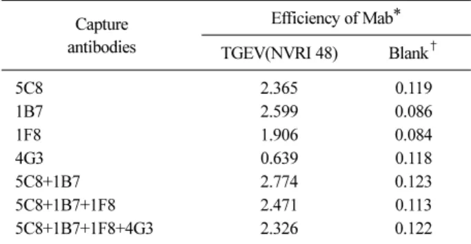
Table 2. Efficiency of sandwich ELISA for detection of trans- missible gastroenteritis virus
Capture antibodies
Efficiency of Mab*
TGEV(NVRI 48) Blank†
5C8 2.365 0.119
1B7 2.599 0.086
1F8 1.906 0.084
4G3 0.639 0.118
5C8+1B7 2.774 0.123
5C8+1B7+1F8 2.471 0.113
5C8+1B7+1F8+4G3 2.326 0.122
*Efficiency of Mab in sandwich ELISA was expressed as the absorbance (450 nm) of ELISA reaction done with Mab or cell culture fluid (Blank†) coated on the plate.
test, the 8H6 was found to produce neutralizing anti- body (Table 1).
Purification of the Mab for conjugation
The Mab, 8H6 was purified using FPLC to conjugate with HRP. As shown in Fig. 2, after passing through the column, the amount of protein was peaked at frac- tions, 18 and 19 corresponding to be 1.7 mg/mL and 1.0 mg/mL, respectively. The two fractions were mixed and conjugated with HRP and used for the conjugated antibody.
Selection of the Mab for antigen capture
The reactivity of the newly produced three (1B7, 1F8, 4G3) and the reported one (5C8) (Chang et al, 1995)
Mabs to TGEV was investigated by ELISA. As a result, 1B7 was the most suitable for capturing antigen as shown in Table 2, followed by 5C8, 1F8 and 4G3. On the oth- er hand, when ELISA was performed by mixing various monoclonal antibodies, only the combination of two monoclonal antibodies of 5C8 and 1B7 showed high re- activity, but the other mixed solutions showed lower re- activity than that of Mab alone. Therefore, in the sub- sequent ELISA, a mixture of 5C8 and 1B7 was selected as a monoclonal antibody to capture the antigen.
TGEV detection efficiency of the sandwich ELISA
The developed ELISA showed no cross-reactivity with
diarrheal agents other than TGEV, i. e., PEDV, porcine
rotavirus, Escherichia coli and Clostridium perfringens,
as shown in Fig. 3. In addition, the newly developed
test method proved to be able to detect TGEV in vari-
Fig. 4. Detection of TGEV by sandwich ELISA in fecal specimens from pig infected with TGEV experimentally.
Fig. 3. Reactivity of produced Mab toward cell-cultured trans- missible gastroenteritis virus and other enteric pathogens in sandwich ELISA. 1. NVRI 48 (107.0TCID50/mL), 2. NVRI 41 (106.25TCID50/mL), 3. WP (106.0TCID50/mL), 4. Miller (103.25TCID50/mL), 5. Purdue (106.5TCID50/mL), 6. PED Japanese vaccine strain (105.0TCID50/mL), 7. PED Wey strain (105.0TCID50/mL), 8. Rota, OSU (106.0TCID50/mL), 9. E. coli, 10. C. perfringens, 11. Culture medium.
Table 3. Comparison of sandwich ELISA and polymerase chain reaction (PCR) for detection of TGEV in fecal specimens
Sandwich ELISA
PCR
Positive Negative Total
Positive 5 3 8
Suspected 1 0 1
Negative 1 15 16
Total 7 18 25