Development of a Quantitative Sandwich Enzyme-Linked Immunosorbent Assay for Detecting the MPT64 Antigen
of Mycobacterium tuberculosis
Mijung Ji,
1,2Byungki Cho,
2Young Shik Cho,
2Song-Yong Park,
3Sang-Nae Cho,
4Bo-Young Jeon,
5and Byoung-Su Yoon
11Department of Life Science, College of Natural Science, Kyonggi University, Suwon; 2Biotech Laboratory, Standard Diagnostics., Yongin;
3Division of Biological Science and Technology, College of Science and Technology, Yonsei University, Wonju;
4Department of Microbiology, Yonsei University College of Medicine, Seoul;
5Department of Biomedical Laboratory Science, College of Health Sciences, Yonsei University, Wonju, Korea.
Received: July 31, 2013 Accepted: August 24, 2013
Co-corresponding authors: Dr. Bo-Young Jeon, Department of Biomedical Laboratory Science, College of Health Sciences, Yonsei University, 1 Yeonsedae-gil, Wonju 220-710, Korea.
Tel: 82-33-760-5108, Fax: 82-504-841-5108 E-mail: [email protected] and Dr. Byoung-Su Yoon,
Department of Life Science,
College of Natural Science, Kyonggi University, 154-42 Gwanggyosan-ro, Yeongtong-gu, Suwon 443-760, Korea.
Tel: 82-31-249-5943, Fax: 82-31-243-1707 E-mail: [email protected]
∙ The authors have no financial conflicts of interest.
© Copyright:
Yonsei University College of Medicine 2014 This is an Open Access article distributed under the terms of the Creative Commons Attribution Non- Commercial License (http://creativecommons.org/
licenses/by-nc/3.0) which permits unrestricted non- commercial use, distribution, and reproduction in any medium, provided the original work is properly cited.
Purpose: Tuberculosis (TB) is a major infectious disease and is responsible for two million deaths annually. For the identification and quantitation of Mycobacte- rium tuberculosis (M. tuberculosis), a causative agent of TB, a sandwich enzyme- linked immunosorbent assay (ELISA) against the MPT64 protein of M. tuberculo- sis, an antigen marker of the M. tuberculosis complex, was developed. Materials and Methods: The MPT64 protein was expressed, and anti-MPT64 monoclonal antibodies were prepared. A sandwich ELISA was established using recombinant MPT64 protein and anti-MPT64 monoclonal antibodies. The sandwich MPT64 ELISA was evaluated using reference and clinical mycobacterial strains. Results:
The sandwich MPT64 ELISA detected MPT64 protein from 2.1 ng/mL to 250 ng/
mL (equivalent to 1.7×10⁴ CFU/mL and 2.0×10⁶ CFU/mL). All 389 clinical M. tu- berculosis isolates tested positive in the sandwich MPT64 ELISA (sensitivity, 100%), and the assay showed no cross reactivity to any tested nontuberculous my- cobacterial strain (specificity, 100%). Conclusion: The sandwich MPT64 ELISA is a highly sensitive and quantitative test for MPT64 protein, which can identify M. tuberculosis.
Key Words: Mycobacterium tuberculosis, MPT64 protein, sandwich ELISA
INTRODUCTION
Tuberculosis (TB) is a major infectious disease that infects one-third of the world’s population. Mycobacterium tuberculosis (M. tuberculosis), a cause of TB, infects 30 million people every year and causes 8 million new TB cases and 3 million deaths annually.1,2 The situation has been worsened by the appearance of multi- drug or extended drug-resistant M. tuberculosis strains, as well as by the combina- tion with human immunodeficiency virus infection.3
Diagnosis of TB relies mainly on microbiological tests, such as smear microsco-
Of the clinical isolates, 231 clinical M. tuberculosis iso- lates grown on 3% Ogawa medium (Asan Pharmaceuti- cal., Seoul, Korea) and 158 clinical M. tuberculosis strains grown in the BacT/ALERT Automated System (BioMéri- eux, Durham, France) were used in this study. All clinical NTM isolates were grown on 3% Ogawa medium. All clinical isolates were identified by Ziehl-Neelsen staining, the AdvanSure TB/NTM real-time PCR kit (LG life sci- ence, Seoul, Korea), and REBA Myco-ID® (M&D, Wonju, Korea).
PCR amplification and cloning of mpt64 of M. tuberculosis
The mpt64 gene was amplified by PCR using oligonucle- otide primers designed to include an EcoRI restriction en- zyme site at the 5’ end and an XbaI restriction enzyme site at the 3’ end. The sequences of the primers were 5’-GAA TTC GCG CCC AAG ACC TAC TGC GAG-3’ (EcoRI/
MPT64-F) and 5’-TCT AGA CTA GGC CAG CAT CGA GTC GAT C-3’ (XbaI/MPT64-R). The amplified mpt64 gene was ligated into the pT7 Blue vector (Novagen, Darm- stadt, Germany), and their sequences were confirmed.
Expression and purification of recombinant MPT64 The mpt64 gene was ligated into the pMAL-p2x expression vector (New England Biolabs, Beverly, MA, USA), and MPT64 protein was expressed using E. coli TB-1 (Invitro- gen, San Diego, CA, USA). The recombinant MPT64 pro- tein was purified using affinity chromatography with an amylose resin column (New England Biolabs) and analyzed by sodium dodecyl sulfate-polyacrylamide gel electrophore- sis and a Western blot assay using mouse polyclonal anti-M.
tuberculosis antibody, which was kindly provided by Prof.
S.N. Cho (Yonsei University, Seoul, Korea).
Production of anti-MPT64 monoclonal antibodies Ten eight-week-old female BALB/c mice (Orient Bio, Seongnam, Korea) were immunized intraperitoneally (i.p.) three times at two-week intervals with 40 µg of recombi- nant MPT64 protein emulsified in incomplete Freund’s ad- juvant (Sigma-Aldrich Co., St. Louis, MO, USA). Spleen cells were isolated and fused with SP2/0 myeloma cells at a ratio of 5:1 in the presence of polyethylene glycol 1500 (Roche Diagnostics GmbH, Mannheim, Germany). The hybridomas were selected in HAT medium (hypoxanthine- aminopterin-thymidine medium) and screened by measur- ing their binding activity to recombinant MPT64 protein by py and culturing of M. tuberculosis. Smear microscopy is a
classical and fast diagnostic method, which detects acid- fast bacilli using Ziehl-Neelsen stain, but its sensitivity is low.4,5 Culturing of mycobacteria is the gold standard for diagnosing TB; however, it takes three to six weeks to form colonies.6-8 Many molecular methods have been developed to identify M. tuberculosis; however, they are used in only a limited number of diagnostic laboratories due to their high costs.9-11
Therefore, it is essential to develop a simple and rapid as- say, which can identify M. tuberculosis and differentiate M.
tuberculosis from nontuberculous mycobacteria (NTM) in cases of contamination by fast-growing NTM.2,11 Moreover, it is necessary to quantitate M. tuberculosis to monitor the therapeutic effects of antimycobacterial drugs.
The MPT64 antigen is a major secretory protein of M. tu- berculosis, and has been shown to differentiate the M. tu- berculosis complex from NTM.12 An immunochromato- graphic assay targeting MPT64 antigen (MPT64 ICA) was developed and is a very simple and rapid test for identify- ing M. tuberculosis.13 However, MPT64 ICA requires more sensitivity to detect M. tuberculosis in cultured specimens, and is not useful for assessing M. tuberculosis bacilli.13,14 Recently, Liu, et al.15 established sandwich enzyme-linked immunosorbent assay (ELISA) against MPT64 using poly- clonal antibody, but its detection level was not high.
Therefore, in this study, in order to develop a highly sen- sitive and quantitative assay for M. tuberculosis, we estab- lished a sandwich ELISA for the MPT64 protein of M. tu- berculosis using expressed MPT64 protein and prepared anti-MPT64 monoclonal antibodies, which can quantify the amount of MPT64 protein and differentiate M. tuberculosis from other mycobacteria. The sensitivity and specificity of this assay were evaluated using reference and clinical my- cobacterial strains.
MATERIALS AND METHODS
Bacterial strains and growth conditions
M. tuberculosis H37Rv (American Type Culture Collec- tion) was used as a reference strain, and was also used for cloning of the MPT64 protein. Five reference strains of M.
tuberculosis, 46 NTM reference strains, 389 clinical M. tu- berculosis isolates, and 64 clinical NTM isolates, including 12 M. abscessus isolates, 25 M. avium isolates, and 27 M.
intracellulare isolates, were used for this study (Table 1).
de, Denmark) were coated with anti-MPT64 monoclonal antibody in a suitable concentration and incubated at 4°C overnight. After blocking with non-fat dry milk, recombi- nant MPT64 protein in phosphate buffered saline (PBS) was added and incubated for 2 h at 37°C. Subsequently, wells were washed four times and incubated with other horseradish peroxidase (HRP)-conjugated anti-MPT64 monoclonal antibodies for 1 h at 37°C. Finally, after six washes, 3,3ʼ,5,5ʼ-Tetramethylbenzidine (TMB) substrate was added to the wells, the plates were incubated for 20 min in the dark, and absorbance was read at 450 nm after stopping the reaction with 2.5 N H2SO4. For generation of a standard curve, 1.0 µg/mL to 1000 µg/mL of recombinant MPT64 protein was used in the sandwich ELISA. The de- tection limit of the assay was defined as the mean value of blank plus three times its standard deviation.
indirect ELISA. Highly reactive hybridomas were enriched in ascetic fluid from BALB/c mice pretreated with 1.0 mL of Pristance (Aldrich, Milwaukee, WI, USA), and the im- munoglobulins were purified by chromatography on a pro- tein G-Sepharose 4B flow (Amersham Bioscience, Piscat- away, NJ, USA).
Sandwich enzyme-linked immunosorbent assay for MPT64 protein
Initially, anti-MPT64 monoclonal antibodies were screened for their reactivity to recombinant MPT64 protein, and highly reactive anti-MPT64 monoclonal antibodies were tested for their suitability for the sandwich ELISA. The op- timum dilutions of these reagents were selected by checker- board titration. Next, the sandwich ELISA was performed as follows: briefly, 96-well microtiter plates (Nunc, Roskil-
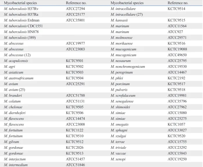
Table 1. List of Mycobacterial Strains
Mycobacterial species Reference no. Mycobacterial species Reference no.
M. tuberculosis H37Rv ATCC27294 M. intracellulare KCTC9514
M. tuberculosis H37Ra ATCC25177 M. intracellulare (27)
M. tuberculosis Erdman ATCC35801 M. kansasii KCTC9515
M. tuberculosis CDC1551 M. marinum ATCC11564
M. tuberculosis HN878 M. marinum ATCC927
M. tuberculosis (389) M. molmoense ATCC29571
M. abscessus ATCC19977 M. morikaense KCTC9516
M. abscessus ATCC23003 M. mucogenicum KCTC19088
M. abscessus (12) M. mucogenicum ATCC49650
M. acapulcensis KCTC9501 M. neoaurum ATCC25795
M. agri KCTC9502 M. nonchromogenicum ATCC19530
M. asiaticum KCTC9503 M. peregrinum ATCC14467
M. austroafricanum KCTC9504 M. phlei KCTC2192
M. avium ATCC25291 M. porcinum KCTC9517
M. avium (25) M. pulveris KCTC9518
M. branderi ATCC51788 M. scrofulaceum ATCC19981
M. celatum ATCC51131 M. senegalense ATCC35796
M. chelonae KCTC9505 M. shimoidei ATCC27962
M. diernhoferi KCTC9506 M. simiae ATCC15080
M. flavescens ATCC14474 M. simiae ATCC25275
M. flavescens ATCC23008 M. smegatis KCTC1057
M. fortuitum KCTC1122 M. sphagni ATCC33027
M. fortuitum KCTC9510 M. szulgai KCTC9520
M. gilvum KCTC9512 M. terrae ATCC15755
M. gordonae KCTC2026 M. triviale ATCC23292
M. gordonae KCTC9513 M. vaccae ATCC15843
M. interjectum ATCC51457 M. xenopi ATCC19250
M. intermedium ATCC51846
ATCC, American Type Culture Collection; KCTC, Korean Collection for Type Culture.
( ): Number of strains.
antibody as a detection antibody for MPT64 protein, which produced minimal background, as well as the highest sensi- tivity and accuracy for the quantification of the MPT64 pro- tein. The optimal concentrations of the capture antibody 1A4 and detection antibody 2E9 were 2 µg/mL and 4 µg/
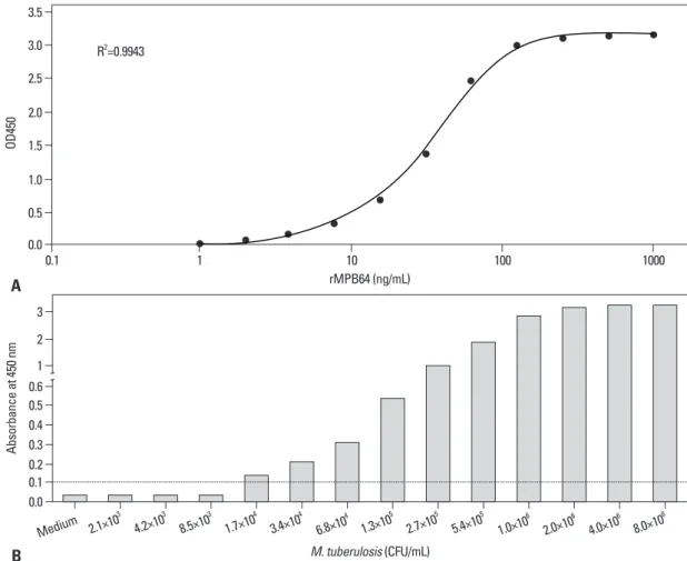
mL, respectively, and the secondary antibody was diluted to 1:20000. A typical calibration curve for quantification of the MPT64 protein was generated by plotting the concen- tration of recombinant MPT64 protein versus the absor- bance, which yielded a coefficient of determination (R2) of 0.9943 (Fig. 2A). The lower limit of detection of this assay was 2.1 ng/mL, equivalent to an absorbance value of 0.102, which was the mean signal of the blank plus three times its standard deviation, and this assay was linear over the range of 3.8 to 250 ng/mL. Therefore, this assay might be a high- ly sensitive assay to quantify the MPT64 protein of M. tu- berculosis.
Detection limit of MPT64 sandwich ELISA and MPT64 ICA
To evaluate the ability of the MPT64 sandwich ELISA to detect secreted MPT64 protein from growing M. tuberculo- sis, the sequential culture supernatants of M. tuberculosis H37Rv were tested. The MPT64 sandwich ELISA detected levels from 1.7×10⁴ CFU/mL of M. tuberculosis as posi- tive, based on a cut-off value of 0.102, which is equivalent to 2.1 ng/mL (Fig. 2B). The upper limit of detection of the Evaluation of MPT64 sandwich ELISA using clinical
Mycobacterium isolates
To determine the detection limit of the MPT64 sandwich ELISA, a series of diluted culture suspensions of M. tuber- culosis H37Rv were applied to the MPT64 sandwich ELI- SA, which was counted by inoculation onto Middlebrook 7H10 agar (Difco, Detroit, MI, USA) plates. To determine the sensitivity and specificity of the tests, 389 clinical M. tu- berculosis isolates, five M. tuberculosis reference strains, 46 NTM reference strains, and 64 clinical NTM isolates were tested (Table 1). Three or four colonies from 3% Oga- wa medium were collected and suspended in 200 µL of PBS, and then 100 µL of the suspension was used as a sam- ple. The samples were classified as positive when the ab- sorbance value was greater than or equal to twice the absor- bance value of the negative control.
Statistical analysis
Statistical analysis was performed using Microsoft® Of- fice Excel (2010) and the GraphPad Prism (version 4.0) program.
RESULTS
Cloning and expression of the mpt64 gene
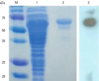
The gene coding for the MPT64 protein was amplified from the M. tuberculosis H37Rv genome by PCR, and the sequence was confirmed (data not shown). The expressed recombinant MPT64 protein has a molecular weight of ap- proximately 65 kDa and is reactive with the mouse poly- clonal anti-M. tuberculosis antibody (Fig. 1).
Characterization of anti-MPT64 protein monoclonal antibodies
Prepared hybridomas were screened for their ability to se- crete monoclonal antibodies against the MPT64 protein.
Two antibodies (1A4 and 2E9) from screened hybridomas showed the highest reactivity in the immunoassay (data not shown). The immunoglobulin isotype of both antibodies belongs to the IgG1 subclass having the lambda light chain (data not shown).
Establishment of a sandwich ELISA for MPT64 protein A sandwich ELISA for the MPT64 protein was established using a 1A4 monoclonal antibody as a capture antibody for the MPT64 protein and HRP-conjugated 2E9 monoclonal
Fig. 1. Analysis of expression of the MPT64 protein in TB-1 cells. TB-1 cells were inducted with 1 mM IPTG for 4 h, and the cell lysates were analysed by SDS-PAGE, after which the gel was stained with Coomassie blue (lanes 1 to 2) or analysed by Western blot with a mouse polyclonal anti-M. tuber- culosis antibody (lane 3). Lane M: pre-stained protein molecular weight markers. Lane 1: cell lysate of TB-1 cells. Lane 2: purified recombinant MPT64. Lane 3: Western blot analysis with anti-MPB64 antibody. TB, tuber- culosis; SDS-PAGE, sodium dodecyl sulfate-polyacrylamide gel electro- phoresis; IPTG, isopropyl β-D-1-thiogalactopyranoside.
20 25 35 50 75
kDa M 1 2 3
Sensitivity and specificity of the MPT64 sandwich ELISA
The sensitivity and specificity of these tests were evaluated using 51 reference strains of M. tuberculosis and NTMs, 389 clinical M. tuberculosis isolates, and 64 clinical NTM iso- MPT64 sandwich ELISA was 2.0×10⁶ CFU/mL (OD450=
3.15), which is equivalent to 253 ng/mL. The MPT64 sand- wich ELISA could quantitatively measure the concentration of MPT64 protein in the culture supernatant, which was correlatively related to the numbers of M. tuberculosis.
Fig. 2. Calibration curve of the developed sandwich ELISA for MPT64 protein (A), and detection limits of the assay (B). The calibration curve was obtained from the mean value of six sandwich ELISAs with the anti-MPT64 antibodies Mab-1A4 and Mab-2E9 under optimized conditions (A). Culture supernatants of M. tuberculosis H37Rv in Middlebrook 7H9 broth with enrichment were serially diluted and ap- plied to the test. The cut-off of the MPT64 sandwich ELISA is indicated by a dotted line. ELISA, enzyme-linked immunosorbent assay;
CFU, colony-forming unit; OD, optical density.
rMPB64 (ng/mL) 0.0
0.5 1.0 1.5 2.0 2.5 3.0 3.5
OD450
0.1 1 10 100 1000
A
R2=0.9943
M. tuberulosis (CFU/mL)
B
Table 2. Comparison of the Results Obtained by the MPT64 Sandwich ELISA and the Lmmunochromatograph- ic TB Ag MPT64 Rapid Test for M. tuberculosis and Other Nontuberculous Mycobacterial Strains
Species (no. of isolates) MPT64 sandwich ELISA
No. of positive (%) No. of negative (%)
M. tuberculosis (5)* 5 (100) 0 (0)
M. tuberculosis (231)† 231 (100) 0 (0)
M. tuberculosis (158)‡ 158 (100) 0 (0)
NTMs (46)* 0 (0) 46 (100)
M. abscessus (12)† 0 (0) 12 (100)
M. avium (25)† 0 (0) 25 (100)
M. intracellulare (27)† 0 (0) 27 (100)
NTM, nontuberculous mycobacteria; ELISA, enzyme-linked immunosorbent assay; TB, tuberculosis.
*Reference mycobacterial strains.
†Clinical isolates grown on 3% Ogawa medium.
‡Clinical isolates grown in the BacT/ALERT Automated System.
0.0 0.1 0.2 0.3 0.4 0.5 0.6 1 2 3
Absorbance at 450 nm
Medium 2.1×10³ 4.2×10³ 8.5×10³ 1.7×10⁴ 3.4×10⁴
6.8×10⁴ 1.3×10⁵ 2.7×10⁵ 5.4×10⁵
1.0×10⁶ 2.0×10⁶ 4.0×10⁶ 8.0×10⁶
of anti-mycobacterial drugs, as well as to the more rapid screening of anti-mycobacterial drug candidates.
In conclusion, a MPT64 sandwich ELISA, developed us- ing recombinant MPT64 protein and anti-MPT64 monoclo- nal antibodies, was shown to be a highly sensitive and specif- ic assay for the identification of M. tuberculosis via detection of the MPT64 protein, and may be a quantitative assay for measuring the MPT64 protein of M. tuberculosis.
ACKNOWLEDGEMENTS
This work was supported by a grant of the Korean Health Technology R&D, Ministry of Health & Welfare, Republic of Korea [HI10C1708 (A101750), SNC], by a grant of the Korea Food and Drug Administration, Health & Welfare, Republic of Korea (BYJ, 13Fre04) and Biotech Laboratory of Standard Diagnostics, Republic of Korea.
REFERENCES
1. Raviglione MC, Snider DE Jr, Kochi A. Global epidemiology of tuberculosis. Morbidity and mortality of a worldwide epidemic.
JAMA 1995;273:220-6.
2. Morris K. Global tuberculosis control amid the world economic crisis. Lancet Infect Dis 2009;9:144-5.
3. Snider DE Jr, Castro KG. The global threat of drug-resistant tuber- culosis. N Engl J Med 1998;338:1689-90.
4. Jenkins SG. Evaluation of new technology in the clinical microbi- ology laboratory. Diagn Microbiol Infect Dis 1995;23:53-60.
5. Thongraung W, Chongsuvivatwong V, Pungrassamee P. Multilev- el factors affecting tuberculosis diagnosis and initial treatment. J Eval Clin Pract 2008;14:378-84.
6. Chakravorty S, Sen MK, Tyagi JS. Diagnosis of extrapulmonary tuberculosis by smear, culture, and PCR using universal sample processing technology. J Clin Microbiol 2005;43:4357-62.
7. Hillemann D, Rüsch-Gerdes S, Richter E. Application of the Ca- pilia TB assay for culture confirmation of Mycobacterium tuber- culosis complex isolates. Int J Tuberc Lung Dis 2005;9:1409-11.
8. Rodrigues C, Vadwai V. Tuberculosis: laboratory diagnosis. Clin Lab Med 2012;32:111-27.
9. Cho SN, Brennan PJ. Tuberculosis: diagnostics. Tuberculosis (Ed- inb) 2007;87 Suppl 1:S14-7.
10. Pathak D, Chakravorty S, Hanif M, Tyagi JS. Lysis of tubercle ba- cilli in fresh and stored sputum specimens: implications for diag- nosing tuberculosis in stored and paucibacillary specimens by PCR. BMC Microbiol 2007;7:83.
11. Magana-Arachchi D, Perera J, Gamage S, Chandrasekharan V.
Low cost in-house PCR for the routine diagnosis of extra-pulmo- nary tuberculosis. Int J Tuberc Lung Dis 2008;12:275-80.
12. Brent AJ, Mugo D, Musyimi R, Mutiso A, Morpeth S, Levin M, et al. Performance of the MGIT TBc identification test and meta- analysis of MPT64 assays for identification of the Mycobacterium
lates, including 12 clinical M. abscessus isolates, 25 clinical M. avium isolates, and 27 clinical M. intracellulare isolates (Table 2). In the MPT64 sandwich ELISA, all five reference M. tuberculosis strains and all 389 clinical M. tuberculosis isolates, including 231 M. tuberculosis isolates grown on 3% Ogawa media and 158 M. tuberculosis grown in the liquid culture system, were detected as positive, equating to a sensitivity of 100%. All NTM strains, including 46 refer- ence NTM strains (39 species) and 64 clinical NTM iso- lates (3 species), generated negative results in the MPT64 sandwich ELISA.
DISCUSSION
In this study, we developed a highly sensitive and quantita- tive sandwich ELISA for the MPT64 protein of M. tubercu- losis using anti-MPT64 monoclonal antibodies. This MPT64 sandwich ELISA could measure the quantity of secreted MPT64 protein from M. tuberculosis, which differentiates M. tuberculosis from NTM. The lower limit of detection of the MPT64 sandwich ELISA was 2.1 ng/mL, and this assay could detect 1.7×10⁴ CFU/mL of M. tuberculosis. Recently, Liu, et al.15 developed an MPT64 ELISA using polyclonal antibodies, and its lower limit of detection was 10 ng/mL, which is approximately five times higher than ours. A pos- sible explanation for this is that the anti-MPT64 monoclo- nal antibodies used for the sandwich ELISA might have a higher affinity than polyclonal antibodies. The MPT64 ICA could detect 1×10⁵ CFU/mL of M. tuberculosis as posi- tive.13,14 This suggests that the MPT64 sandwich ELISA might be a more sensitive assay than MPT64 ICA for the de- tection of MPT64 protein. The sensitivity and specificity of the MPT64 sandwich ELISA were both 100%. These results seem to be comparable to or more sensitive than MPT64 ICA, which has a reported sensitivity of 92-99% and a speci- ficity of 97-100%.12,13,15 This indicates that the MPT64 pro- tein is highly specific for the M. tuberculosis complex and is therefore a comparably more sensitive target analyte.16 In addition, the MPT64 sandwich ELISA has an advantage over the MPT64 ICA, in that the MPT64 sandwich ELISA is able to quantify the MPT64 protein of M. tuberculosis, which is significantly correlated with the count of M. tuber- culosis (R2=0.998, p<0.001). This would be very useful for measuring counts of M. tuberculosis, because it takes three to six weeks for enumeration of M. tuberculosis and also could contribute to the monitoring of the therapeutic effects
Dis 2011;15:1475-7.
15. Liu Z, Zhu C, Yang H, Hu H, Feng Y, Qin L, et al. Clinical value of ELISA-MPT64 for the diagnosis of tuberculous pleurisy. Curr Microbiol 2012;65:313-8.
16. Hasegawa N, Miura T, Ishii K, Yamaguchi K, Lindner TH, Merritt S, et al. New simple and rapid test for culture confirmation of My- cobacterium tuberculosis complex: a multicenter study. J Clin Mi- crobiol 2002;40:908-12.
tuberculosis complex in liquid culture. J Clin Microbiol 2011;49:
4343-6.
13. Park MY, Kim YJ, Hwang SH, Kim HH, Lee EY, Jeong SH, et al.
Evaluation of an immunochromatographic assay kit for rapid identification of Mycobacterium tuberculosis complex in clinical isolates. J Clin Microbiol 2009;47:481-4.
14. Ang CF, Cajucom MA, Kim Y, Bang H, Lee H, Cho SN, et al.
Evaluation of a rapid assay for identification of Mycobacterium tuberculosis grown in solid and liquid media. Int J Tuberc Lung