정공피으로부터 항염증물질의 연구
윤용갑·채규윤*·이경관*·록란잔밭*·백승화**,#
원광대학교 한의과대학 방제학교실, *자연과학대학 생명나노학부, 한의과대학 방제학교실
**한의학전문대학원 한약자원개발학과
(Received August 13, 2009; Revised December 20, 2009; Accepted December 27, 2009)
Studies on the Antiinflammatory Compounds from Sorbus Commixta
Young Gab Yun, Kyu Yun Chai*, Kyung Kwan Lee*, Lok Ranjan Bhatt* and Seung Hwa Baek**,#
Department of Oriental Medical Prescription, School of Oriental Medicine,
*Division of Nanobiochemistry, College of Natural Sciences, and **Department of Herb! al Resources, Professional Graduate School of Oriental Medicine, Wonkwang University, Iksan 570-749, Korea
Abstract
— Sakuranetin, prunetin and dihydroquercetin were isolated from the methanol extract of Sorbus commixta by 1D/2D-NMR and LC-MASS spectrometry. Medicating these compounds to RAW264.7 cell that was pre-treated by lipopolysaccharide revealed anti-inflammatory effects that greatly inhibited the production of NO (nitric oxide) and PEG2 (prostaglandin E2), which are well known to cause the expression of iNOS (inducible nitric oxide synthase) and COX-2 (Cyclooxygenase-2). These results suggest that these compounds can be used as stable anti-inflammatory materials.
Keywords □
Sorbus commixta, sakuranetin, prunetin, dihydroquercetin, anti-inflammatory effects, NO, PEG2
정공피는장미목장미과의낙엽소교목의일종인마가목중산 마가목의경피이며
,
열매와나무껍질은약재로사용되고있으며,
鎭咳
(
진해),
祛痰(
거담),
利水(
이수),
止渴(
지갈),
强壯(
강장)
의효능
,
咳嗽(
해수),
기관지염,
폐결핵,
水腫(
수종),
胃炎(
위염),
신체허약등을치료하는데사용되어왔다
.
1)면역세포가세균,
바이러스 등을포함한미생물또는생체의이물질,
외부물질에노출되면,
면역세포가활성화되고
,
활성화된면역세포에서염증반응에원인이되지않는많은인자를분비하게됨으로써염증반응을가 속시킨다
.
세포는세균에함유되어있는lipopolysaccharide(LPS)
로자극될때
cyclooxygenase-2
의발현으로염증성매개물질인prostaglandin E2(PGE2)
를과량으로생성하게 된다.
2-5)Cyclo- oxygenase(COX)
는arachidonic acid
에서prostanoid
로전환시키는효소로알려졌으며
, COX-1, COX-2
에의해합성된적은양의prostanoid
는nitric oxide(NO)
와비슷하게염증반응을가속화시 킨다고알려졌다. NO
는혈압조절,
신경전달,
혈액응고,
면역기능,
혈관확장및항균작용등의역할을하는것으로알려져있으며
,
포유동물의세포에서생성되는매우작은분자량의물질이다
.
이는여러조직과세포로부터
L-arginine
으로부터nitric oxide syn- thase(NOS)
에의해서합성된다.
2)지금까지알려진NO
생성효소로는
constitutive NOS(c-NOS)
와inducible NOS(iNOS)
의두종류이며
,
이중cNOS
는Ca
2+/calmodulin
의존성이며,
단시간 동안소량의NO
를생성하여정상적인생리기능을담당하고있다
.
여기에신경세포에존재하는neuronal NOS(nNOS)
와내피세포가존재하는
endothelial constitutive NOS(eNOS)
가속한다. iNOS
는세포내Ca
2+의농도에비의존성이며, macrophage
및heaptocyte
등에존재한다.
대식세포가interferon-
γ(INF-
γ)
또는LPS
로자극할때,
생체내에서감염,
염증등의자극에의해서, L-arginine
으로부터NOS
에의한대사과정에서NO
를생성하여,
종양세포를죽이거나미생물에의한감염을방어하여생체를지 키는중요한역할을하고있다
.
그러나NO
가필요이상으로생 성되면,
쇼크에의한혈관확장,
염증반응으로유발되는조직손상,
돌연변이및신경조직의손상등을일으켜생체에유해한작용을 나타낸다
.
3-5)이러한증상에대한치료제의개발이요구되어왔으 며,
최근에천연물로부터NO
생성저해제를찾으려는연구가많이진행되고있다
.
이와동시에iNOS
에의한NO
생성을저해하 는새로운염증치료제의개발에대한연구도활발하게진행되고#본논문에관한문의는저자에게로
(
전화) 063-850-6225 (
팩스) 063-850-6225
(E-mail) [email protected]
있는추세이다
.
특히대식세포는생체내에서감염증등의자극에의해서
, L-arginine
을NOS
에의해대사하여NO
를생성하여종양세포를죽이거나미생물에의한감염을방어하여
,
생체를지 키는중요한역할을하고있다.
6,7)본연구에서는정공피로부터생리활성물질을분리하여생물검정을얻었기에보고합니다
.
실험방법
시약및기기
정공피추출및
open columm
분리에서사용된용매는GR
급을사용하였으며
,
물질의분리에는25 TLC plastic sheets(20×
20 cm) silica gel 60 F254(MERCK), 25 TLC plates 5×10 cm RP-18 F254S(MERCK), silica gel 60(0.015~0.140 mm) (MERCK), silica gel 60(0.040~0.063 mm; MERCK), octade- cyl-functioalized silica gel(Aldrich), lipophilic sephadex LH-20 (SIGMA)
등충진제와Phenomenex
사의Luna 5u C18(2)(250×
21.20 mm 5 micron) HPLC column
과YMC
사의Hydrosphere C18(250×20 mm, I. D. S-5
µm, 12 nm) HPLC column, EYEL4 rotary vacuum evaporator, Vision Workstation per- septive biosystems HPLC
등을사용되었다.
순수한물질의확 인은ESI-MS
을얻기위해, Micoromass Quatro LC
을사용하였으며
, NMR spectrum
은JEOL Eclipse 500 FT-NMR spec- trometer(500 MHz)
를사용하였으며, NMR
용매는methanol-
d4, DMSO-
d6, chloroform-
d 내부표준물질은tetramethylsilane (TMS)
를사용하였다.
생리활성물질분리
정공피
1.2 kg
을methanol 1
l에넣고일주일동안상온에서추출한후
,
여과하여추출물18.84 g(1.6%)
을얻었다.
이를3
차 증류수에현탁시켜,
노르말헥산으로3
회추출하여,
추출물0.812 g(4.3%)
을얻었으며,
같은추출방법으로에틸아세테이트추출물9.406 g(49.9%)
을얻었다.
에틸아세테이트층(3 g)
을메탄올(5 m
l)
을넣어녹인후
,
실리카겔(150 g)
로충진된flash column
에넣어
,
에틸아세테이트, methylene chloridde,
메탄올을혼합하여 극성이증가되는순으로gradient
를걸어,
분획1(3.3 mg),
분획2(30.2 mg),
분획3(183.6 mg),
분획4(316.0 mg),
분획5(54.9 mg),
분획6(54.6 mg),
분획7(79.5 mg),
분획8(1.7 mg),
분획9 (7.3 mg),
분획10(76.0 mg),
분획11(49.3 mg),
분획12(39.8 mg),
분획13(107.2 mg),
분획14(515.0 mg),
분획15(121.0 mg),
분획
16(63.2 mg),
분획17(34.7 mg),
분획18(35.7 mg),
분획(80.0 mg)
을얻었다.
분획물중에서생리활성이있다고판단된분 획3(183.6 mg),
분획4(316.0 mg),
분획6(54.6 mg)
을 역상HPLC
를이용하여,
화합물1, 2
및3
를얻었다.
ESI-MS
과1H,
13C-NMR data
와HMBC, HMQC, HMCOSY
를이용하여
,
분자구조를규명하여, sakuranetin(1),
8-10)prunetin (2)
11,12)과dihydroquercetin(3)
13,14)을얻었다.
세포배양
생쥐의대식세포
(murine macrophage Raw 264.7 cell line)
를10% FBS(heat inactivated), 1% L-glutamine, nonessential amino acids
및1% antibiotic/antimycotic(100 U/m
lof penicil- lin, 25.0
µg/m
lof amphotericin D, and 100.0
µg/m
lof strep- tomycin)
등이 포함된RPMI 1640
배지에 넣고37% CO
2incubator
에서24
시간배양하였다.
세포생존율측정
Prunetin, sakuranetin
및dihydroquercetin
에 대한RAW 264.7
세포주의세포생존율을MTT(2-[4,5-dimethylthiazol-2-yl]- 2,5-diphenyltetrazolium bromide)
방법을이용하여분석하였다. RAW 264.7
세포주를96-well plate
에well
당1×10
4가되도록seeding
한다음, IFN-
γ/LPS
및약제를24
시간처리하고새로운 배지에50.0
µg/m
l이되도록, MTT
를첨가하여2~4
시간동안반응시켰다
. MTT
와생존세포로부터 생성된보라색불용성formazan
을DMSO
로용해하여, 595 nm
파장에서흡광도를측 정하고대조군과비교하여백분율(%)
로표시하였다.
Nitric oxide(NO)의농도의측정
Prunetin, sakuranetin
및dihydroquercetin
을DMSO
에녹여저장용액을만들었으며
, Sarcoma
생성된NO
의양은세포배양액중에존재하는
NO
2-의형태로서griess
시약(1% sulfanilamide, 0.1% N-(1-naphthyl)-ethylenediaminedihydrochloride in 2.5%
phosphoric acid solution)
을이용하여측정하였다.
이것을희석하여적정용액을만든후
LPS
를1
µg/m
l처리하여, 18
시간동 안배양시킨다.
배양액중100
µl를취하고,
동량의griess
시약을넣고실온에서
10
분간방치한다음, ELISA plate reader
를이용하여
, 540 nm
에서 흡광도를측정하였다. NO
2-는sodium nitrite
를표준용액으로측정하였으며,
각측정치는5~8
µM
의NO
2-을함유하는cell-free
배양액의흡광도를이용하여보정하 였다.
Western blotting
Prunetin, sakuranetin
및dihydroquercetin
과LPS
에의해자 극된세포를PBS(phosphatebuffered saline)
으로세척한다음, cell lysis buffer(50 mM Tris, pH 8.0, 110 mM NaCl, 5 mM EDTA, 1% Triton X-100, PMSF 100.0
µg/m
l)
로용혈시킨다음,
원심분리하여단백질용액을얻는다
. Bradford
분석방법을이용하여정량한다음
, 30.0
µg
을취하여동량의sample buffer(125
mM Tris pH 6.8, 4% SDS 20% glycerol, 10% 2-mercaptoet-
hanol)
를혼합한다음, 95
oC
에서5
분동안가열하여단백질변성을유도하였다
.
변성된단백질을12% acrylamide gel
에서전기영동을 수행한 다음
, nitrocellulose membrane(Amersharm Pharmacia
사)
으로전위시키고, 5% skim milk/TBS-T
로상온에서
1
시간동안반응시켜비특이적인항체반응을억제시켰다. Cox- 2
과iNOS
및β-actin
에대한1
차항체(primary antibody)
를3%
skim milk/TBS-T
에서1 : 1,000
으로희석하여, membrane
과상온에서
1
시간30
분동안반응시키고세척한다음, Anti-mouse, rabbit
혹은goat IgG conjugated horseradish peroxidase
이차 항체(secondary antibody)
를1% skim milk/TBST
에서1 : 5,000
으로희석하여
, membrane
과상온에서1
시간동안반응시킨다. TBS-T
로세척한다음, ECL reagent kit(Amersham, Bucking- hamshire, England)
로발색시킨다음, X-ray film
에감광시는방법으로분석하였다
.
Cyclooxygenase-2(COX-2)농도의측정
Prunetin, sakuranetin
및dihydroquercetin
을 농도별로RAW 264.7
세포에1
시간전처리한다음, LPS
로자극하고24
시간 후에 배양액을회수하여 후
, ELISA kit(enzyme linked immunosorbant assay, R&D System)
를이용하여분석하였다.
모든분석과정은
R&D System
에서제공된방법에준하여측정 하였다.
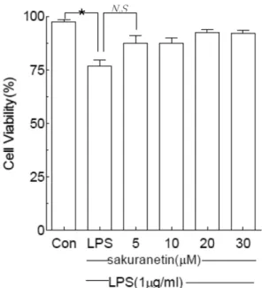
Fig. 1 −
Effects of sakuranetin on the cell viability of in RAW 264.7 cells (2.5×10
5/m l ). Cells were incubated with or without LPS (1
µg/m l ) for 24 h in the presence or absence of sakuranetin at indicated concentrations. The cell viability was determined by the MTT method. One of three blots is shown and each bar represents the mean±SD from three separate experiments. * P <0.005, N.S : not significant.
Fig. 3 −
Effects of dihydroqueroetin on the cell viability of in RAW 264.7 cells (2.5×10
5/m l ). Cells were incubated with or without LPS (1
µg/m l ) for 24 h in the presence or absence of dihydroqueroetin at indicated concentrations. The cell viability was determined by the MTT method. One of three blots is shown and each bar represents the mean±SD from three separate experiments. * P <0.005, N.S : not significant.
Fig. 2 −
Effects of prunetin on the cell viability of in RAW 264.7 cells (2.5×10
5/m l ). Cells were incubated with or without LPS (1
µg/m l ) for 24 h in the presence or absence of prunetin at indicated concentrations. The cell viability was determined by the MTT method. One of three blots is shown and each bar represents the mean±SD from three separate experiments. * P <0.005, N.S : not significant.
통계분석
대조군과각시료들로부터얻은실험 결과들은
mean±S.D.
값으로표시하였고
,
각실험결과로부터ANOVA(analysis of variance)
를구한후Duncan's multiple range test
를이용하여각군의평균간의유의성을검정하였다
.
일반적으로, p
값이
0.05
이하인 것만통계적으로유의성이있는것으로 검증하였다
.
실험결과 및 고찰
Prunetin, sakuranetin및dihydroquercetin의세포생존률 에미치는영향
Prunetin, sakuranetin
및dihydroquercetin
을540 nm
파장에 서흡광도를측정한결과,
대조군을100%
로했을경우, LPS
로 자극한대조군은75%
의세포생존률을나타냈으며,
생리활성물질과
LPS
를처리한군은90%
이상의세포생존률이관찰되었 다. 5.0~30.0
µM
생리할성물질의농도사이에서는세포생존률 에큰영향을미치지않는플라보노이드계열의화합물임을확인 하였다(Figs. 1, 2
와3).
15)Prunetin, sakuranetin및dihydroquercetin에대한Nitric oxide(NO)의농도측정
Prunetin, sakuranetin
및dihydroquercetin
이RAW 264.7
세포에서
LPS
의유도에의한NO
생성에미치는영향에대해알아보기위하여
, RAW 264.7
세포(1×10
6cells/m
l)
를주입하고, 5
시간후에
5.0~30.0
µM
생리활성물질의농도로2
시간동안전처리한다음
, LPS(1.0 mg/m
l)
를처리하여18
시간배양하였다.
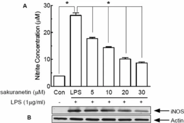
Fig. 4 −
Effects of sakuranetin on NO production and iNOS protein expression in RAW 264.7 cells (2.5×10
5/m l ). (A) Cells were incubated 24 h for NO assay or (B) 18 hr for iNOS western blot with medium, NO concentration was determined by griess reagent for the determination of intracellular iNOS protein, Western immunoblot analysis was carried out as described in materials and methods. One of three blots is shown and each bar represents the mean±SD from three separate experiments. * P <0.005.
Fig. 6 −
Effects of dihydroquercetin on NO production and iNOS protein expression in RAW 264.7 cells. (A) Cells (2.5×10
5/ m l ) were incubated 24 h for NO assay or (B) 18 h for iNOS Western blot with medium, NO concentration was determined by griess reagent. For the determination of intracellular iNOS protein, western immunoblot at analysis was carried out as described in materials and methods. One of three blots is shown and each bar represents the mean±SD from three separate experiments. * P <0.005.
Fig. 5 −
Effects of prunetin on NO production and iNOS protein expression in RAW 264.7 cells (2.5×10
5/m l ). (A) Cells were incubated 24 h for NO assay or (B) 18 h for iNOS western blot with medium, NO concentration was determined by griess reagent. For the determination of intracellular iNOS protein, western immunoblot at analysis was carried out as described in materials and methods. One of three blots is shown and each bar represents the mean±SD from three separate experiments. * P <0.005.
그결과생리활성물질이처리되지않고
LPS
로자극한대조군은23.6
µM
의아질산염을생성시킨반면, 5.0~30.0
µM
생리활성물질을처리한실험군은
8.0~22.0
µM
의아질산염을생성시켰다.
이러한 연구결과에의하면
, prunetin, skuranetin
및dihydro- quercetin
는LPS
로활성화된RAW 264.7
세포에서NO
생성을억제하는것으로판단된다
(Figs. 4, 5
과6).
15)Prunetin, sakuranetin및 dihydroquercetin에대한 iNOS 의측정
Prunetin, sakuranetin
및dihydroquercetin
과1.0
µg/m
lLPS
가처리된
RAW 264.7
세포를용해시킨후, Bradford
법으로분석한결과에의하면
,
농도의존적으로감소되는현상을관찰할 수가있었으며,
이들생리활성물질은대조군과비교할때에30
µg/
m
l에서발생하는NO
의50%
이상감소하는것으로나타났다.
이러한연구결과는
prunetin, sakuranetin
및dihydroquercetin
이항염증제로서활용될가치가있는것으로사료된다
(Figs. 4, 5
과6).
15)Prunetin, sakuranetin 및 dihydroquercetin에 대한 Cyclooxygenase-2(COX-2)의측정
PGE2
생성에직접적으로영향을 미치는COX-2
의발현을Western immunoblot analysis
방법과ELISA
방법으로조사하 였다.
그연구결과에의하면, COX-2
단백질발현이농도의존적 으로억제되는사실을증명하였으며, LPS
로자극된대조군을기준으로했을때
, 30.0
µM
생리활성물질을처리했을때, 50%
의 감소율이관찰되었다.
이러한연구결과에 의하면, prunetin, sakuranetin
및dihydroquercetin
이항염증제로서활용될가치가있는것으로사료된다
(Figs. 7, 8
과9).
15)결 론
정공피에서는처음으로분리된
prunetin, sakuranetin
및dihy- droquercetin
은RAW 264.7
세포에서농도의존적으로iNOS
의발현
, NO
의생성과COX-2
의발현을억제하여, PGE2
생성이억 제됨을 관찰되었다.
이러한 연구결과에 의하면, prunetin, sakuranetin
및dihydroquercetin
이염증성질환을예방하거나치료할가능성을시사하고있다
.
감사의 말씀
본연구는
2008
년도원광대학교교비연구비지원에의하여이Fig. 9 −
Effects of dihydroquercetin on (A) COX-2 activity and (B) COX-2 protein expression in RAW 264.7 cells (1×10
6/m l ).
Cells were incubated with or without LPS (1
µg/m l ) for 18 h in the presence or absence of dihydroquercetin.
Western immunoblot analysis was carried out as described in materials and methods. One of three blots is shown and each bar represents the mean±SD from three separate experiments. * P <0.005.
Fig. 7 −
Effects of sakuranetin on (A) COX-2 activity and (B) COX- 2 protein expression in RAW 264.7 cells (1×10
6/m l ). Cells were incubated with or without LPS (1
µg/m l ) for 18 h in the presence or absence of sakuranetin. Western immunoblot analysis was carried out as described in materials and methods. One of three blots is shown and each bar represents the mean±SD from three separate experiments. * P <0.005.
Fig. 8 −
Effects of prunetin on (A) COX-2 activity and (B) COX-2 protein expression in RAW 264.7 cells (1×10
6/m l ). Cells were incubated with or without LPS (1
µg/m l ) for 18 h in the presence or absence of prunetin. Western immunoblot analysis was carried out as described in materials and methods. One of three blots is shown and each bar represents the mean±SD from three separate experiments.
* P <0.005.
루어졌으며
,
이에감사드립니다.
참고문헌
1) Bae, K. : The Medicinal Plants of Korea, Kyo-Hak Publishing Co., Seoul, Korea, p. 236 (2000).
2) Chun, H. T., Pae, H. O., Choi, B. M., Billiar, T. R. and Kim, Y. M. : Nitric oxide as a bioregulator of apoptosis. Biochem.
Biophys. Res. Commun.
282, 1075 (2001).
3) Brieva, A., Guerrero, A., Alonso-Lebrero, J. L. and Pivel, J. P. : Inmunoferon®, a glycoconjugate of natural origin, inhibits LPS-induced TNF-
αproduction and inflammatory responses.
Int. Immunopharmacol.
1, 1979 (2001).
4) Wang, Y., Vodovotz, Y., Kim, P. K., Zamora, R. and Billiar, T. R. : Mechanisms of hepatoprotection by nitric oxide. Ann. N. Y.
Acad. Sci.
962, 415 (2002).
5) Galla, H. J. : Nitric oxide, NO, an intercellular messenger Angew. Chem. Int. Ed. Engl.
32, 378 (1993).
6) Narumi, S., Finke, J. H. and Hamilton, T. A. : Interferon gamma and interleukin 2 synergize to induce selective monokine expression in murine peritoneal macrophages. J. Biol. Chem.
265
, 7036 (1990).
7) Cox, G. W., Melillo, G., Chattopadhyay, G., Mullet, D., Fertel, R. H. and Varesio, L. : Tumor necrosis factor-alpha-dependent production of reactive nitrogen intermediates mediates IFN- gamma plus IL-2-induced murine macrophage tumoricidal activity J. Immunol.
149, 3290 (1992).
8) Lee, S. M. and Lee, C. G. : Isolation and gas chromatographic analysis of lupenone and lupeol from Sorbus Cortex. Anal. Sci.
Technol.
12, 136 (1999).
9) Rakwal, R., Hasegawa, M. and Kodama, O. : A methyl- transferase for synthesis of the flavanone phytoalexin sakuranetin in rice leaves. Biochem. Biophys. Res. Comm.
222, 732 (1996).
10) You, M. J., Kim, B. M., Bhatt, L. R., Chai, K. Y. and Baek, S. H. : Inhibitory effect of sakuranetin on (1,3)-
β-glucan synthase. Orien. Pharm. Exp. Med. submitted.
11) Peng, T., Tu, Y. Q., Deng, Y. and Zhang, X. : Studies on chemical constituents of Primula sikkmensis. Zhong Yao Cai.
31