• 검색 결과가 없습니다.

Candida albicans urinary tract infection in a Shih Tzu dog with immune-mediated hemolytic anemia

N/A
N/A
Protected

Academic year: 2021

Share "Candida albicans urinary tract infection in a Shih Tzu dog with immune-mediated hemolytic anemia"

Copied!
3
0
0

로드 중.... (전체 텍스트 보기)

전체 글

(1)

pISSN 2466-1384 eISSN 2466-1392 大韓獸醫學會誌 (2017) 第 57 卷 第 2 號 Korean J Vet Res(2017) 57(2) : 139~141 https://doi.org/10.14405/kjvr.2017.57.2.139

139

<Case Report>

Candida albicans urinary tract infection in a Shih Tzu dog with immune-mediated hemolytic anemia

In-Sung Jang

1

, Won-Kyoung Yoon

1,2

, Changbaig Hyun

1,

*

1

Section of Small Animal Internal Medicine, College of Veterinary Medicine, Kangwon National University, Chuncheon 24341, Korea

2

Guardian Angel Veterinary Hospital, Anyang 14112, Korea

(Received: February 4, 2017; Revised: April 25, 2017; Accepted: June 5, 2017)

Abstract: An 8-year-old castrated male Shih Tzu dog (weighing 7.0 kg) presented with anemia and lethargy. Initial diagnosis indicated immune-mediated hemolytic anemia. During therapy, a secondary urinary infection, probably due to the immune suppressive therapy, was diagnosed. Subsequent diagnostic tests, including urinalysis and urine culture, indicated candidal cystitis. Despite ketoconazole therapy for candidal cystitis, the dog died suddenly. A Candida albicans infection was confirmed upon postmortem evaluation. Prolonged immunosuppressive therapy might be the cause of this infection. This is the first case report describing a Candida albicans urinary tract infection accompanied by hemolytic anemia in a dog in Korea.

Keywords: Candida albicans, dog, candidal cystitis, immune-mediated hemolytic anemia, urinary tract infection

Candida (C.) albicans is an opportunistic fungal pathogen that causes urogenital infections in dogs, although it is typi- cally part of the normal flora in the gastrointestinal, urogeni- tal, and upper respiratory tracts in dogs [9, 11]. The contributing factors involved in a C. albicans infection in dogs include immunosuppressive diseases, long-term steroid therapy, mucosal damages, and long-term catheterization in the lower urinary system. Although a C. albicans urinary tract infection (UTI) has been well documented in dogs and cats [3, 4, 9], a UTI associated with immune-medicated hemolytic anemia (IMHA) has never been reported in dogs. This case report described a C. albicans UTI in a dog, which occurred because of IMHA and a subsequent immunosuppressive therapy.

An 8-year-old castrated male Shih Tzu dog (weighing 7.0 kg) presented with major complaints of abrupt exercise intolerance and marked depression. According to the refer- ring vet, the dog was treated for chronic dermatitis from demodecosis and otitis externa. At presentation, the dog showed pale mucosa and delayed capillary refill time (> ~3.0 sec), suggesting anemia. His heart rate, rectal temperature, and systolic blood pressure were 120 bpm/min, 38.0

o

C and 135 mmHg, respectively. A routine complete blood cell count (CBC; IDEXX LaserCyte Dx Hematology Analyzer; IDEXX Laboratories, USA) revealed hypochromic regenerative ane- mia (hemoglobin 7.7 g/dL; reference range [RI], 12.4–19.1 g/

dL; hematocrit 22.2%; RI, 29.8–57.5%; reticulocyte 1.37%;

RI, 0–1%) and leukocytosis (white blood cells 26.3 × K/µL;

RI, 5.4–15.3 × K/µL) with segment neutrophilia (neutrophils 23.3 × K/µL; RI, 1.2–6.8 × K/µL). A blood cytological exami- nation revealed spherocytosis, anisocytosis, and polychroma- sia, indicating regenerative anemia (Fig. 1). A saline red blood agglutination test confirmed a positive reaction. Blood chem- istry analysis (Vetscan; ABAXIS, USA) indicated increased blood urea (40 mg/dL; RI, 7–27 mg/dL) and total bilirubin (1.6 mg/dL; RI, 0–0.9 mg/dL). Based on laboratory tests, the dog was tentatively diagnosed with IMHA and hospitalized for intensive therapy. The dog was initially treated with pred- nisolone (1 mg/kg, per orally [PO], q12h, Solondo; Yuhan, Korea), azathioprine (2 mg/kg, PO, q24h, Imuran; Samil pharmaceuticals, Korea), danazol (5 mg/kg, PO, q12h, Dana- zol cap; Young Poong Pharmaceuticals, Korea), and ursode- oxycholic acid (15 mg/kg, PO, q24h, Ursa; Daewoong Phar- maceuticals, Korea). Despite intensive treatment with immu- nosuppressive drugs, the CBC performed on the next day showed worsening of anemia and leukocytosis (hematocrit 18.7%; hemoglobin 6.2 g/dL; white blood cells 42.6 × K/µL).

Therefore, a blood transfusion was performed with packed red blood cells (112 mL) to increase the dog’s hematocrit lev- els. After the blood transfusion, the anemia improved (hema- tocrit 29%; hemoglobin 9.4 g/dL), although leukocytosis deteriorated (56.4 × K/µL). After the transfusion, the dog was administered medication including prednisolone (2 mg/kg, PO,

*Corresponding author

Tel: +82-33-250-8681, Fax: +82-33-244-2367

E-mail: hyun5188@kangwon.ac.kr

(2)

140 In-Sung Jang, Won-Kyung Yoon, Changbaig Hyun

q12h), azathioprine (2 mg/kg, PO, q24h), danazol (5 mg/kg, PO, q12h), famotidine (15 mg/kg, PO, q24h, Famotidine tab;

Withus Pharmaceutical, Korea), biphenyl dimethyl dicarbox- ylate (8.3 mg/kg, PO, q24h, Lefotil tab; CMG Pharm, Korea), milk thistle extract (35 mg/head, PO, q24h, Silymarin Tab;

Sinil pharmaceuticals, Korea), and ursodeoxycholic acid (15 mg/kg/day, PO, q24h). After 20 days of pharmacother- apy, the dog still showed clinically significant anemia (hema- tocrit 19.4%; hemoglobin 6.5 g/dL; white blood cells 45.6 × K/µL). Therefore, azathioprine was replaced with cyclospo- rine (7 mg/kg, PO, q24h, Cypol; Chong Kun Dang pharma- ceuticals, Korea). During treatment with cyclosporine, the dog started showing side effects (i.e., vomiting, depression, anorexia, or paleness) and more severe anemia (e.g., hemat- ocrit 16.6%; hemoglobin 5.4 g/dL). Blood chemistry also found increased renal and hepatic enzymes (blood urea 40 mg/dL; alkaline phosphatase 2,028 U/L; RI, 20–150 U/L;

alanine transaminase 412 U/L; RI, 10–118 µ/L). To improve his anemic condition, a blood transfusion was repeated with packed red blood cells (80 mL). Although the dog was less anemic after therapy (hematocrit 33.2%; hemoglobin 11 g/

dL), his urine was suddenly cloudy. The urine was collected and analyzed. Further urinalysis, including a sediment test, revealed aciduria (pH 6), leukocyturia (mostly neutrophils and macrophages), proteinuria (3+), hemoglobinuria, and funguria (Fig. 2B). Fungal pseudohyphae were found in the urine and thus submitted for a fungal culture confirmation to a commercial veterinary lab (NEODIN Vetlab; Korea). To interrogate the extent of the urinary tract infection, an abdominal ultrasonographic examination was performed in the kidneys and urinary bladder. Ultrasonography revealed pel- vic dilation, an irregularly thickened corticomedullary junction in the kidneys, and markedly increased echogenecity of the renal pelvis (Fig. 3A). The wall of the urinary bladder was intact but thickened (Fig. 3B). There was hyperechogenic sediment (arrow head) inside the bladder (Fig. 3B).

The tentative diagnosis was candidal cystitis and the dog was treated with intermittent bladder washing with Tris-EDTA

solution (TrizEDTA; Dechra, USA) and with oral medica- tions, including cephalexin (30 mg/kg, q12h, Cephalexin cap;

KOLON Pharma, Korea), ketoconazole (10 mg/kg, q12h, Spike tab; JW Pharmaceutical, Korea) and ursodeoxycholic acid (15 mg/kg, q24h). Cyclosporin was again replaced with azathioprine (2 mg/kg, PO, q24h). However, the dog died suddenly after 6 days of therapy at home. A postmortem examination was declined owing to refusal from the owners.

A C. albicans infection was finally confirmed by a fungal culture 10 days after the dog’s death.

This case report described a secondary yeast infection (C.

albicans) due to immunosuppressive therapy against IMHA in a dog. Candida spp. are saprophytic yeasts. Three Can- dida species, including C. albicans, C. parapsilosis, and C.

tropicalis, are pathogenic to dogs and occasionally colonize the skin, oral cavity, and urogenital, and gastrointestinal tracts in dogs [10]. Prolonged immunosuppression from several disease conditions can develop into candidiasis in dogs [3, 4, 8, 9]. Even though infections can be confined to the skin, dogs with systemic candidiasis can present more general symptoms, including peritonitis and chronic cystitis [1], as seen in this case. Although one recent study has reported candiduria from chronic cystitis in a dog [1], this is the first case study describing secondary candidal cystitis from immu- nosuppressive therapy. Six Candida spp. were isolated in one Fig. 1. Blood smear from this dog. There were numerous sphero-

cytes (arrows), nucleated red blood cells (arrow head) due to immune-mediated hemolytic anemia. Diff-Quick stain. 1,000×.

Fig. 2. Urine from this case. (A) Cloudy sediment (black arrow) after centrifugation of urine. (B) Microscopic examination found pseudohyphae and budding yeast (white arrow) indicating Can- dida infection. 1,000×.

Fig. 3. Abdominal ultrasonography of this case. (A) There was

pelvic dilation (arrow head) and irregularly thickened corti-

comedullary junction in the left kidney. The echogenecity of

renal pelvis was markedly increased. (B) The wall of urinary

bladder was intact but thickened. There was hyperechogenic

sediment (arrow head) inside bladder.

(3)

Candida albicans in a dog 141

recent retrospective study with 20 dogs and cats with can- didal urinary infections. C. albicans was the most common species of candida isolated in this study. The pre-existing dis- eases in that study group were diabetes mellitus (n = 4), non- urogenital neoplasia (n = 3), and noncandidal urogenital disease (n = 14), while the medications were antibiotics (n = 16) and corticosteroids (n = 6). All animals had a cer- tain degree of local or systemic immune suppression, owing to pre-existing diseases. This retrospective study concluded that proper management for pre-existing conditions should be performed prior to medical treatment against a candidal uri- nary infection [9]. The cause of our case might be long-term administration of steroid and immune suppressive drugs to control clinical signs related to IMHA. According to the lit- erature [5], candidal UTIs in dogs and cats have rarely caused typical clinical signs related to a lower UTI, includ- ing dysuria, pollakiuria, and hematuria, but have occasion- ally caused yellow-whitish clotting in urine. Therefore, it often led to clinicians missing a potential candidal infection in animals with diabetes mellitus or extraneous steroid over- dose. In this case, a diagnosis of candidal infection was made in a later stage of disease (after clinical signs had worsened) because the dog did not show any clinical signs related to a UTI. An accidental finding of cloudy urine forced the authors to perform urinalysis and a urine culture. An earlier interven- tion against candidal cystitis might result in a more favor- able outcome after antifungal therapy in this case.

Although the recommended therapeutic protocol against candidal infections in dogs has yet been established, the Infectious Diseases Society of America (IDSA) has proposed several therapeutic protocols against human systemic can- didal infection [2]. According to the IDSA, an intravenous injection of amphotericin B and an intravenous or oral administration of fluconazole were the most effective against systemic candidal infections in humans [9]. A therapy with fluconazole had fewer side effects than that with amphoteri- cin B in humans [6]. Fluconazole has also been recom- mended for therapy of candidiasis in dogs [5, 8]. Treatment with fluconazole is generally required for 2 to 3 weeks in dogs. One recent study reported that an intermittent bladder infusion with clotrimazole was successful for treatment of candiduria in a dog [4]. The authors expected a more favor- able outcome if the fluconazole was used to treat a candidal infection in our case.

In conclusion, this case report described a secondary C.

albicans UTI resulting from IMHA in a dog. Although a C.

albicans UTI has been well documented in dogs and cats, a UTI associated with IMHA has never been reported in dogs.

Since the clinical signs from a C. albicans UTI in dogs are mostly obscure, a regular routine urinalysis with a urine cul- ture is required for dogs with immune suppression.

References

1. Choi US. [Practical Guide to Diagnostic Cytology of the Dog and Cat]. 1st ed. pp. 351, OKVET, Daejeon, 2012.

Korean.

2. Edelmann A, Krüger M, Schmid J. Genetic relationship between human and animal isolates of Candida albicans. J Clin Microbiol 2005, 43, 6164-6166.

3. Forward ZA, Legendre AM, Khalsa HD. Use of intermittent bladder infusion with clotrimazole for treatment of candiduria in a dog. J Am Vet Med Assoc 2002, 220, 1496-1498.

4. Humphrey MJ, Jevons S, Tarbit MH. Pharmacokinetic evaluation of UK-49,858, a metabolically stable triazole antifungal drug, in animals and humans. Antimicrob Agents Chemother 1985, 28, 648-653.

5. Jin Y, Lin D. Fungal urinary tract infections in the dog and cat: a retrospective study (2001-2004). J Am Anim Hosp Assoc 2005, 41, 373-381.

6. Karlowsky JA, Zhanel GG, Klym KA, Hoban DJ, Kabani AM. Candidemia in a Canadian tertiary care hospital from 1976 to 1996. Diagn Microbiol Infect Dis 1997, 29, 5-9.

7. Manolakaki D, Velmahos G, Kourkoumpetis T, Chang, Y, Alam HB, De Moya MM, Mylonakis E. Candida infection and colonization among trauma patients. Virulence 2010, 1, 367-375.

8. Pappas PG, Rex JH, Sobel JD, Filler SG, Dismukes WE, Walsh TJ, Edwards JE. Guidelines for treatment of candidiasis. Clin Infect Dis 2004, 38, 161-189.

9. Pressler BM. Candidiasis and rhodotorulosis. In: Greene CE (ed.). Infectious Diseases of the Dog and Cat. 4th ed. pp.

666-671, Saunders, Philadelphia, 2012.

10. Pressler BM, Vaden SL, Lane IF, Cowgill LD, Dye JA.

Candida spp. urinary tract infections in 13 dogs and seven cats: predisposing factors, treatment, and outcome. J Am Anim Hosp Assoc 2003, 39, 263-270.

11. Warren NG, Hazen KC. Candida, Cryptococcus, and other

yeasts of medical importance. In: Murray PR, Baron EJ,

Pfaller MA, Tenover FC, Yolken RH (eds.). Manual of Clinical

Microbiology. 7th ed. pp. 1184-1199, ASM press, Washington

DC, 1999.

수치

Fig. 3. Abdominal ultrasonography of this case. (A) There was pelvic dilation (arrow head) and irregularly thickened  corti-comedullary junction in the left kidney

참조

관련 문서

This book contains hundreds of complete, working examples illustrating many common Java programming tasks using the core, enterprise, and foun- dation classes APIs.. Java Examples

However, recent reports suggested that dynamin has been implicated in altering cell membrane shape during cell migration associated with cytoskeleton

Although Korea has been playing a leading role in the development of fusion power generation and Korea is playing a leading role in the world stage,

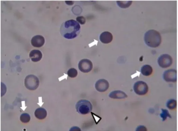
Background : Janus tyrosine kinase 2 (JAK2) V617F mutation has been described in a high proportion of patients with Philadelphia

Purpose: We investigated recent aspects of causative organism and antimicrobial susceptibility of urinary tract infections(UTI).. Materials and Methods:

Since then, studying to steppe routes has been made in various fields, allowing a balanced understanding of the role of nomadic imperial history, which

• Common polymorphism in MAO-A gene resulting in relatively low-activity enzyme, has been associated with increased risk for violent or antisocial behavior as well as for

(A) Interestingly, this has often been the case with English animal expressions.. As you may have observed, they make irregular twists and turns in their