교신저자 : 김 승 현
서울특별시 성동구 행당동 17 한양대학교 의과대학 신경과학교실
TEL) 02-2290-8371, FAX) 02-2290-8377, e-mail) [email protected]
체성감각 유발 전위에서 m o n t a g e에 대한 개념
동아대학교 의과대학 신경과학교실, 한양대학교 의과대학 신경과학교실*
차 재 관・김 승 현*
The Concepts of Montage in Somatosensory Evoked Potentials.
Jae-Kwan Cha, M.D., Seung-Hyun Kim M.D.*
Department of Neurology, College of Medicine, Dong-A University, Pusan, Korea Department of Neurology, College of Medicine, Hanyang University, Seoul, Korea*
- Abstract -
Although somatosensory evoked potentials(SSEPs) have been utilized as the useful diagnostic tools in evaluating the wide variety of pathological conditions, such as focal lesions affecting the somatosensory pathways, demyelinat- ing diseases, and detecting the clinically occult abnormality, their neural generators is still considerably uncertain. To appreciate the basis for uncertainties about the origins of SSEPs, consider criteria that must be met to establish a causal relationship between activity in a neural structure and a spine/ scalp-recorded potential. Electrode locations and channel derivations for SSEPs recordings are based on two principles:(1) the waveforms are best recorded from electrode sites on the body surface closest to the presumed generator sources along the somatosensory pathways, and(2) studies of the potential-field distribution of each waveform of interest dictate the best techniques to be used. In this article, authors will describe followings focused on ;(1) the concepts of near field potentials(NFPs) and far field potentials(FFPs) - the voltage of NFPs is highly dependent upon recording electrode position, FFPs are unlike NFPs in that they are widely distributed, their latencies and amplitudes are independent of recording electrode.(2) appropri- ate montage settings to detect the significant potentials in the median nerve and posterior tibial nerve SSEPs(3) neural generators of various potentials(P9, N13, P14, N18, N20, P37) and their clinical significance in interpretating the results of SSEPs. Especially, Characteristics of N18(longduration, small superimposed inflection) suggested that N18 is a complex wave with multiple generators including brainstem structures and thalamic nuclei. And N18 might be used as the parameter of braindeath. Precise understanding on these facts provide an adequate basis utilizing SSEPs for numerous clinical purposes.
서 론
유발전위(evoked potential)이란 외부자극에 대한 생 체에서 얻어지는 전기적 신호로서, 자극에 의해 얻어진 유발전위는 일정한 시간에 나타나는 특징이 있지만 진폭 이 매우 작아, 자발적으로 발생하는 정상 뇌파 및 여러 잡파와 혼합되어 나타나므로 그대로는 볼 수가 없어 일 정한 시간에 나타나는 전위만을 선택하여 증폭시키는 동 시에 잡파는 제거하는 평균화 과정( a v e r a g i n g )을 거쳐 야 한다.1 이와 같은 평균화 과정을 거치면서 얻어지는 전위는 자극종류, 자극조건, 증폭기의 조건, 기록 조건 등의 비생리적인 요인과 환자의 신체조건등과 같은 생리 적 요인에 결과가 매우 상이하게 나타날 수 있다. 따라서 각 검사실 마다 이에 대한 조건을 표준화한 상태에서 얻 어진 정상결과를 이용하여 결과를 분석하는 것이 필수적 이지만 이와 같은 조건 설정 외에 얻고자 하는 파형을 적 절히 얻기 위해서는 환자의 상황에 따라 적절한 m o n- t a g e의 설정을 하여야 하는 것이 중요한 관건이 된다.
체성감각 유발전위( S E P )는 생리적 혹은 전기적 자극을 이용하여 말초신경의 Ia fiber를 통해 proximal plexus, dorsal root ganglion, 척수의 후주(posterior column), 내측융대(medial lemniscus)를 거쳐 thalamic radia- t i o n에 이르는 경로를 비교적 객관적으로 검사할 수 있는 방법으로,2체성감각 신경의 경로에 따라 얻어지는 파형을 적절히 얻고 이를 정확하게 분석하기 위해서는 m o n t a g e 설정에 대한 정확한 개념을 알아야 된다고 생각된다. 이에 본 고에서는 임상에서 흔히 이용되는 체성감각 유발전위 검사시 고려하여야 할 montage 설정 방법 및 각 전위의 유발기( g e n e r a t o r )에 대해 알아 본 후 임상적 의의에 대 해 기술하고자 한다.
Montage 설정의 기본원리 및 응용
유발 전위 검사를 임상적으로 적절하게 적용 할 수 있 고 이를 분석하기 위해서는 m o n t a g e의 설정을 어떻게 하느냐에 따라 달려 있을 만큼 적절한 m o n t a g e의 설정 은 검사 분석의 가장 중요한 사항이라고 할 수 있다.
전기자극을 시행한 후 뇌표피 전극에서 얻어진 유발 전위는 회백질에서 유래하는 postsynaptic poten- t i a l ( P S P )과 백질부의 신경섬유 경로에서 발생하는 전 위로 크게 대별된다. 백질부에서 유래하는 활성 전위는 다시 near field potential(NFP)과 far field poten- t i a l ( F F P )로 나뉘는데, 전류가 흐르며 탈분극을 일으 킨 국소 부위에서 기록한 신호를 near field poten- tial(NFP) 이라고 하고, 신경주행을 따라 활성 전위가
흐르면서 신경조직을 싸고 있는 주위조직의 변화 (impedance 등의 차이)에 의해 발생하는 전위를 f a r field potential(FFP) 이라고 한다.4 - 1 0 전자는 탈분극 이 일어나는 신경 자체에서 유래하는 것이므로 기록전 극의 위치의 변동에 의해 진폭 및 잠복기의 변동이 일어 난다. 그러나 후자의 경우 기록전극의 위치에 따라 진 폭, 파형, 잠복기에 변화 없이 일정하게 관찰되는 특징 이 있어 stationary potential이라고도 한다. 또한 회 백질 부위의 P S P에 의한 전위도 N F P과 F F P의 개념 이 적용되기도 하나 백질부에서 유래하는 전위보다는 명확한 구분이 되지 않는다.
F F P에 대한 원리는 처음 뇌간 청각유발 전위( B A E P ) 에서 기술되었는데, BAEP에서 나타나는 모든 파형들은 실제로 뇌간의 깊은 곳에서 유발되는데 기록은 유발기에 서 멀리 떨어진 s c a l p에서 이루어지므로 전형적인 F F P 의 모델이라 하겠다.1 1 초기의 F F P의 고전적인 개념은 첫째로 F F P은 넓게 분포하며, 둘째로 F F P는 기록 전극 의 위치에 상관없이 일정한 잠복기를 가지며, 셋째로 F F P는 언제나 양극성(positive polarity)를 나타난다고 정의하였다.1 1
체성감각 유발전위에서 F F P이 나타나는 기전은 크게 3가지로 나눌 수 있는데 첫째로 활성전위가 신경조직을 따라 흐르는 중간에 갑자기 주위조직의 부피( v o l u m e ) 이 변하는 경우와, 둘째로 신경조직이 진행을 하다가 갑 자기 진행방향이 바뀌는 경우, 셋째로 신경조직을 싸고 있는 주변조직의 저항이 바뀌는 경우이다.1 2
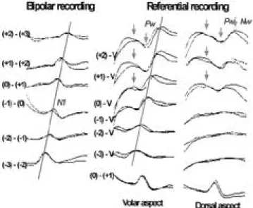
1 9 8 6년 K i m u r a등1 3의 연구는 정중신경에서 활성전 위가 흐르면서 수부( h a n d )에서 팔( a r m )로 부피가 갑 자기 변하는 손목( w r i s t )부위에서 어떻게 F F P가 형성 되는지를 잘 보여 주었는데 Fig. 1에서 손목을 0기점으 로 잡고 원위부로 1.5cm 간격으로 -1, -2, -3 위치에 전극을 부착하고 근위부에서도 마찬가지로 1.5cm 간격 으로 +1, +2, +3 위치에 전극을 부착한후 정중신경에 대해 정방향( o r t h o d r o m i c )으로 감각신경전도 검사를 시행할 때 bipolar recording을 시행할 경우에는 손끝 에서 근위부로 활성전위가 진행함에 따라 잠복기가 길어 지는 전형적인 N F P들의 특성을 나타내나, 접지전극 (reference electrode)을 정중신경의 진행과는 상관없 는 다섯 번째 손가락 즉 referential recording을 한 경 우에는 N F P뿐만 아니라 손목을 기점으로 잠복기가 기 록 전극에 상관없이 나타나는 일종의 far field poten- t i a l을 관찰할 수 있다. 이 F F P를 더욱 잘 관찰하기 위 해 손등에서 기록한 경우에는 이전에 보인 N F P는 소실 되고 잠복기가 일정한 F F P들을 관찰할 수 있어 F F P가 신경조직을 싸고있는 주위조직의 부피변화에 의해 발생 할 수 있음을 보여 주었다(Fig. 1).
본 고에서 설명하고자 하는 체성감각 유발전위의 FFP 및 N F P에 대한 개념을 확인하기 위해서 저자들
은 정중신경을 자극 한 후 활성전극은 10-20 체계에 의 한 두피의 여러 부분에서 r e f e r e n c e는 자극 반대측의 상지에 부착한 후 전위를 기록하여 보았다. Fig. 2에서 보는 바와 같이 자극 후 얻어진 파형 중 P9, N10, P11, N12, P13, 그리고 N 1 8은 기록전극의 위치에 관 계없이 일정한 잠복기가 일정한 특징을 보여 F F P에 해 당하며 C 3에서는 다른 부위에서 얻어지는 파형 외에 N 2 0이 추가로 관찰되었는데 이는 다른 부위에서는 관 찰되지 않는 N F P이라고 할 수 있다.
이와 같은 상황은 경추 전위에서도 관찰 할 수 있는데 Fig. 3과 같이 N13 전위는 r e f e r e n c e의 위치의 변화에 따라 그 p o l a r i t y가 변화되어 N F P의 특성을 나타내나 N 1 2는 p o l a r i t y의 변화가 없어 전형적인 F F P의 특성 을 보인다고 할 수 있다. 실제 임상에서 많이 이용하는 정중신경의 체성감각 유발전위에서 대표적인 F F P의 하
나인 P9 전위는 활성전위가 E r b’s 지점을 통과하는 시 기에 주변조직의 부피가 팔에서 몸통으로 이행하면서 큰 차이를 보이면서 나타나는데, Fig. 4와 같이 F F P의 기본적인 특징의 하나인 넓게 전위들이 분포하고, 전극 의 위치에 상관없이 잠복기가 일정한 형태를 보이고 있 다. 그러나 앞에서 언급한 F F P의 고전적 3가지 개념과 는 달리 언제나 양극성만 보이는 것이 아니라 접지전극 의 위치에 따라서는 음극성도 나타날 수 있음을 보여주 어 F F P의 개념중 극성에 대한 개념은 새로이 바꾸어 졌다. FFP를 쉽게 이해할 수 있는 쉬운 예는 아마도 심 전도( E K G )를 상상해보면 된다. 일반적으로 E K G는 신 체의 여러 곳에서 측정을 하여도 그 잠복기나 모양이 변 하지 않는다. 이것은 결국 E K G가 F F P의 형태로 전달 됨을 알 수 있다.
체성감각 전위검사시 위에서 언급한 N F P와 F F P을 잘 기록하기위해 필요한 m o n t a g e의 설정의 조건은 다 음과 같다.
1. FFP을 정확하게 관찰할 수 있는 m o n t a g e가 최소 한 하나가 있어야 하며
2. 관심이 되는 N F P을 얻기 위한 montage setting 이 있어야 하는 데 cortical potential을 얻기 위해서는 scalp-scalp derivation의 m o n t a g e가 필수적이며, cervical NNF potential을 얻기 위한 m o n t a g e가 있 어야 한다.
3. 그 외에 임상적으로 의미가 있을 파형 (예 P 1 3 / 1 4 , N 1 8등)을 얻기 위한 montage 의 설정이 필수적이라고 하겠다.
체성감각 유발전위에서 m o n t a g e의 설정
체성감각 유발전위에서 적절한 m o n t a g e의 설정은 위 에서 언급한 N F P과 F F P을 분석 가능한 것이어야만 한 다. Fig. 5는 N F P과 F F P의 개념을 설명하기 위한 monatge setting의 개념적인 그림으로서 체성감각 유 발전위 검사시 scalp-noncephalic reference (channel 1, 2, 3)를 사용할 경우 F F P은 모든 두피영역( c h a n n e l Figure 1. Sensory nerve potentials across the wrist recorded bipo -
larly(left), or referentially from volar(middle) or dorsal(right) aspect with distal reference.
Figure 2. The differences of near field potential(N20) and far field potentials(P9,N10,P11,N12,P13) in scalp-scalp montage and scalp-noncephalic montage.
Figure 3. Median nerve SEPs recorded from a ring of electrodes about the neck using noncephalic reference site.
1 -Fz, channel 2-C4)에서 기록되나 체감각피질부위 ( C 3 )에서는 F F P외에 N F P도 동시에 관찰되게 된다.
즉 Fig. 5의 channel 1, 2에서는 noncephalic refer- e n c e에 해당하는 elbow 지점에 r e f e r e n c e를 둘 경우 F F P에 해당하는 P9, N10, P11, N12, P14, N18전위 들이 모두 나타나게 되나 channel 3에서는 F F P에 해당 하는 P9, N10, P11, N12, P14, N18전위 외에 N F P 에 해당하는 N20, P23도 관찰되게 된다. 따라서 Fig. 5 의 channel 4와 같이 scalp-scalp montage (C3-C4) 를 설정함으로 두 scalp electrode에서 공통적으로 갖는 P9, P11, P13등의 F F P는 상쇄되고 순수한 N F P들만 이 기록된다. 이와 같은 현상은 Fig. 1을 참조하여도 동 일한 결과를 볼 수 있다.
American EEG Society14에서 권장하는 m o n t a g e
는 감각대뇌에서 발생되는 N F P을 측정하기 위한 scalp-scalp montage, FFP들을 측정하기 위한 s c a l p - noncephalic montage, central conduction time을 분석할 수 있는 spine-scalp montage, 그리고 E r b’s 전위를 잘 기록할 수 있는 m o n t a g e등 4 channel을 기 준으로 하나 최근의 유발전위 기기들이 4 channel 이상 인 것을 고려할 때 각 파형을 잘 관찰할 수 있는 부가적 인 m o n t a g e설정을 하는 것이 좋다고 생각된다.
따라서 저자들의 검사실에서 기본적으로 설정하는 정 중신경 체성감각 유발 전위의 m o n t a g e는 AEEG soci- e t y의 추천 m o n t a g e외에 두피에서 F F P을 좀더 관찰 하기 위한 c h a n n e l을 하나 더 설정하고 있으며( c h a n- nel 5) 경추부의 N F P에 대한 정보를 얻고자하는 montage (channel 6)를 하나더 추가로사용하고 있다.
이를 비교하여 보면 Table 1과 같다.
후경골 체성감각 유발전위의 경우에는 정중신경 체성감 각 유발전위에 비해서는 두피에서 얻어지는 F F P에 대한 m o n t a g e에 대한 연구는 적은 편이다. 두피에서 s c a l p - Figure 4. Distibution of P9s in response to stimulation of the left median at the wrist.
Figure 5. Basic concept of FFP and NFP in Median Nerve SEP
Table 1. Comparison of montage ; AEEG society guideline and HYUH & Dong-A montage in median nerve SEP
AEEG Society guideline HYUH and DongA Montage Ch 1 ; C4’-Erbc Ch 1 ; C3’-Fz(NFP) Ch 2 ; C4’-Fz Ch 2 ; C4’-Erbc(FFP) Ch 3 ; C5S-Fz Ch 3 ; C5S-Fz(N/P 13) Ch 4 ; Erbi-Erbc Ch 4 ; Erbi-Erbc(Erb)
Ch 5 ; C5S-Ant neck(N13) Ch 6 ; Fz-Erbc(FFP)
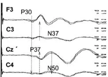
scalp montage에 의해 기록되는 P 3 7은 C z와 c e n t r a l parasagittal 부위에서 가장 크게 관찰되고 전후좌우 부 위에서는 진폭의 현저한 저하를 보이는 N F P의 특징을 나타낸다. 자극 반대쪽의 central parasagittal 부위보다 자극 동측에서 큰 진폭을 갖는 이유는 두피에서 기록할 때 자극 동측에서 반대측 보다 d i p o l e이 크게 형성되기 때문이다.1 5 - 1 7P T S E P에서 N F P와 F F P의 특징을 잘 관 찰하기 위해서는 적어도 하나이상의 s c a l p - n o n c e p h a l i c r e f e r e n c e와 2개의 scalp-scalp montage가 있어야 하는 데 저자들은 Table 2와 같이 설정한다. 최근 저자들은 P T S E P에서 scalp-ear lobe montage를 이용하여 M N S E P의 P 1 4에 해당하는 P 3 0을 비교적 쉬운 방법으 로 얻음으로 향후 P T S E P의 새로운 m o n t a g e의 방법으 로 개발되리라 기대된다(Fig. 6).
체성감각 유발전위에서 파형들의 유발기
1) P9
P 9전위는 활성전위가 brachial plexus volley를 지 나면서 생기는 F F P의 일종으로, N10은 근위부 p l e x u s 를 지나면서 생기는 것으로 생각된다.1 8특히 P9 전위가
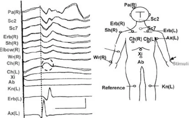
brachial plexus 부근을 활성전위가 지나다 주변조직의 부피의 변화에 의해 생기며 P 9과 같은 F F P가 주변조직 에 변화에 의해 생긴다는 것은 Fig. 7에서 보듯이 체성 감각 유발전위를 시행하는 중에 brachial plexus 부근 의 견관절(shoulder joint)을 움직이면 P 9의 잠복기가 확연히 변하는 것을 관찰하면 쉽게 이해할 수 있다.1 9
2) N13
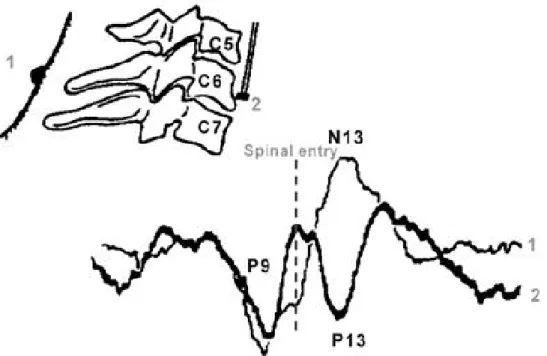
경추 척수에서의 활성전위가 진행하면서 발생되는 전위 로 생각되며 통상적으로 전기 생리 검사실에서 c e r v i c a l cord(C5)-scalp(Fz) montage에서 측정하므로 c o r d에 서 발생하는 N13 NFP과 s c a l p에서 발생하는 P13 FFP 가 합쳐져 형성된 전위이므로 순수하게 척수에서 발생하 는 전위가 아님을 알 수 있다. 그러므로 실제 경추 척수의 이상이 있는 환자에서 s c a l p에서 발생된 P 1 3에 의해 정 상소견으로 나올 가능성도 있다. 이를 보완하기 위해서는 목뒤에 활성전극을 부착하고 thyroid cartilage 부근에 r e f e r e n c e를 설정하면 순수한 척수에서 발생하는 전위를 얻을 수 있다. 이 이론적 근거로는 Fig. 8과 같이 정중신 경 체성감각 유발전위를 검사시 목뒤와 식도내에 전극들 을 부착하면 목뒤 전극에서는 음극성의 전위를 얻고 목의 앞부분에선 양극성의 동일한 잠복기를 갖는 전위를 얻게 되므로 자연히 목뒤와 목앞에서 m o n t a g e를 설정하면 순 수하게 경추 척수에서 발생하는 N13 전위를 잘 관찰하게 된다2 1.
기존의 P 1 3의 개념대신 1 9 9 1년 R e s t u c c i a와 M a u - g u i e r e등2 2은 syringomyelia 환자 중 통각과 온도감각 이 저하되고 위치, 진동감각이 보존된 환자들에서 F i g . 9와 같이 N 1 3은 소실되나 P 1 4가 보존되는 현상을 보 고함으로 N 1 3의 유발기가 통각과 온도각의 진행경로와 관계가 있음을 보고하였으나 이에 대해서는 보다 많은 연구가 필요하다.
Table 2. Comparison of montage ; AEEG society guideline and HYUH & Dong-A montage in posterior tibial nerve SEP
AEEG Society guideline HYUH Montage
Ch 1 ; Cz’-FPz(NFP) Ch 1 ; Cz’-PFz(NFP) Ch 2 ; T12S-r4cm T12S Ch 2 ; T12S-ICc
Ch 3 ; L3S-r4cm L3S Ch 3 ; L5S-ICc
Ch 4 ; PF-Kn Ch 4 ; PF-Kn
Ch 5 ; Cz’-elbow(FFP) Ch 6 ; C4’-PFz(NFP)
Figure 7. Wave form changes of P9 with right median nerve stimu - lation in different arm positions.
Figure 6. Scalp somatosensory evoked potentials to the right tibial nerve stimulation(ear lobe reference). All Channels showed P30 potential.
3) P14
P14 전위는 scalp-noncephalic montage에서 관찰되 는 F F P의 일종으로 P 1 3이 경추 척수가 유발기라면 P 1 4 는 foramen magnum위의 내측융대(medial lemnis- c u s )가 그 유발기로 알려져있다.2 3 통상적으로 s c a l p - noncephalic montage에서는 P 1 3과 P 1 4가 한 개의 파 형으로 융합되어 나타나거나 혹은 bifid 형태로 관찰된다.
그러므로 척수 전위와 뇌간에서 발생되는 전위를 잘 구분 할 수 없다. 이런 단점을 극복하기 위해 1 9 9 6년 W a g n e r 등2 4은 scalp-nasopharyngeal montage를 사용할 경우
순수한 foramen magnum 위에서 유발되는 P 1 4전위를 관찰할 수 있다. 이것은 nasopharygeal 전극이 P13 전 위를 기록할 수 있는 위치이므로 s c a l p에서의 P 1 3을 서 로 상쇄되므로 가능하다. 그러나 nasopharyngeal 전극 을 통상적인 전기 생리 검사실에서 사용하기는 어려우므 로 최근에는 n a s o p h a r y n g e a l전극과 같은 위치에 있는 ear lobe전극을 사용하면 순수한 P14 전극을 구할 수 있 다. 저자들의 검사실에서 예비적으로 시행한 연구결과 (Fig. 10)에서 보면 scalp-noncephalic montage에서는 P 1 3 / 1 4가 하나의 파형으로 융합된 형태를 보이는데 이 것의 잠복기가 posterior neck-anterior neck에서 얻는 척수 전위에 비해 늦으나 scalp-nasopharyngeal mon- t a g e에서 얻는 P 1 4와 유사하게 나타나 ear lobe 접지전 Figure 9. Abnormal somatosensory evoked potential in patients
with syringomyelia(pain, temperature sensory loss, pre - serve vibration and position). Absent N13 with normal P14 and N20.
Figure 10. Somatosensory evoked potential to right median nerve stimulation.
P13/14 on channel 1-12.9 msec.
N13 on channel 2-11.2 msec.
P14 on channel 3-13.5 msec.
Figure 8. Superimposed averaged somatosensoy evoked potential to median nerve stimulation, recorded in front(electrode 2) or behind vertebra(electrode 1).
극을 이용하면 특히 cervico-medullary junction에 병변 을 의심하는 환자들에서 P 1 4의 존재 유무를 통해 비교적 쉽게 진단 할 수 있으리라 생각된다.
4) N18
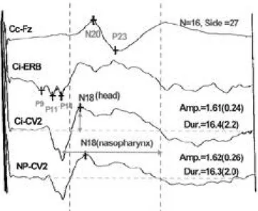
N18 전위는 최근까지 그 유발기에 대해 잘 알려져 있 지 않았다. 이전까지는 N 1 8의 유발기로 N 1 8의 반응기간 ( d u r a t i o n )이 길어 여러 가지 피질하 핵들과 시상의 핵들 이 관여하리라 추측하였다2 5 - 2 9. 그러나 1 9 9 6년 S o n o o등3 0 이 내측 연수 증후군(medial medullary syndrome) 환 자에서 P 1 4는 소실되나 N 1 8이 보존되는 소견을 증명하 여 N 1 8의 유발기가 연수 이하임을 제시하였다. 또한 1 9 9 9년 김등3 1이 nasopharyngeal 전극을 연수의 복부 위치에서 접근시키고 정상인에서 s c a l p에서 얻은 N 1 8과 관찰한 결과 두 전위가 차이가 없어 N 1 8의 유발기가 연 수 근처임을 간접적으로 증명하였고(Fig. 11), 특히 이 연구에서 뇌교-연수 접합부에 뇌출혈이 있는 환자에서 P 1 4는 소실되나 N 1 8은 보존되어 이 전위의 유발기가 뇌 교-연수 접합부 아래임을 간접적으로 증명하였다( F i g . 12). 최근의 보고에서 N 1 8의 유발기는 설상핵( c u n e a t e n u c l e u s )로 추측되는데 이 가설이 맞는다면 향후 N 1 8 전위는 체성감각 유발전위에서 뇌사(brain death)의 중 요한 판단 지표로 사용될 수 있다고 생각된다.
결 론
체성감각 유발 전위에서 NFP, FFP의 개념 그리고 m o n t a g e의 설정과 유발기들에 대한 이해는 검사의 민
감도를 증진시키고 임상적 유용성에 큰 도움이 되리라 생각된다.
향후 체성감각 유발 전위에서 P14, P30, N18과 같 은 F F P들에 대한 보다 많은 연구를 기울인다면 뇌자기 공명영상에서 발견하기 힘든 하부 뇌간부 그리고 척수- 연수 접합부(cervico-medullary junction)의 이상 여 부를 진단하는데 큰 도움이 되리라 생각되며, 뇌사환자 의 객관적 판정에 도움이 될 것이다.
참고문헌
01. Chiappa KH. Evoked potential in clinical medicine. 3rd ed.
Raven Press: New-York, 1997;1-2.
02. Chiappa KH. Evoked potential in clinical medicine. 3rd ed.
Raven Press: New-York, 1997;341-342.
03. Anziska BJ and Cracco RQ. Short latency SEPs to median nerve stimulation. Electroenceph. Clin. Neurophysiol. 1981;
52:531-539.
04. Brazier MAB. A study of electrical fields at the surface of the head. Electroencephalogr. Clin. Neurophysiol. 1949;2:38-52.
05. Woodbury WJ. Potential in volume conductor. In: Neurophy- siology, 2nd ed, Philadelphia, Saunders, 1965;85-91.
06. Emerson RG, Seyal M, Pedely TA. Somatosensory evoked potentials following median nerve stimulation. I. The cervical components. Brain. 1984;10:169-182.
07. Kimura J, Mitsudome A, Yamada T, Dickins QS. Stationary peaks from moving source in far field recording. Electroence- phalogr. Clin. Neurophysiol. 1984;58:351-361.
08. Kimura J, Mitsudome A, Yamada T, Beck DO, Dickins QS.
Field distribution of antidromically activated digital nerves.
Neurology 1986;33:1164-1169.
09. Leuders H, Lesser R, Hahns J, Little J, Klem G. Subcortical evoked potentials to median nerve stimulation. B r a i n. 1983;
Figure 11. Somatosensory evoked potential to the right median nerve stimulation. Cc: contralateral cortex to stimula - tion, Ci: ipsilateral cortex to stimulation, CV2: 2nd cer - vical vertebra, NP: nasopharyngeal electrode.
Figure 12. Somatosensory evoked potential in patient with ponto- medullary junction hemorrhage. P13/14 was not recorded. but N18 was preserved.
106:341-372.
10. Nakanishi T. Action potential recorded by fluid electrodes.
Electroenceph. Clin. Neurophysiol. 1983;55:114-115.
11. Jewett DL and willsion JS. Auditory evoked potential far-field averaged from the scalp of humans. Brain. 1971;94:681-696.
12. Dumitru D and Jewett DL. Far-field potentials. Muscle and Nerve. 1993;16:237-254.
13. Kimura J, Ishida T, Sujuki S, et al. Far-field recording of the junctional potential generated by median nerve volley at the wrist. Neurology. 1986;36:1451-1457.
14. Chatrian et al. American Electroencephalographic Society Guidelines for Clinical Evoked Potential Studies. Journal of Clinical Neurophysiology. 1984;1:3-53.
15. Cruse R, Klem G, Lesser RP, Leuders H. Paradoxical lateral- ization of the cortical potentials evoked by stimulation of the posterior tibia nerve. Arch Neurol. 1982;39:222-225.
16. Kakigi R and Shibasaki H. Scalp topography of the short la- tency somatosensory evoked potential following posterior tib- ial nerve stimulation in man. Electroenceph. Clin. Neuro-phys - iol. 1983;56:430-437.
17. Seyal M, Emerson RG, Pedely TA. Spinal and early scalp recorded components of the somatosensory evoked potential following stimulation of the posterior tibial nerve. Electroen- ceph. Clin. Neurophysiol. 1983;55:323-331.
18. Jones SJ. Short latency potentials recorded from the neck and scalp following median nerve stimulation in man. Electroen- ceph. Clin. Neurophysiol. 1977;43:853-863.
19. Hashimoto S, Kawamura J, Segawa Y, Yamamoto T, Naka- mura M. Possible model for generation of P9 far field poten- tials. Muscle and Nerve 1992;15:106-110.
20. Emerson RG and Pedely TA. Effects of cervical spinal cord lesions on early components of the median nerve somatosen- sory evoked potential. Neurology. 1986;336:20-26.
21. Desmedt JE and Cheron G. Prevertebral recording of subcorti- cal somatosensory evoked potential in man; The spinal P13 component and dual nature of the spinal generators. Electroen- cephalogr. Clin. Neurophysiol. 1981;52:257-275.
22. Restuccia D and Mauguiere F. The contribution of median nerve SEPs in the functional assessment of the cervical spinal cord in syringomyelia. Brain 1991;114:361-379.
23. Desmedt JE and Cheron RG. Central somatosensory conduc- tion in man. Electroenceph. Clin. Neurophysiol. 1980;50:382- 403.
24. Wagner W. Scalp, ear lobe and nasopharyngeal recordings of he median nerve somatosensory evoked P14 potential in coma and brain death. Brain 1996;119:1507-1521.
25. Desmedt JE, Cheron G. Non-cephalic reference recording of early somatosensory potentials to finger stimulation in adult or aging normal man. differentiation of widespread N18 and con- tralateral N20 from the prerolandic P22 and N30 components.
Electroenceph clin Neurophysiol 1981;52:553-570.
26. Mauguiere F, Desmedt JE. Bilateral somatosensory evoked potentials in four patients with long standing surgical hemi- spherectomy. Ann Neurol 1989;26:724-731.
27. Urasaki E, Wada S, Kadoya C, Yokota A, Matsuoka S, Shima F. Origin of scalp far-field N18 of SSEPs in response to medi- an nerve stimulation. Electroenceph clin Neurophysiol 1 9 9 0 ; 77:39-51.
28. Urasaki E, Wada S, Kadoya C, Tokimura T, Yokota A, Yama- moto S et al. Amplitude abnormalities in scalp far-field N18 of SSEPs to median nerve stimulation in patients with midbrain- pontine lesion. Electroenceph clin Neurophysiol 1992;84:232- 242.
29. Urasaki E, Uematsu S, Lesser RP. Short latency somatosenso- ry evoked potentials recorded around the human upper brain- stem. Electroenceph clin Neurophysiol 1993;88:92-104.
30. Sonoo M, Shimpo T, Genba K, Masaki T, Satuka M, Mannen T. Origin of upper cervical N13(ucN13) and widespread N18 in the median nerve SEP. Electroenceph clin Neurophysiol 1990;76:106.
31. 김대현,차재관,김상호,김재우.체성감각 유발전위중N18 Far- field 전위의 비강 인두를 통한 접근. 대한신경과학회지. 1999;
17:112-116.