Address for correspondence;
Young H. Sohn, M.D., Ph.D.
Department of Neurology, Yonsei University Medical Center, 250 Seongsan-ro, Seodaemun-gu, Seoul 120-752, Korea Tel: +82-2-2228-1600 Fax: +82-2‐393-0705 E-mail: [email protected]
Review
뇌자기자극을 이용한 운동신경계의 신경생리학적 평가
중앙대학교 의과대학 신경과학교실1, 연세대학교 의과대학 신경과학교실2
신혜원
1
․손영호2
Neurophysiological Evaluation of the Motor System Using Transcranial Magnetic Stimulation
Hae-Won Shin, M.D., Ph.D.
1
, Young H. Sohn, M.D., Ph.D.2
Department of Neurology, Chung-Ang University College of Medicine
1, Seoul; Department of Neurology, Yonsei University College of Medicine
2, Seoul, Korea
Received 31 May 2011; accepted 2 June 2011.
Transcranial magnetic stimulation (TMS) is a non-invasive tool used to study aspects of human brain physiology, including motor function and the pathophysiology of various brain disorders. A brief electric current passed through a magnetic coil produces a high-intensity magnetic field, which can excite or inhibit the cerebral cortex. Although various brain regions can be evaluated by TMS, most studies have focused on the motor cortex where motor evoked potentials (MEPs) are produced.
Single-pulse and paired-pulse TMS can be used to measure the excitability of the motor cortex via various parameters, while repetitive TMS induces cortical plasticity via long-term potentiation or long-term depression-like mechanisms. Therefore, TMS is useful in the evaluation of physiological mechanisms of various neurological diseases, including movement disorders and epilepsy. In addition, it has diagnostic utility in spinal cord diseases, amyotrophic lateral sclerosis and demyelinating diseases. The therapeutic effects of repetitive TMS on stroke, Parkinson disease and focal hand dystonia are limited since the duration and clinical benefits seem to be temporary. New TMS techniques, which may improve clinical utility, are being developed to enhance clinical utilities in various neurological diseases.
Key Words: Transcranial magnetic stimulation, Motor system, Neurophysiology, Neurological disease
서 론
뇌자기자극은 비침습적으로 사람 뇌피질의 신경생리학 적인 특징과 변화를 측정할 수 있는 도구이다.
1비침습적 인 뇌피질의 자극에 대한 역사는 1980년 초 뇌전기자극
(transcranial electrical stimulation, TES)으로 올라간다.
2뇌전
기자극은 높은 전압의 순간적인 전기자극을 이용하여 운
동피질을 자극하는 방법으로 운동유발전위 (motor evoked
potential)를 발생시킨다. 이 방법은 운동피질의 특성을 조
사하는 데에 유용하였으나 두피의 통증섬유를 함께 자극
하기 때문에 통증이 수반되는 점이 한계였다. 5년 뒤 소개
된 뇌자기자극은 자기코일 외부에 형성되는 자기장으로
뇌신경을 자극하는 방법으로 통증이 없다는 점에서 뇌전
기자극을 대신하게 되었다.
3뇌자기자극은 자기코일에 순
간적으로 높은 전압을 흘려 보내 자기장을 발생시켜 자기
장의 방향에 평행하게 전류를 발생시키는 방법으로 뇌피
Figure 2. (A) Circular coil, (B) Eight coil.
Figure 1. A high current electrical pulse produces a mag- netic field perpendicularly to the plane of the coil and then an electrical field is induced perpendicularly to the magnetic flow.
8질을 국소적으로 흥분시킬 수 있다.
4운동피질을 자극할 때에 이에 대한 반응으로 나타나는 해당 근육의 운동유발 전위를 통하여 운동피질의 흥분성(excitability)을 수치화할 수 있다.
5,6뇌자기자극은 다양한 변수를 통하여 시간에 따 른 뇌피질의 흥분 또는 억제상태를 조사할 수 있으며 뇌피 질 위치의 기능적지도화와 일시적 기능병변 재현 등의 방 법으로 뇌피질의 신경생리에 대한 연구에 널리 쓰이고 있 다.
1,5또한 반복뇌자기자극을 통하여 신경질환에서 뇌의 흥분성의 변화를 유도하여 치료적인 효과를 기대할 수 있 다
7. 여기에서는 운동신경계의 뇌자기자극에 대해 전반적 으로 살펴보고 임상적인 적용에 대하여 알아보고자 한다.
본 론
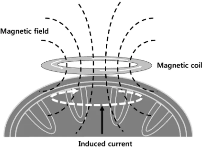
1. 뇌자기자극의 원리와 신경생리학적 배경
두피에 자기코일을 위치시키고 전류를 흘려보내면 두개 골을 통과하는 자기장이 형성되어 자기장 영역의 변화로 와상전류가 유도되고 이는 신경막을 통과하여 전위를 형 성하게 된다(Figure 1).
8전류가 흐르도록 만들어진 자기코 일은 크게 원형코일과 팔자코일이 있다(Figure 2). 원형코 일은 약 14 cm의 반경으로 코일의 정중앙에서 최고 전류 가 흐르고 두피아래 1.5-2.0 cm의 신경세포까지 흥분시킬 수 있다. 팔자코일은 두 개의 원형코일이 편평하게 결합된 형태로 각각의 코일반경은 약 9 cm이다. 팔자코일에서는 각각의 코일의 중앙에서 최고 강도의 자기장이 흐르게 되 어 결국 두 코일이 만나 지점의 아래에서 최고점이 되고 원형코일에 비하여 뇌신경을 보다 국소적으로 자극할 수
있는 장점이 있다 . 자기장은 코일과 멀어질 때 급속도로 그 강도가 떨어진다 . 팔자코일을 사용하였을 때에 자극이 가능한 효과적인 자기장영역은 최대 3-4 cm 정도이다. 뇌 전기자극과 비교하여 다른 점은 뇌자기자극에 의하여 유 도되는 전류는 코일의 표면에 평행하게 흐르므로 뇌피질 의 평행한 뇌섬유를 따라서 흥분된다는 점이다 (Figure 1).
8전극을 연수의 피라미드 또는 뇌척수의 등가쪽에 위치 시키고 운동피질을 한번 자극하게 되면 피질억제기간 후 에 높은 주파수의 몇 개의 파동을 관찰할 수 있다.
9가장 처음 나타나는 파동은 피라미드로를 따라서 빠르게 전달 된 신호로 피라미드로를 ‘직접(direct)’ 자극했다는 의미에 서 ‘D’ 파동이라고 부른다. ‘D’ 파동 이후 나타나는 몇 개 의 파동은 피라미드로 신경의 신경연접간 활성을 통해 전 달된 파동으로 간접적으로 자극되었다는 의미에서 ‘I’파동 이라고 부른다 . 뇌전기자극은 주로 ‘D’ 파동을 유발하는 반면에 뇌자기자극은 ‘D’ 파동과 ‘I’ 파동을 모두 유발하나 임계점 부근의 자극 강도에서는 ‘I’ 파동만을 유발한다.
10이는 자기장 흐름의 방향이 뇌피질에 평행하므로 피질-피 질간 또는 피질하 시냅스간 활성을 통해 간접적으로 피라 미드로를 자극하기 때문이다.
2. 뇌자기자극으로 측정하는 운동신경계의 변수
1) 중추전도시간(central conduction time)
뇌자기자극을 통하여 중추전도시간을 측정할 수 있다.
중추전도시간은 운동유발전위의 잠복기(latency)에서 말초 신경전달 및 신경-근 접합부 전달시간을 제외한 시간이다.
말초신경전달시간은 척수에서 근육까지의 경로인 F-파를 측정하는 방법과 척수를 직접 자극하는 방법이 있다. 척수 레벨을 직접 자극하는 경우 전각세포에서 3 cm 떨어진 지 점이 자극되기 때문에 이보다 근위부에 병변이 있는 경우 에는 정상 소견을 보일 수 있다. F-파는 검사하는 근육에 해당하는 말초신경을 역치상(suprathreshold) 자극 강도로
A B
Figure 3. The central conduction time can be obtained as MEP latency (A)-[F(B)+M(C)-1)/2].
8MEP; motor evoked potential.
Figure 4. Recruitment curve shows the growth of MEP size along with increasing stimulation intensity. MEP; motor evoked potential, MT 1 mV; motor threshold 1 mV.
자극하는데 이 자극은 정방향으로는 말초운동신경을 통하 여 근육에 짧은 잠복기 파형을 형성하고(M-파) 역방향으 로는 척수를 통하여 운동원신경을 거쳐 돌아오는 방향 으로 긴 잠복기를 가지는 F-파를 형성한다. 전체 말초운동 전달시간은 (F+M-1)/2 (1은 알파운동신경원에서의 지체시 간을 의미한다.) 로 계산할 수 있다. 따라서 중추전도시간 은 [운동유발전위 잠복기–(F+M-1)/2]로 나타낼 수 있다 (Figure 3).
82) 단일뇌자기자극(single-pulse TMS) (1) 운동역치(Motor threshold)
운동역치는 해당근육의 운동유발전위를 발생시킬 수 있는 최소 자극강도이다 . 안정운동역치(resting motor threshold)는 10번의 자극 중에서 5번 이상에서 50 uV 이상의 운동전위 가 유발되는 자극 강도로 정의하며 최대 자극강도의 1%
간격으로 변화시키면서 측정한다. 활성운동역치(active motor threshold)는 약하게 근육에 힘을 준 상태(최대수축의 5-10%
정도)에서 역시 10번의 자극 중 5번 이상에 100 uV 이상의 운동전위가 유발되는 자극강도로 정의한다. 이완근육 운 동역치는 자극지점의 중심에 있는 신경세포의 흥분성을 반영하는 지표로서 전압개폐 Na
+통로(voltage-gated Na
+channel)관련 약물을 처치하였을 때에 그 값이 변하므로 세포막의 흥분성을 반영하는 지표로 알려져 있다.
11(2) 운동유발전위 점증곡선(MEP recruitment curve) 점증곡선은 자극강도를 증가시킴에 따라서 나타나는 운
동유발전위의 변화를 나타낸다(Figure 4). 점증곡선의 의미 가 무엇인지는 아직 정확하게 밝혀져 있지 않으나, 아마도 자극강도가 커질수록 자극부위의 중심에 있는 신경의 활 성에 주변신경의 활성이 더해지는 현상을 반영한다고 생 각하고 있다.
8따라서 자극부위의 중심에 있거나 매우 흥 분성이 높은 신경은 낮은 자극강도에서 활성이 되며 자극 부위에서 멀리 있거나 흥분성이 높지 않은 신경은 높은 자극 강도에서 활성이 될 것이다. 점증곡선의 변화는 높은 자극 강도에서 뚜렷하게 나타나므로 점증곡선은 흥분성이 낮거나 주변부에 위치한 신경의 흥분성을 대변한다고 할 수 있다.
1(3) 무반응기(silent period)
자발적으로 근육을 수축하면서 근전도를 활성화 시킨
상태에서 운동피질에 뇌자기자극을 가하게 되면 지속되는
근전도활성이 일시적으로 중지되는데 이를 무반응기라고
한다. 무반응기는 중등도로 힘을 준 상태(최대 근육 수축
강도의 5-10%) 에서 역치상자극강도로 뇌자기자극을 가하
는 방법을 사용한다.
8무반응기지속시간은 통상적으로 뇌
자기자극이 가해진 시점에서부터 무반응기가 끝나고 다시
근전도 활성화가 나타나는 순간까지의 시간으로 정한다
(Figure 5).
8,12,13무반응기는 두 부분으로 나누어 지는데 초
반부는 척수의 무반응기를 반영하고 후반부가 뇌피질의
억제기능을 대변하며 이를 피질무반응기(cortical silent period)
라고 별도로 부르기도 한다. 피질무반응기는 앞으로 설명
할 긴간격피질내억제(long interval intracortical inhibition)
기전과 함께 뇌피질의 GABA
B관련 억제기능을 대변하는
현상이라고 알려져 있다.
14Figure 5. The SP can be measured from TMS trigger [a]
to reoccurrence of EMG activity [b].
8SP; silent period.
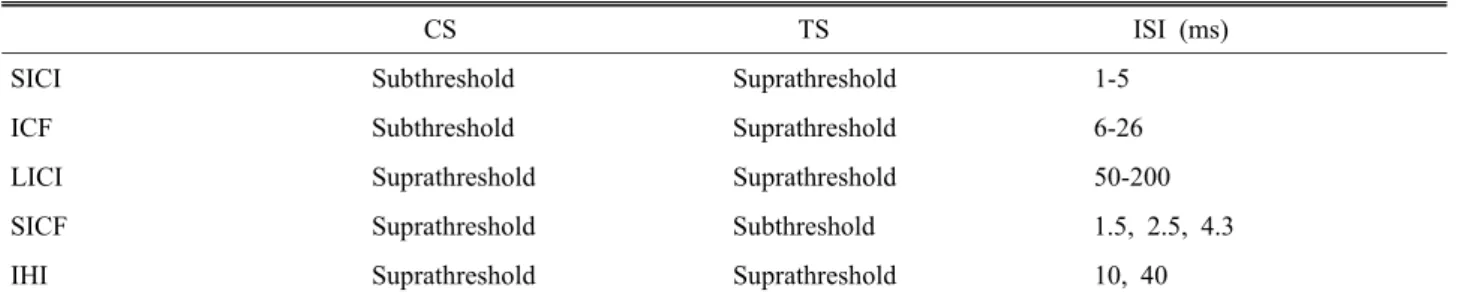
Table 1. Stimulation intensity and intersimulus intervals for paired-pulse TMS
CS TS ISI (ms)
SICI Subthreshold Suprathreshold 1-5
ICF Subthreshold Suprathreshold 6-26
LICI Suprathreshold Suprathreshold 50-200
SICF Suprathreshold Subthreshold 1.5, 2.5, 4.3
IHI Suprathreshold Suprathreshold 10, 40
SICI; short-interval intracortical inhibition, ICF; intracortical facilitation, LICI; long-interval intaracortical inhibition, SICF;
short-interval intracortical facilitation, IHI; interhemispheric inhibition, ISI; interstimulus interval.
3) 쌍자기자극(paired-pulse)
쌍자기자극방법은 실험자극(test stimulus)전 선행자극 (conditioning stimulus)을 주었을 때 운동유발전위가 변하는 정도를 실험자극만을 적용했을 때 발생하는 운동유발전 위의 비로 나타내는 방법이다. 실험자극이나 선행자극의 강도, 그리고 실험자극과 선행자극사이의 시간간격에 따 라서 운동유발전위는 증가하기도 하고 감소하기도 한다.
이를 통하여 운동피질내 또는 운동피질간의 억제와 촉진 기전을 조사할 수 있다(Table 1).
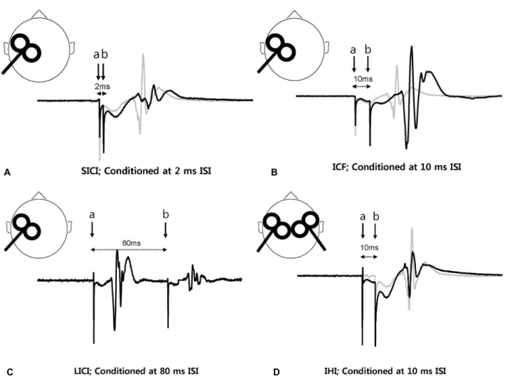
(1) 짧은간격 피질내억제(short interval intracortical inhibition) 와 피질내촉진(intracortical facilitation)
역치하자극강도(subthreshold stimulation intensity)로 운동 피질을 자극하면 사이신경세포(interneuron)를 흥분시켜서 자극간시간(interstimulus interval) 1-5 ms에서는 실험자극에 의한 운동유발전위를 억제하고, 자극간격 8-20 ms에서는 실험자극에 의한 운동유발전위를 촉진시킨다 (Figure 6A, B).
15이를 각각 짧은간격 피질내억제와 피질내촉진이라고 부른 다. 피질내억제와 피질내촉진은 역치하선행자극(통상적으
로 70-90%의 안정운동역치 또는 90%의 활성운동역치) 과 역치상실험자극을 사용하여 측정하며, 그 수치는 선행 자극을 적용하지 않은 운동유발전위를 선행자극을 적용한 후 발생한 운동유발전위로 나눈 퍼센트 값으로 나타낸다.
짧은간격 피질내억제는 GABA
A작용제에 의해 증가하고,
16피질내촉진은 glutamate 길항제에 의해 억제되기 때문에
17각각 운동피질의 GABA
A수용체관련 억제작용 그리고 glutamate 수용체관련 촉진작용의 기전을 반영하는 현상으 로 추측하고 있다. 짧은간격 피질내억제는 자극간격 1 ms 와 2.5 ms에서 각각 최대로 나타나는데 1 ms에서의 짧은 간격 피질내억제는 GABAa 작용제에 영향을 받지 않는 것 으로 미루어 보아 두 위상(phase)은 서로 다른 기전의 피질 억제현상이거나 1 ms의 짧은간격 피질내억제는 피질척수 로의 불응기를 반영하는 것으로 추측하고 있다 .
(2) 긴간격 피질내 억제(long-interval intracotrical inhibition) 선행자극을 역치상자극강도로 정하고 역시 역시상자극 강도의 실험자극를 가하게 되면 실험자극에 의한 운동유 발전위가 억제되는 현상을 관찰할 수 있다 (Figure 6C). 이 를 긴간격 피질내억제 라고 한다. 긴간격 피질내억제는 실 험자극 50-200 ms 전에 선행자극을 가할 때 관찰할 수 있 다.
18간격 피질내억제는 baclofen을 전 처치 하였을 때에 증가하는 것으로 미루어 보아 피질내의 GABA
B매개 억제 현상을 반영한다고 추측을 하고 있다.
19무반응기는 GABA
B신경전달 물질과 관련된 뇌내억제기전의 시간적인 측면을 담당하는 반면 긴간격 피질내억제는 억제 정도 (진폭)을 반 영한다고 할 수 있다.
20(3) 짧은간격 피질내촉진(short-interval intracortcial facilitation)
짧은간격 피질내촉진은 I-파간의 상호작용을 반영하는
현상이다. 짧은간격 피질내억제나 피질내촉진현상과 달리
짧은간격피질내촉진은 선행자극강도를 역치상으로 설정
하고 실험자극을 역치하로 설정하였을 때에 운동유발전위
Figure 6. Paired-pulse TMS. ISI is demonstrated as interval from conditioning stimulation (a) to test stimulation (b). (A) SICI; MEP size is suppressed (black line) with subthreshold conditioning stimulation compared to that in test stimulation alone (grey line) at ISI 2 ms. (B) ICF; MEP size is facilitated (black line) with subthreshold conditioning stimulation com- pared to that in test stimulation alone (grey line) at ISI 10 ms. (C) LICI; MEP of the test stimulation is suppressed compared to that of the suprathreshold conditioning stimulation at ISI 80 ms. (D) IHI; MEP size is suppressed (black line) when the contralateral motor cortex is simulated with suprathreshold conditioning stimulation (grey line). TMS; transcranial magnetic stimulation, SICI; short interval intracortical inhibition, MEP; motor evoked potential, ISI; interstimulus interval, ICF; intra- cotrical facilitation, LICI; long interval intracortical inhibition, IHI; interhemispheric inhibition.
촉진 현상이 자극간시간 1.1-1.5 ms, 2.3-2.9 ms, 4.1-4.4 ms 에서 관찰된다.
21자극간시간 1.5 ms에서의 촉진현상은 I-파 사이의 간격과 유사한 것으로 미루어 보아 짧은간격 피질내 촉진의 근원은 I-파 형성을 담당하는 신경구조에서 기인할 것이라고 추정한다. 1.5 ms의 정점 이후 나타나는 두 부분의 운동유발전위 정점 역시 첫 번째 정점과 같이 후반부에 나타나는 I-파간의 상호작용을 반영하는 현상일 것으로 추측하고 있다.
(4) 반구간 억제(interhemispheric inhibition)
양측 뇌반구의 운동피질은 뇌량 (corpus callosum)으로 연
결이 되어 있다 .
22,23양측 운동피질은 뇌량을 통하여 서로 흥분성을 증가시키기도 하고 억제시키기도 한다.
24,25쌍자 기자극방법을 이용하여 반구간 억제 (interhemispheric inhibition) 를 측정할 수 있으며 이는 뇌량의 기능을 반영한다.
23역치 상선행자극을 반대편 운동피질에 주었을 때에 자극간격 6-50 ms사이에서 실험자극에 의한 운동유발전위가 억제된 다(Figure 6D).
24(5) 짧은간격 구심성억제(short-latency afferent inhibition)와 긴간격 구심성억제(long-latency afferent inhibition) 말초신경에 선행자극을 가하고 운동피질에 뇌자기자극을
A B
C D
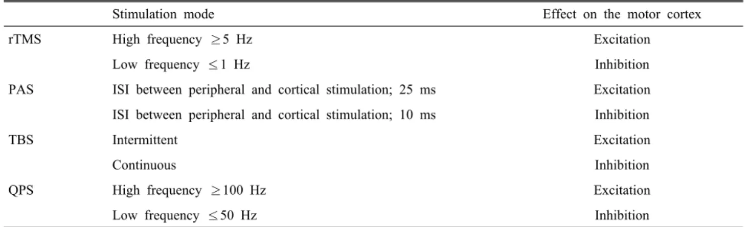
Table 2. TMS methods for inducing cortical plasticity
Stimulation mode Effect on the motor cortex
rTMS High frequency ≥5 Hz Excitation
Low frequency ≤1 Hz Inhibition
PAS ISI between peripheral and cortical stimulation; 25 ms Excitation
ISI between peripheral and cortical stimulation; 10 ms Inhibition
TBS Intermittent Excitation
Continuous Inhibition
QPS High frequency ≥100 Hz Excitation
Low frequency ≤50 Hz Inhibition
rTMS; repetitive transcranial magnetic stimulation, PAS; paired associative stimulation, TBS; theta burst stimulation, QPS;
quadripulse stimulation, ISI; interstimulus interval.
Figure 7. MEPs recorded from the ADM during index finger flexion (circle) is suppressed compared with baseline MEPs recorded during rest.
31MEP; motor evoked potential, ADM;
abductor digiti minimi, FDS; flexor digitorum superficialis, EIP; extensor indicis proprius, FDI; first dorsal interosseus.
가할 때 운동유발전위가 억제되는 현상을 관찰할 수 있다.
자극간격이 20 ms일 때와 200 ms일 때에 억제현상을 보이며 각각 짧은간격 구심성억제, 긴간격 구심성억제라고 한다.
26짧은간격 구심성억제는 아세티콜린 길항제인 scopolamine 을 전처치했을 때 억제되므로 콜린성신경경로의 치밀도를 조사 하는 데에 이용할 수 있다.
27짧은간격 구심성억제는 알츠하이머병 환자에서 감소하고 아세티콜린에스터라제 억제제인 rivastigmine을 투약하였을 때에 정상화 된다.
28-30긴간격구심성억제와 관련된 정확한 기전은 아직 확실하지 않으나 짧은간격구심성억제와는 다를 것이라고 추측한다.
4) 자동-유발 뇌자기자극(self-triggered TMS)- 주변억제현상(surround inhibition)
원하는 동작을 정확하게 하기 위해서는 해당 동작을 위한 신경네트워크가 선택적으로 활성화되어야 하는 반면에 원 하지 않는 동작에 대한 운동은 억제가 되어야 한다. 이를 위해서는 해당 동작과 관련 없는 주변 신경부에 대한 억제 기전이 필요한데 이를 주변억제현상(surround inhibition) 이 라고 한다.
31자동-유발 뇌자기자극방법은 특정한 동작에 대한 근전도 활성에 반응하여 뇌자기자극이 가해지도록 설정하고 해당 동작을 수행하면서 뇌자기자극이 가해지는 동안 해당동작과 관련 없는 주변근육의 운동유발전위를 측정하는 방법으로 주변억제의 신경생리학적 현상을 관찰 할 수 있다. 검지손가락을 구부리는 동작을 수행하는 동안 이 동작과는 관련이 없는 주변근육인 새끼벌림근(abductor digiti minimi)에서는 척수의 활성화를 반영하는 F-파의 진 폭은 증가하는 반면에 운동유발전위는 억제되는 현상을 관찰하였고 이는 정상적으로 정확한 동작을 수행하기 위 해 피질수준에서 작용하는 억제기전이 존재한다는 사실을 반영한다(Figure 7).
3. 반복뇌자기자극
반복뇌자기자극은 특정주파수의 뇌자기자극을 통하여 뇌피질의 가소성(plasticity) 를 유도하는 방법이다(Table 2).
5Hz 이상의 높은 주파수 자극을 가하게 되면 뇌피질의 흥
분성이 증가하는 반면에
320.2-1 Hz의 낮은 주파수로 반복
자극하면 감소한다.
33반복뇌자기자극이 뇌피질의 가소성
을 유도하는 기전은 아직 확실하지 않다. 흥분성을 증가시
키는 것은 장기상승작용(long-term potentiation, LTP), 감소
시키는 것은 장기하강작용(long-term depression, LTD)과 관
련되었을 것으로 추정하고 있다.
5최근 뇌피질의 가소성을 유도하는 반복뇌자기자극의 새로운 방법이 몇 가지 소개 되었다. 고전적인 장기상승작용 모델은 유발전위를 일으 키지 않는 정도의 낮은 자극강도로 구심성섬유와 원심성 섬유의 동시자극을 반복적으로 가하게 되면 신경경시냅스 간 강화를 통하여 유발전위를 일으키게 된다는 가설로 뇌 의 가소성을 유도하는 기전으로 잘 알려져 있다.
34,35이 가 설을 모델로 하여 구심성자극으로 말초신경 자극을, 원심 성자극으로 뇌피질자극을 동시에 하는 말초신경-피질 연 계자극(paired associative stimulation)를 시행하였을 때에 운 동유발전위가 증가됨을 증명하였다.
36말초정중신경에서 뇌피질까지 평균 도달시간이 25 ms라는 전제하에 정중신 경을 전기자극한 후 25 ms 후에 뇌피질을 자극하게 되면 두 자극이 동시에 가해지게 되고 운동유발전위는 항진 된다. 말초신경자극과 뇌피질자극의 자극간 시간을 10 ms 으로 하는 경우 운동피질자극이 정중신경 신호에 선행하 고 운동유발전위는 감소한다.
37쎄타돌발파자극(theta burst stimulation, TBS)은 높은 주파수의 돌발파자극(통상적으로 50 Hz)을 약 5 Hz로 반복적으로 가하는 방법으로 10초의 중단기와 2초 지속자극을 반복하는 경우(중단 돌발파 자극, intermediate TBS) 뇌피질의 흥분성을 증가시키고, 40초 이 상 지속적으로 자극하게 되면(지속 돌발파자극, continuous TBS) 흥분성을 감소시킨다.
38가장 최근 소개된 반복뇌자 기자극방법으로는 사연속파자극(quadripulse stimulation)으 로 자극간시간 1.5 ms의 4회 연속자극을 100 Hz 이상의 빈 도로 시행했을 때에 운동피질의 흥분성이 증가하고 50 Hz 이하의 빈도로 시행하면 억제된다.
39사연속파자극으로 인 한 운동피질 흥분성의 변화에 대한 기전은 정확히 알려져 있지 않으나 다른 자극방법과 유사하게 뇌신경 시냅스의 장기상승작용이나 장기하강작용과 연관이 되어 있을 것으 로 추측하고 있다.
4. 뇌자기자극의 진단적 이용
뇌자기자극을 신경학적 질환의 진단에 이용하려는 많은 연구에도 불구하고 현재까지 뇌자기자극의 진단적 적응증 은 넓지 않다.
1) 척수병변
뇌자기자극을 진단적으로 유용하게 이용할 있는 대표적 인 질환이 척수병변이다.
척수병변이 있을 때 중추전도시간이 연장되는 것으로 척수병변의 진단에 도움을 줄 수 있고 중추전도시간의 연 장 정도를 비교하여 병변이 가장심한 위치를 추측할 수 있
다. 따라서 중추전도시간은 척수변병의 진단 및 병변의 위 치를 찾는 데에 척수 MRI와 함께 보조적으로 이용할 수 있는 방법이다.
402) 근위축측상경화증(amyotrophic lateral sclerosis, ALS) 근위축측상경화증에서는 말초신경 운동유발전위시간 및 중추전도시간이 연장되는데
41척수변병과 구별하기 위해 서는 등세모근에서 운동유발전위와 중추전도시간을 측정 하는것이 도움이 된다 .
42근위축측상경화증에서 피질내 억 제에 대한 결과는 연구마다 차이가 있다. 대부분 피질의 피질내 억제현상이 감소되는 것으로 알려져 있는데, 휴지 기가 단축되거나 짧은간격 피질내억제가 감소된다.
41,433) 다발성경화증
다발성 경화증에서는 병변이 피질척수로를 침범한 경우 중추전도시간이 연장되며 임상적인 증상이 없더라도 피질 척수로를 침범했음을 시사하는 소견이 될 수 있다.
44중추 전도시간 연장은 재발 -완화형다발경화증보다 진행형다발 경화증에서 더 뚜렷하게 관찰되며,
45임상증상이 있는 경 우 증상의 심한 정도와 상관관계를 보이므로 가치 있는 변 수이다.
46,474) 뇌졸중
심한운동마비가 있는 뇌졸중환자에서는 운동유발전위 가 관찰되지 않을 수 있고 경한운동마비가 있는 환자에서 운동유발전위가 감소되고 잠복기가 연장될 수 있으며 운 동역치가 증가한다. 뇌졸중에서 뇌자기자극은 기능적인 회복에 대한 예후를 예측하는 데에 도움을 준다. 급성기 뇌졸중환자에서 운동유발전위가 존재하는 경우 좋은 예후 인자가 될 수 있고 운동유발전위의 잠복기연장의 회복이 마비가 있는 손의 기능회복의 정도와 연관이 있다고 알려 져 있다.
485. 신경질환의 생리학적 기전의 규명
뇌자기자극은 신경계질환의 생리학적 기전을 규명하는 데에 유용하게 이용할 수 있다.
1) 이상운동질환
뇌자기자극은 신경계질환 중에서 이상운동질환의 기전
을 밝히는 데에 많은 기여를 하고 있다. 파킨슨병과 근긴
장이상증 및 헌팅턴병에서 뇌자기자극을 이용한 생리학적
기전이 비교적 잘 알려져 있다. 파킨슨병에서는 운동역치
의 변화는 없고 운동유발전위 역시 정상인과 비교하여 차
이를 보이지 않는다.
49,50피질무반응기는 단축되고 짧은간 격피질내억제 현상은 감소하나 긴간격 피질내억제현상은 증가한다.
49,50근긴장이상증에서도 파킨슨병환자에서와 비 슷한 소견을 관찰할 수 있다.
49헌팅턴병에서는 피질무반 응기가 연장되어 있고 그 정도는 증상의 심한 정도와 비례 한다고 알려져 있다.
49이러한 소견은 이상운동질환 각각 의 특이한 현상을 반영하기보다는 기저핵과 운동피질을 연결하는 운동회로의 병적인 변화로 인한 운동피질의 억 제-흥분성 변화를 반영한다. 기저핵은 원하는 운동을 정확 하게 선택하고 그 운동과 관련이 없는 다른 움직임을 담당 하는 주변부를 억제하여 정확한 운동을 수행하도록 하는 기능을 한다.
51파킨슨병과 근긴장이상증에서는 주변부억 제기능에 장애가 발생하게 되어 정확한 운동조절의 장애 를 초래한다. 뇌자기자극방법 중 자동-유발 뇌자기자극방 법을 이용하여 주변억제의 정도를 측정하였을 때에 근긴 장이상증과 파킨슨병환자에서는 정상인에서 관찰되는 주 변억제 현상이 관찰되지 않았다.
52,53이는 주변억제현상이 정상적으로 작용하지 않는 것이 근긴장이상증이나 파킨슨 병의 운동조절기능장애를 일으키는 신경생리학적인 기전 임을 보여주는 결과로 기저핵의 기능저하를 반영한다.
최근에는 국소 손근긴장이상증(focal hand dystonia) 환자 에서 말초신경-피질 연계자극에 의한 뇌피질가소성 유도 의 정도가 정상인에 비하여 과도하게 증가되어 있다는 사 실을 통하여 운동피질의 과도한 가소성이 근긴장이상의 발생과 관련이 있음을 증명한 바가 있다.
54,55또, 레보도파 복용 후 이상운동증이 발생한 파킨슨병 환자에서 말초신 경-피질 연계자극에 대하여 뇌피질의 가소성이 과도하게 증가한다는 사실을 통하여 레보도파에 의해 발생하는 이 상운동증의 기전 역시 뇌의 가소성의 이상과 관련이 있음 을 알 수 있었다.
562) 간질 및 항경련제
뇌자기자극으로 간질환자의 운동피질 흥분성의 변화에 대한 측정이 가능하다. 특발성전신간질(idiopathic generalized epilepsy) 환자에서 운동역치는 감소되어 있고 피질무반응 기는 연장되어 있다.
57,58이는 간질로 인한 운동피질 흥분 성의 증가를 나타내는 것으로 이해할 수 있다.
59근간대성 간질환자 에서는 운동피질의 흥분성-억제성 기전의 부조 화를 나타낸다피질내억제기전은 감소되어 있는 반면 피질 내촉진기전의 변화는 없고 자극간시간 100-150 ms에서 피 질 억제가 소실되어 있고 50 ms에서 오히려 촉진현상이 관찰되었다.
60-62뇌자기자극은 항경련제의 기전을 밝히는 데에 도움을
줄 수 있다 . Gabapentin의 투여가 피질내촉진을 감소시키 고 피질내억제현상을 증가시킨다는 사실을 통하여 이 약 제가 GABA의 신경내전달을 항진시키는 기전으로 항경련 효과를 나타낸다는 것을 알 수 있었다.
16,63Levetiracetam은 운동역치 , 피질무반응기 및 피질내억제와 촉진현상에 모 두 영향을 미치지 않는 대신에 운동전위 점증곡선의 기울 기를 감소시킨다 .
64이를 통하여 levetiracetam 은 다른 항경 련제와는 다른 기전으로 운동피질의 흥분성을 억제하는 효과를 가진다는 것을 알 수 있다.
6. 뇌자기자극의 치료적인 이용
반복뇌자기자극은 뇌피질의 가소성을 유도하여 뇌피질 의 흥분성을 증가시키거나 감소시키므로 뇌피질의 흥분성 의 변화로 인하여 운동조절에 이상을 초래하는 질환에서 치료적 효과를 기대할 수 있다.
1) 파킨슨병
정상인 및 파킨슨병 환자에서 운동피질 또는 등가쪽앞 전뇌(dorsolateral prefrontal cortex)를 반복뇌자기자극 할 때 도파민의 분비가 증가한다는 뇌영상소견을 통하여 반복뇌 자기자극의 파킨슨병에 대한 효과를 기대할 수 있다.
65여 러 뇌자기자극연구에서 고주파 반복뇌자기자극을 시행하 였을 때에 파킨슨병 환자의 운동증상이 약 10-30% 호전됨 을 증명하였다.
66단발 세션으로는 운동증상이 호전되는 시간이 너무 짧아 다발 세션 반복뇌자기자극으로 지속적 인 운동효과를 기대할 수 있다. 그러나 파킨슨병의 운동증 상은 도파민 제제에 매우 효과가 뛰어나기 때문에 반복뇌 자기자극으로 기대할 수 있는 운동증상의 호전 정도는 제 한적이라고 할 수 있다. 레보도파에 의한 이상운동증은 진 행된 파킨슨 환자에서 약물치료의 후기 운동합병증 중의 한가지로 약물로 조절이 힘든 증상이다. 피킨슨병의 이상 운동증은 레보도파의 오랜 복용에 의한 운동피질-기저핵 을 연결하는 운동회로 가소성의 과도한 증가와 관련이 있 다.
67이 가설을 바탕으로 저주파반복뇌자기자극을 통하여 뇌피질의 흥분성을 억제하여 레보도파에 의한 이상운동증 을 호전시킬 수 있을 것이라는 이론적인 가설을 최근 몇몇 연구에서 증명하였다.
68,692) 근긴장이상
근긴장이상증 환자에서 공통적으로 관찰되는 신경생리
학적인 소견은 뇌피질 억제기전의 감소이다. 기능적뇌영
상촬영에서도 근긴장이상증 환자에서 뇌피질의 다양한 부
위에서 활성이 증가해 있음을 증명하였다.
70근긴장이상증
환자의 신경병리학적 기전은 아직 정확하지 않지만 직업 특이 근긴장이상(task specific dystonia) 환자의 경우 과도한 운동수행으로 인한 뇌피질가소성의 증가와 연관이 있을 것으로 추정한다.
54,55최근의 말초신경-피질 연계자극을 통 한 결과는 이를 뒷받침하는 소견으로 근긴장이상증 환자 에서 반복뇌자기자극은 뇌피질의 비정상적인 흥분성 증가 및 가소성을 정상화시키고 이를 통하여 환자의 임상적인 호전도 이끌어낼 수 있으리라는 기대를 받아 왔다.
71,72그 러나 현재까지 근긴장환자에서의 반복뇌자기자극의 효과 에 대한 연구는 이를 증명하기에 불충분하다. 근긴장이상 증의 뇌피질의 벼화는 단순한 억제기전의 감소가 아니라 가소성의 과도한 발달에 의한 것이므로 단순반복뇌자기자 극으로 뇌피질의 기능적인 변화와 함께 임상적인 효과도 기대하는 것이 어려운 것이 아닌가 추측한다. 최근 개발된 다양한 반복뇌자기자극 프로토콜을 이용하여 근긴장이상 증에 대한 뇌자기작극의 치료적 효용성이 증명되기를 기 대해 볼 수 있겠다.
3) 뇌졸중
뇌졸중으로 인한 운동마비는 자연경과와 더불어 적극적 인 재활치료가 증상의 호전에 중요한 역할을 한다. 이러한 치료효과에 반복뇌자기자극을 통하여 운동피질의 흥분성 을 증가시키게 되면 운동마비를 더 빨리 효과적으로 호전 시킬 수 있을것이라는 것이 뇌졸중에서의 반복뇌자기자극 의 치료적 가설의 출발이다. 운동마비증상이 있는 팔 다리 의 반대편 운동피질에 고주파 반복뇌자기자극을 가하게 되면 운동피질의 흥분성이 증가하고 운동마비의 임상적 호전을 가져올 수 있다.
73,74운동마비의 호전을 위해서 손 상을 받은 뇌피질의 반대편 운동피질을 자극할 후도 있다.
뇌량은 정상적으로 반대편 운동피질을 억제하는 역할을 통하여 한쪽 팔다리를 움직일 때 반대편 팔다리의 불필요 한 움직임이 일어나지 않도록 하여 양쪽 팔다리가 각각 서 로 다른 운동수행을 할 수 있도록 하는 역할을 한다. 뇌졸 중이 발생하게 되면 정상적으로 작용하던 반대편 운동피 질에서의 억제기전이 운동마비의 회복을 방해할 수 있을 것이다. 따라서 반대편 운동피질을 억제하게 되면 손상받 은 운동피질의 기능적 회복에 도움이 될 수 있을 것이라는 것이 반대측 운동피질을 치료 목표로 삼는 연구의 가설이 다. 실제 저주파자극을 사용하여 뇌졸중이 발생한 반대편 운동피질을 자극하였을 때에 운동마비의 호전에 도움이 되었다는 임상적인 효과를 증명한 바가 있다.
75반복뇌자 기자극은 급성뇌졸중 환자와 만성뇌졸중 환자 모두에서 효과가 있는데, 급성기의 경우 운동마비를 호전시키는 데
에 도움을 줄 수 있고 만성뇌졸중 환자의 경우 운동조절 정교성의 증가와 운동학습효과의 향상효과를 기대할 수 있다.
73,74결 론
![Figure 3. The central conduction time can be obtained as MEP latency (A)-[F(B)+M(C)-1)/2]](https://thumb-ap.123doks.com/thumbv2/123dokinfo/5005778.305497/3.892.466.805.740.1002/figure-central-conduction-time-obtained-mep-latency-f.webp)