저작자표시-비영리-변경금지 2.0 대한민국 이용자는 아래의 조건을 따르는 경우에 한하여 자유롭게 l 이 저작물을 복제, 배포, 전송, 전시, 공연 및 방송할 수 있습니다. 다음과 같은 조건을 따라야 합니다: l 귀하는, 이 저작물의 재이용이나 배포의 경우, 이 저작물에 적용된 이용허락조건 을 명확하게 나타내어야 합니다. l 저작권자로부터 별도의 허가를 받으면 이러한 조건들은 적용되지 않습니다. 저작권법에 따른 이용자의 권리는 위의 내용에 의하여 영향을 받지 않습니다. 이것은 이용허락규약(Legal Code)을 이해하기 쉽게 요약한 것입니다. Disclaimer 저작자표시. 귀하는 원저작자를 표시하여야 합니다. 비영리. 귀하는 이 저작물을 영리 목적으로 이용할 수 없습니다. 변경금지. 귀하는 이 저작물을 개작, 변형 또는 가공할 수 없습니다.
Ajou University Graduate School
Science in Medicine Major
Jung E Park
Master's Thesis of Science in
Medicine
Repetitive Transcranial Magnetic
Stimulation for Limb-kinetic Apraxia in
Parkinson’s Disease
Repetitive Transcranial Magnetic
Stimulation for
Limb-kinetic Apraxia in
Parkinson’s Disease
Byung Gon Kim, MD, PhD, Advisor
I submit this thesis as the
Master's thesis of Science in Medicine.
February, 2018
Ajou University Graduate School
Science in Medicine Major
The Master's thesis of Jung E Park in Medicine
is hereby approved.
Thesis Defense Committee President
김병곤 Seal
Member 홍지만 Seal
Member 주인수 Seal
Ajou University Graduate School
ABSTRACT
Background and Objective: Apraxia, defined as inability to perform skilled or learned movements, is frequently seen in neurodegenerative diseases such as Parkinson’s disease, corticobasal syndrome and Alzheimer's dementia. Apraxia is further classified into subtypes such as limb-kinetic, ideomotor or ideational apraxia. Limb-kinetic apraxia, characterized as difficulty making precise, independent and coordinated finger and hand movements, leads to impaired dexterity and has been shown to affect activities of daily living in PD patients. To date, there is no effective treatment for limb-kinetic apraxia. We aim to report the effects of repetitive transcranial magnetic stimulation (rTMS), a noninvasive brain stimulation method on limb-kinetic apraxia in patients with Parkinson's disease.
Methods: Eight patients diagnosed with Parkinson’s disease underwent rTMS. Patients performed sequential unbuttoning and buttoning of a standardized gown they wore for assessment of limb-kinetic apraxia. A 20-minute rTMS session of the left primary motor cortex (M1) was performed (10 Hz frequency, stimulation intensity of 80% resting motor threshold, 10 seconds/train and 20 trains) in the medication-ON state.
Results: Eight PD patients (M:F=1:1, mean age 71.1 years, SE 2.5 years) underwent rTMS with no adverse events. Buttoning and unbuttoning time was found to be significantly reduced at 24 hours post-rTMS (mean change:
ii
-22%, SE: 6%), compared to the medication-ON state. No significant change was noted immediately following the rTMS session.
Conclusion: Our results suggest that high-frequency rTMS of the left M1 may be effective in limb-kinetic apraxia, lending support to the need for future long-term studies to further determine if rTMS is truly efficacious in the treatment of this disorder.
Keywords: apraxia, limb-kinetic, transcranial magnetic stimulation, motor cortex, Parkinson’s disease
TABLE OF CONTENTS
ABSTRACT ··· ⅰ
TABLE OF CONTENTS ··· ⅲ
LIST OF FIGURES ··· ⅳ
LIST OF TABLES ··· ⅴ
LIST OF ABBREVIATIONS ··· ⅵ
Ⅰ. INTRODUCTION ··· 1
Ⅱ. MATERIALS AND METHODS ··· 3
A. Initial assessment ··· 3
B. Electromyography (EMG) ··· 3
C. Transcranial magnetic stimulation (TMS) ··· 4
D. Post-intervention assessment ··· 4
Ⅲ. RESULTS ··· 5
Ⅳ. DISCUSSION ··· 8
V. CONCLUSION ··· 11
BIBLIOGRAPHY ··· 12
국문 요약 ··· 15
iv
-LIST OF FIGURES
FIGURE 1. Upper Extremity Bradykinesia Scores and Time for Sequential
LIST OF TABLES
vi
-LIST OF ABBREVIATIONS
PD: Parkinson’s disease
rTMS: repetitive transcranial magnetic stimulation MEP: motor-evoked potential
RMT: resting motor threshold
UPDRS: Unified Parkinson Disease Rating Scale AST: apraxia screen of TULIA
TULIA: test of upper limb apraxia MMSE: mini-mental status examination EMG: electromyography
FDI: first dorsal interosseous muscle tDCS: transcranial direct current stimulation PAS: paired associative stimulation
TBS: theta-burst stimulation M1: primary motor cortex ADL: activities of daily living
I. INTRODUCTION
Humans go through their daily lives performing a variety of movements. Movements can be largely divided into those requiring tools (i.e., transitive movements) and those that do not (i.e., intransitive movements)(1). Some examples of the former include cooking and cleaning with the use of tools. Examples of the latter include daily activities such as cleansing, grooming and dressing. Many of these activities require multi-step sequential actions, and sometimes fine motor skills in order for the movements to be carried out successfully(2). It is needless to say that the ability to perform such actions and thereby carry out one’s activities of daily livings (ADLs) is integral to one’s functional independence.
Praxis is defined as the ability to perform skilled or learned movements(3). While the definition itself is relatively simple, various components are involved, such as the transformation of visual and somatosensory information into movements, and for transitive movements, semantic knowledge of objects(2). Hugo Liepmann, a neurologist who studied patients with apraxia proposed a motor engram with information flowing from posterior brain regions to anterior regions(4). To date, anatomical and functional neuroimaging studies have also shown various brain regions to be relevant in praxis, including the left posterior parietal, temporal, premotor and motor cortices(5-10).
Apraxia refers to the inability to carry out praxis movements in the absence of elementary motor, sensory or coordination deficits that could serve as the primary cause(11). Apraxia is seen in a variety of neurological disorders such as dementia,
2
-stroke and Parkinsonism(12). This phenomenon can be further classified into subtypes such as ideomotor, ideational and limb-kinetic apraxia(12). Limb-kinetic apraxia is the loss of ability to make precise, independent but coordinated limb movements and has been noted to be present in Parkinsonism, affecting ADLs in this patient population(13, 14). To date, there is no effective treatment available for limb-kinetic apraxia.
Transcranial direct current stimulation (tDCS), a type of noninvasive brain stimulation have been used to study and treat apraxia (15, 16). Some improvement of ideomotor apraxia in patients with corticobasal syndrome using anodal tDCS has been reported(15). In this study, we report the effects of high-frequency repetitive transcranial magnetic stimulation (rTMS) on limb-kinetic apraxia in patients with Parkinson’s disease (PD).
Ⅱ. MATERIALS AND METHODS
A. Initial assessment
We studied eight right-handed patients fulfilling the UK Parkinson’s Disease Society Brain Bank criteria for PD. Prior to the intervention, all patients were clinically assessed via part III of the Unified Parkinson Disease Rating Scale (UPDRS) in the medication-OFF and ON state. The medication-OFF state was defined as discontinuation of levodopa and dopamine agonist medication for approximately 12 hours, and ON was defined as 30 minutes to 1 hour since medication intake. For assessment of limb-kinetic apraxia, patients were asked to perform sequential unbuttoning and buttoning of their hospital gown they wore. The gown had five buttons aligned vertically (button diameter: 1.9mm, 9.5 cm inter-button distance). The Apraxia Screen of TULIA (Test of upper limb apraxia)(AST) was also performed to evaluate for the presence and severity of ideomotor apraxia(17). This bedside screening tool is comprised of 12 items that include eight transitive gestures, three intransitive gestures and one meaningful gesture.
B. Electromyography (EMG)
Following initial assessment, patients in the medication-ON state underwent standard EMG procedures for TMS. Patients sat relaxed in a chair, with both hands resting on their lap. The ground electrode was placed over the dorsum of the right wrist. EMG recordings were obtained from the right first dorsal interosseous (FDI) muscle, using surface Ag-AgCl electrodes. The EMG signal was amplified using an
EMG machine (Synergy, Natus Neurology, Middleton, WI, USA) and
4
-C. Transcranial magnetic stimulation (TMS)
A figure-of-8, double 70 mm TMS coil was placed tangentially on the left primary motor cortex (M1), at a 45 degree angle to the anteroposterior axis and with the handle pointing posteriorly, corresponding to the right upper extremity. The motor hotspot was determined based on the motor-evoked potential (MEP) evoked in the right FDI muscle. The resting motor threshold (RMT: the minimum stimulation intensity producing MEP amplitude of ≥50 µV, in 5 out of 10 trials) was obtained by delivering single TMS pulses. Once the RMT was determined, a 20-minute rTMS session of the left primary motor cortex (M1) was performed (10 Hz frequency, stimulation intensity of 80% RMT, 10 seconds/train, total of 20 trains, 50 second inter-train interval).
D. Post-intervention assessment
Patients were evaluated both immediately after receiving rTMS and at 24 hours following, via the UPDRS part III, sequential unbuttoning and buttoning and the AST.
Ⅲ. RESULTS
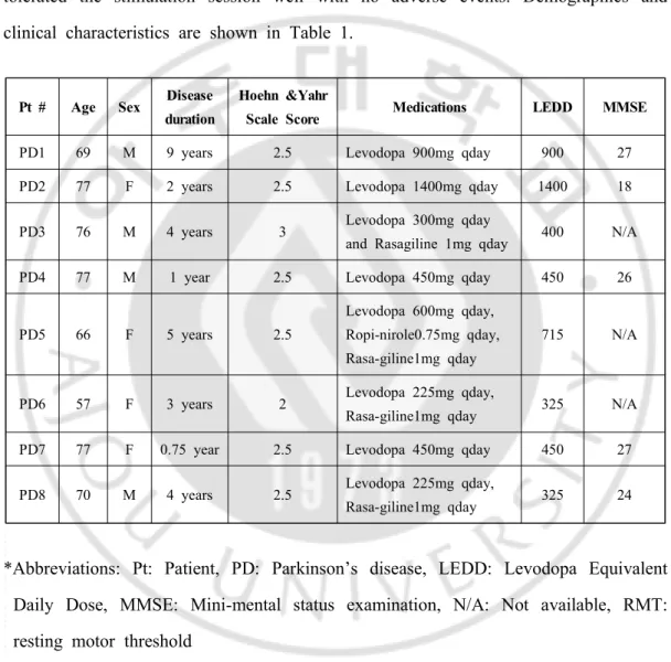
Eight PD patients (M:F=1:1, mean age 71.1 years, standard error: SE 2.5 years) underwent 20 minutes of high-frequency (10 Hz) rTMS of the left M1. All patients tolerated the stimulation session well with no adverse events. Demographics and clinical characteristics are shown in Table 1.
Pt # Age Sex Disease duration
Hoehn &Yahr
Scale Score Medications LEDD MMSE
PD1 69 M 9 years 2.5 Levodopa 900mg qday 900 27
PD2 77 F 2 years 2.5 Levodopa 1400mg qday 1400 18
PD3 76 M 4 years 3 Levodopa 300mg qday
and Rasagiline 1mg qday 400 N/A
PD4 77 M 1 year 2.5 Levodopa 450mg qday 450 26
PD5 66 F 5 years 2.5
Levodopa 600mg qday, Ropi-nirole0.75mg qday, Rasa-giline1mg qday
715 N/A
PD6 57 F 3 years 2 Levodopa 225mg qday,
Rasa-giline1mg qday 325 N/A
PD7 77 F 0.75 year 2.5 Levodopa 450mg qday 450 27
PD8 70 M 4 years 2.5 Levodopa 225mg qday,
Rasa-giline1mg qday 325 24
*Abbreviations: Pt: Patient, PD: Parkinson’s disease, LEDD: Levodopa Equivalent Daily Dose, MMSE: Mini-mental status examination, N/A: Not available, RMT: resting motor threshold
6
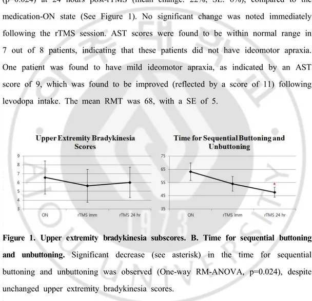
-The mean UPDRS part III score in the medication-ON state was 19.5, with a SE of 3.6. One-way repeated measures analysis of variance (RM-ANOVA) was conducted, showing that the buttoning and unbuttoning time, which was the primary outcome measure for assessment of limb-kinetic apraxia, was significantly reduced (p=0.024) at 24 hours post-rTMS (mean change: 22%, SE: 6%), compared to the medication-ON state (See Figure 1). No significant change was noted immediately following the rTMS session. AST scores were found to be within normal range in 7 out of 8 patients, indicating that these patients did not have ideomotor apraxia. One patient was found to have mild ideomotor apraxia, as indicated by an AST score of 9, which was found to be improved (reflected by a score of 11) following levodopa intake. The mean RMT was 68, with a SE of 5.
Figure 1. Upper extremity bradykinesia subscores. B. Time for sequential buttoning and unbuttoning. Significant decrease (see asterisk) in the time for sequential buttoning and unbuttoning was observed (One-way RM-ANOVA, p=0.024), despite unchanged upper extremity bradykinesia scores.
In summary, our findings suggest that limb-kinetic apraxia in PD improves with high-frequency rTMS of the left M1, with significant benefit seen at 24 hours. The degree of upper extremity bradykinesia, measured as subscores of the UPDRS part
III did not show significant change both immediately and at 24 hours compared to the non-stimulated, ON state. Limb-kinetic apraxia, reflected by the buttoning and unbuttoning time, showed a trend for improvement immediately after rTMS. Significant improvement in limb-kinetic apraxia, however, was found at 24 hours following rTMS, with no improvement in the bradykinesia subscore.
8
-Ⅳ. DISCUSSION
Apraxia is seen in a variety of neurological disorders, notably in neurodegenerative disorders such as PD, Alzheimer’s disease and Parkinsonian disorders(12). While there are different subtypes of apraxia that can even coexist in a single individual, limb-kinetic apraxia involves deficits in fine motor control and is therefore relevant in dexterity. Examples of tasks utilized in assessment of limb-kinetic apraxia include buttoning and coin rotation(13, 14, 18). Limb-kinetic apraxia can negatively affect patients’ quality of life but due to its subtle nature, can go unrecognized despite its undesirable effects on daily functioning(13).
While there are distinct characteristics of different subtypes of apraxia, relevant brain regions are presumed to include the left premotor, motor, posterior parietal and temporal cortices. We looked at the effects of high-frequency rTMS on limb-kinetic apraxia, as this brain stimulation method has been postulated to work in an excitatory fashion, analogous to anodal tDCS, which has been previously found to have beneficial effects on ideomotor apraxia(15, 16). The left M1 was chosen as the stimulation site, as anatomical and functional neuroimaging studies have found this brain region to be involved in praxis(5). The left M1 is also a brain region that is readily identifiable via delivering single TMS pulses to obtain MEP recordings, and therefore has been the most frequently studied target(19). While previous TMS studies targeting the left M1 found effects such as reduced rigidity and improved bradykinesia, we did not find such significant effects in our patient group. We speculate that the significant improvement in limb-kinetic apraxia seen at 24 hours following stimulation, but not immediately, reflects the time required for alteration of cortical excitability induced by high-frequency rTMS.
Our study has several limitations. This was a single-session study performed on a small number of subjects and future studies of a larger scale are necessary to confirm the effects that we observed. However, as previously mentioned, other rTMS studies targeting the left M1 in Parkinson’ disease patients also found significant effects on motor performance after a single session(20), and we adopted stimulation parameters of these and found significant effects on limb-kinetic apraxia at 24 hours. Another limitation of our study is that only active stimulation was used, and therefore a placebo effect cannot be definitively excluded. Lastly, while we assessed for limb-kinetic apraxia using a single modality(buttoning and unbuttoning) that has been used in previous studies, it may be worthwhile to use other modalities as well that also reflect actions commonly performed on a daily basis.
Prior to the advance in neurophysiology, knowledge on praxis or apraxia relied on clinical observations of human subjects(21). With recent advances in application of noninvasive brain stimulation methods, knowledge on the subject of praxis and apraxia has expanded. Noninvasive brain stimulation techniques are also being increasingly used for the treatment of various neurological disorders. These stimulation methods are often well-tolerated with minimal or no side effects, and therefore holds potential to be clinically applicable(22). Examples of widely-used methods include rTMS, theta-burst stimulation (TBS), tDCS and paired associative stimulation (PAS)(23).
TMS in particular has been used to study processes underlying movement such as motor attention, selection and other aspects of judgement that are relevant.
10
-Numerous studies using rTMS have aimed to address both motor and non-motor symptoms of PD, with variable results. Low-frequency rTMS of the left M1 has been found to decrease rigidity, while high-frequency rTMS of the left M1 appears to have an effect on both rigidity and bradykinesia(24-26). High-frequency rTMS and anodal tDCS are analogous in that both are thought to exert a net effect of excitation. Based on the positive effects of anodal tDCS on ideomotor apraxia and our observations of high-frequency rTMS having beneficial effects on limb-kinetic apraxia, we conclude that excitatory stimulation targeted towards brain areas relevant in praxis may be promising in treating apraxic disorders.
V. CONCLUSION
Our results indicate that limb-kinetic apraxia in PD appears to improve with high-frequency rTMS of the left M1, as indicated by reduced time to perform sequential unbuttoning and buttoning 24 hours following stimulation. These findings suggest that high-frequency rTMS of the left M1 may be effective in limb-kinetic apraxia. Noninvasive brain stimulation is being increasingly used for the treatment of various neurological disorders. Our observations have implications for future research directions in the treatment of apraxia using excitatory modes of noninvasive brain stimulation such as rTMS.
12
-BIBLIOGRAPHY
1. Gross RG, Grossman M. Update on apraxia. Curr Neurol Neurosci Rep. 2008;8(6):490-6.
2. Park JE. Apraxia: Review and Update. J Clin Neurol. 2017;13(4):317-24.
3. Goldenberg G. Apraxia and the parietal lobes. Neuropsychologia.
2009;47(6):1449-59.
4. Leiguarda RC, Marsden CD. Limb apraxias: higher-order disorders of
sensorimotor integration. Brain. 2000;123:860-79.
5. Bohlhalter S, Hattori N, Wheaton L, Fridman E, Shamim EA, Garraux G, et al. Gesture subtype-dependent left lateralization of praxis planning: an event-related fMRI study. Cereb Cortex. 2009;19(6):1256-62.
6. Assmus A, Giessing C, Weiss PH, Fink GR. Functional interactions during the retrieval of conceptual action knowledge: an fMRI study. J Cogn Neurosci. 2007;19(6):1004-12.
7. Canessa N, Borgo F, Cappa SF, Perani D, Falini A, Buccino G, et al. The different neural correlates of action and functional knowledge in semantic memory: an FMRI study. Cereb Cortex. 2008;18(4):740-51.
8. Choi SH, Na DL, Kang E, Lee KM, Lee SW, Na DG. Functional magnetic resonance imaging during pantomiming tool-use gestures. Exp Brain Res. 2001;139(3):311-7.
9. Fridman EA, Immisch I, Hanakawa T, Bohlhalter S, Waldvogel D, Kansaku K, et al. The role of the dorsal stream for gesture production. Neuroimage. 2006;29(2):417-28.
10. Ogawa K, Imai F. Hand-independent representation of tool-use pantomimes in the left anterior intraparietal cortex. Exp Brain Res. 2016;234(12):3677-87. 11. Steinman KJ, Mostofsky SH, Denckla MB. Toward a narrower, more pragmatic
view of developmental dyspraxia. J Child Neurol. 2010;25(1):71-81.
12. Wheaton LA, Hallett M. Ideomotor apraxia: a review. J Neurol Sci. 2007;260(1-2):1-10.
13. Foki T, Vanbellingen T, Lungu C, Pirker W, Bohlhalter S, Nyffeler T, et al. Limb-kinetic apraxia affects activities of daily living in Parkinson's disease: a multi-center study. Eur J Neurol. 2016;23(8):1301-7.
14. Quencer K, Okun MS, Crucian G, Fernandez HH, Skidmore F, Heilman KM. Limb-kinetic apraxia in Parkinson disease. Neurology. 2007;68(2):150-1.
15. Bianchi M, Cosseddu M, Cotelli M, Manenti R, Brambilla M, Rizzetti MC, et al. Left parietal cortex transcranial direct current stimulation enhances gesture processing in corticobasal syndrome. Eur J Neurol. 2015;22(9):1317-22.
16. Bolognini N, Convento S, Banco E, Mattioli F, Tesio L, Vallar G. Improving ideomotor limb apraxia by electrical stimulation of the left posterior parietal cortex. Brain. 2015;138:428-39.
17. Vanbellingen T, Kersten B, Van de Winckel A, Bellion M, Baronti F, Muri R, et al. A new bedside test of gestures in stroke: the apraxia screen of TULIA (AST). J Neurol Neurosurg Psychiatry. 2011;82(4):389-92.
18. Foki T, Pirker W, Geissler A, Haubenberger D, Hilbert M, Hoellinger I, et al. Finger dexterity deficits in Parkinson's disease and somatosensory cortical dysfunction. Parkinsonism Relat Disord. 2015;21(3):259-65.
19. Lefaucheur JP, Andre-Obadia N, Antal A, Ayache SS, Baeken C, Benninger DH, et al. Evidence-based guidelines on the therapeutic use of repetitive
transcranial magnetic stimulation (rTMS). Clin Neurophysiol.
2014;125(11):2150-206.
20. Helmich RC, Siebner HR, Bakker M, Munchau A, Bloem BR. Repetitive transcranial magnetic stimulation to improve mood and motor function in Parkinson's disease. J Neurol Sci. 2006;248(1-2):84-96.
14
-21. Pearce JM. Hugo Karl Liepmann and apraxia. Clin Med (Lond).
2009;9(5):466-70.
22. Vonloh M, Chen R, Kluger B. Safety of transcranial magnetic stimulation in Parkinson's disease: a review of the literature. Parkinsonism Relat Disord. 2013;19(6):573-85.
23. Hallett M. Transcranial magnetic stimulation: a primer. Neuron.
2007;55(2):187-99.
24. Lefaucheur JP, Drouot X, Von Raison F, Menard-Lefaucheur I, Cesaro P, Nguyen JP. Improvement of motor performance and modulation of cortical excitability by repetitive transcranial magnetic stimulation of the motor cortex in Parkinson's disease. Clin Neurophysiol. 2004;115(11):2530-41.
25. Gonzalez-Garcia N, Armony JL, Soto J, Trejo D, Alegria MA, Drucker-Colin R. Effects of rTMS on Parkinson's disease: a longitudinal fMRI study. J Neurol. 2011;258(7):1268-80.
26. Khedr EM, Rothwell JC, Shawky OA, Ahmed MA, Hamdy A. Effect of daily repetitive transcranial magnetic stimulation on motor performance in Parkinson's disease. Mov Disord. 2006;21(12):2201-5.
국문 요약 파킨슨병에서의 사지 운동성 실행증을 위한 반복적 경두개 자기 자극 아주대학교 대학원 의학과 박정이 (지도교수: 김 병 곤) 서론 실행증은 숙련되거나 학습된 움직임을 실행하는 행위 (praxis)가 원활히 되지 않는 현상으로, 다양한 신경과적인 질환에서 발견된다. 파킨슨병, 기저핵 증후 군과 같은 파킨슨 증후군, 그리고 알츠하이머 치매가 그 예가 되겠다. 실행증은 세부적으로 나뉘는데, 사지 운동성 실행증, 관념운동성 실행증 그리고 관념성 실행증이 주된 예에 해당된다. 사지 운동성 실행증은 세밀하고 독립적인 손가 락 움직임에 어려움이 생기는 현상으로, 파킨슨병에서 발견이 되며 일상생활 수행에 어려움을 초래하는 것으로 알려져 있다. 현재까지 실행증을 위한 구체 적인 치료법은 아직 미흡한 실정이다. 이 연구를 통해 비침습적 뇌 자극기법에 해당하는 반복적 경두개 자기 자극이 파킨슨병에서 발견되는 사지 운동성 실행 증에 미치는 영향에 대해 알아보고자 했다. 방법 파킨슨병으로 진단된 8 명의 환자가 반복적 경두개 자기 자극을 받았다. 사지 운동성 실행증 평가를 위해 환자들은 착용한 병원 가운의 단추를 차례대로 잠 그고 채우는 동작을 수행하였다. 우측 상지에 상응하는 좌측 운동 피질을 겨냥
16 -한 반복적 경두개 자기 자극을 약효가 있는 상태에서 20분 동안 시행하였다 (자극 빈도 10Hz, 자극 강도 80% 안정 운동 역치, 10초/열, 총 20열). 결과 파킨슨병으로 진단된 8명의 환자가 (남:녀=1:1, 평균 연령 71.1 세, 표준오차 2.5 세) 이상반응 없이 반복적 경두개 자기 자극을 받았다. 반복적 경두개 자기 자 극 24시간 후 약효가 있는 상태에서 측정한 단추 잠그기, 열기 시간은 경두개 자기 자극 이전의 약효가 있는 상태에서 측정한 시간과 비교하여 유의하게 감 소됨이 발견되었다 (평균 변화: 22%, 표준 오차: 6%). 반복적 경두개 자기 자극 시행 직후에는 유의한 차이가 발견되지 않았다. 결론 본 연구 결과는 좌측 운동피질을 겨냥한 고빈도 반복적 경두개 자기 자극이 사 지 운동성 실행증에 효과적일 수 있음을 나타내며, 이러한 효과는 향후 장기적 인 연구를 통해 확인하는 것이 필요할 것으로 사료된다. 주요어: 실행증, 사지 운동성, 경두개 자기 자극, 운동피질, 파킨슨병