371
Rhabdoid tumor of the kidney is a rare and highly aggressive childhood neoplasm, with a mortality rate of 80%.
1,2This tu- mor is characterized by rhabdoid cells with large vesicular nu- clei, prominent nucleoli, and abundant eosinophilic cytoplasm containing globular inclusion bodies reminiscent of rhabdo- myoblasts. Such cells are also encountered in otherwise conven- tional neoplasms of the kidney
1and other organs, which are classified according to the non-rhabdoid, conventional compo- nent.
In general, tumors with rhabdoid features are associated with rapid growth and a poor prognosis. Several reports have indi- cated the significance of rhabdoid features in otherwise typical renal cell carcinoma (RCC), but the independent significance of this component in relation to prognosis has not been thorough- ly evaluated.
3-6To the best of our knowledge, no study regard- ing RCC with rhabdoid feature has been reported in Korea. In this retrospective study, we evaluated the incidence and clinico- pathologic characteristics of RCC with rhabdoid features to clar- ify the prognostic significance and nature of the rhabdoid com- ponent.
MATERIALS AND METHODS Cases and clinical data
The study included 174 consecutive patients with RCC who underwent a radical nephrectomy at Dongsan Medical Center between January 1997 and December 2007. Clinical data were obtained from the medical records and pathology reports. All RCC histological slides were screened for the presence of rhab- doid cells as described by Weeks et al.
1: large epithelioid cells with vesicular nuclei, prominent nucleoli, and large paranuclear intracytoplasmic hyaline globules. Cases with a rhabdoid com- ponent comprising roughly <5% of the tumor volume were excluded. The tumors were typed histologically according to the 2004 World Health Organization classification and graded according to the Fuhrman’s nuclear grading scheme. Further, the pathologic tumor stage was assigned according to the 7th edition of American Joint Committee on Cancer staging manual.
Prognostic Significance and Nature of Rhabdoid Features in Renal Cell Carcinoma
Misun Choe · Ji-Young Park Ilseon Hwang · Sang Pyo Kim
Department of Pathology, Keimyung University School of Medicine, Daegu, Korea
Background: Recent reports have indicated that renal cell carcinoma (RCC) with rhabdoid fea
tures follows an aggressive clinical course. We investigated the prognostic significance and na
ture of the rhabdoid component. Methods: We retrospectively analyzed the incidence and clini
copathologic characteristics of RCC with rhabdoid features in 174 radical nephrectomy cases.
The specimens were examined histologically and immunohistochemically. Results: Twelve of the 174 RCC cases (6.9%) showed rhabdoid features. Histologically, all the tumors with rhabdoid features were of the clear cell type. The presence of rhabdoid features was significantly associat
ed with higher Fuhrman’s nuclear grade and higher pathologic tumor stage at presentation. Among the 12 patients who showed the rhabdoid component, nine (75%) developed metastasis and se
ven (58.3%) died of diseaserelated causes. The presence of rhabdoid features was independent
ly associated with metastasis and diseaserelated mortality. The rhabdoid cells were positive for vimentin; variably positive for pancytokeratin, epithelial membrane antigen, and CD10; and neg
ative for cytokeratin 7, smooth muscle actin, desmin, Ecadherin, and cKit. No case showed loss of integrase interactor1; one was p53 positive, and five were insulinlike growth factor mRNA binding protein 3 positive. The Ki67 labeling index was 125% (mean, 5.5%). Conclusions: The rhabdoid component is an independent prognostic factor for metastasis of RCC; therefore, iden
tification of this component is critical.
Key Words: Carcinoma, renal cell; Kidney; Rhabdoid tumor; Prognosis; Metastasis Received: May 4, 2011
Accepted: July 29, 2011 Corresponding Author Misun Choe, M.D.
Department of Pathology, Keimyung University School of Medicine, 194 Dongsan-dong, Jung-gu, Daegu 700-712, Korea
Tel: +82-53-580-3815, +82-53-250-7036 Fax: +82-53-250-7211
E-mail: [email protected]
Immunohistochemistry
Paraffin-embedded tissue microarrays were used for immu- nohistochemistry. A 5-mm-diameter paraffin core was obtained from a representative area of each tumor with the typical rhab- doid feature and arrayed. The following primary antibodies were used: vimentin (1:4,000, mouse, BioGenex, San Ramon, CA, USA), pan-cytokeratin (pan-CK; 1:2,000, mouse, Dako, Glos- trup, Denmark), epithelial membrane antigen (EMA; 1:2,000, mouse, Dako), cytokeratin 7 (CK7; 1:2,000, mouse, Dako), smooth muscle actin (SMA; 1:2,000, mouse, Dako), desmin (1 :800, mouse, Dako), CD10 (1:100, mouse, Novocastra, Leica Microsystems GmbH, Wetzlar, Germany), E-cadherin (1:1,200, mouse, Zymed, Invitrogen, Carlsbad, CA, USA), c-Kit (1:400, rabbit, Dako), p53 (1:1,000, mouse, Novocastra), insulin-like growth factor mRNA binding protein 3 (IMP3; 1:300, mouse, Dako), integrase interactor-1 (INI1) (1:200, mouse, BD, San Jose, CA, USA), and Ki-67 (1:200, mouse, Novocastra).
The immunohistochemical staining procedures were con- ducted using BenchMark XT with an iVIEW diaminobenzi- dine (DAB) kit (Ventana Medical Systems Inc., Tucson, AZ, USA) except for IMP3 and INI1, which were immunostained using a Lab Vision Autostainer 360 (Thermo Fisher Scientific, Inc., Fremont, CA, USA) with an Ultravision LP kit (LabVi- sion, Fremont, CA, USA). Briefly, the tissue microarray blocks were cut at 4-μm thickness, deparaffinized in xylene, and rehy- drated in a graded series of ethyl alcohol. Endogenous peroxidase activity was quenched by immersing the slides in 3% H
2O
2for 20 minutes. After rehydration with phosphate-buffered saline (pH 7.4), microwave-mediated epitope retrieval was performed for vimentin, EMA, SMA, CD10, E-cadherin, c-kit, p53, IMP3, INI1, and Ki-67, but sections for pan-CK and CK7 were incu- bated in protease. The sections were visualized with DAB and counterstained with hematoxylin. Appropriate positive and ne- gative control sections were also used.
The Ki-67 labeling index (percentage of Ki-67-positive nu- clei) was determined by assessing the area with maximal stain- ing at × 400 magnifications. The remaining results were graded depending on the percentage of positive cells, irrespective of the intensity of the immunoreactive signal, as follows: no positive cells in the rhabdoid component, 0; positive cells in <5% of the rhabdoid component, 1; positive cells in 5-50% of the rhabdoid component, 2; positive cells in >50% of the rhabdoid compo- nent, 3. Cases with grade 0 or 1 were considered negative for expression and those with grades 2 to 3 were defined as posi- tive.
Statistical analysis
The relationship of the presence of rhabdoid features with patient age and tumor size was analyzed with the independent t-test. Fisher’s exact test was used to analyze the relationship between the presence of the rhabdoid component and gender.
The relationships with pathologic tumor stage and Fuhrman’s nuclear grade were analyzed by the chi-square test with linear- by-linear association. After an initial screening for associations with metastasis using a univariate analysis, a multivariate anal- ysis with a logistic regression model was conducted by includ- ing the significant variables in the univariate analysis. A Ka- plan-Meier analysis and the Cox regression model corrected for competing risks were applied to analyze the significant risk fac- tors for disease-related mortality. All statistical analyses were performed using PASW ver. 18.0 (IBM SPSS Inc., Chicago, IL, USA). A p-value of <0.05 was considered statistically signifi- cant.
RESULTS Clinicopathologic characteristics
The male-to-female ratio and age of the 174 patients with RCC were 1.9:1 and 19-82 years (mean, 55 ± 12 years), respec- tively. The tumor size ranged from 1.0 to 17.0 cm (mean, 5.7 ± 2.8 cm). Histologically, 146 (83.9%) tumors were of the clear cell type; others included papillary, chromophobe, Xp11 trans- location, mucinous tubular and spindle cell, and unclassified types. RCCs with rhabdoid features were identified in 12 of the 174 (6.9%) cases (Table 1). All RCCs with rhabdoid features were of the clear cell type, comprising 8.2% of the 146 clear cell-type cases, and the rhabdoid component constituted 5-90%
of tumor volume. Areas with rhabdoid features were closely as- sociated with clear cell areas with conventional morphology or sarcomatoid changes and were almost always accompanied by necrosis, except in one case. Rhabdoid cells were mostly disco- hesive and arranged in diffuse sheets as alveolar structures with thin fibrovascular septae or individual cells scattered among con- ventional tumor cells (Fig. 1). Desmoplasia and a myxoid stro- ma were observed in two and one case, respectively. Fuhrman’s nuclear grade for the rhabdoid component was either 3 (n=2, 16.6%) or 4 (n=10, 83.3%). Multinucleated giant cells in rha- bdoid foci were observed in 10 of the 12 cases (83.3%).
Follow-up data were available for all 146 patients with clear
cell-type RCC (mean duration, 45 ± 31 months; median dura- tion, 37 months; range, 1 to 135 months). At presentation or during follow-up, metastasis developed in 29 of the 146 (19.9%)
patients, including nine (75%) with the rhabdoid component.
Disease-related death occurred in 21 (14.4%) of 146 patients, including seven (58.3%) patients with rhabdoid features.
Table 1. Clinicopathologic summary of renal cell carcinoma cases with rhabdoid features Case No. Age (yr) Sex Size (cm) T stage Nuclear
grade Volumea
(%) Sarcomatoid change/
Necrosis Ki-67 index
(%) Metastasis Outcome
1 61 M 5.7 3b 4 30 +/+ 1 + DOD
2 45 M 7.0 1b 4 20 +/+ 1 + DOD
3 45 M 5.3 1b 3 15 -/+ 2 + DOD
4 65 M 8.0 2a 4 40 -/+ 6 - NED
5 55 M 7.0 1b 4 10 -/+ 1 + AWD
6 63 M 10.5 3a 4 5 -/+ 17 + DOD
7 47 M 8 3a 4 40 -/+ 8 - NED
8 61 M 5.8 1b 4 8 -/- 1 - NED
9 68 M 5.9 3a 4 50 -/+ 1 + AWD
10 47 M 12.5 3a 4 70 -/+ 1 + DOD
11 71 F 6.8 3a 4 60 -/+ 25 + DOD
12 49 M 11.0 3a 3 90 -/+ 2 + DOD
a Volume (%) indicates the volume of rhabdoid component in the entire renal cell carcinoma.
M, male; DOD, dead of disease; NED, no evidence of disease; AWD, alive with metastatic disease; F, female.
A C
B
Fig. 1. Radical nephrectomy specimen showing the (A) close association between rhabdoid cells (upper) and clear cells in renal cell carcino- ma (lower). (B) The rhabdoid cells exhibit vesicular nuclei, prominent nucleoli, and intracytoplasmic hyaline globules, (C) compared with clear cells.
Relationship between rhabdoid features and clinicopathologic parameters
The presence of rhabdoid features was positively correlated with larger tumor size (p=0.002), higher pathologic tumor stage (p=0.001), and higher nuclear grade (p<0.001) of the clear cell-type RCCs. No differences in gender or age distribu- tion were observed between the RCCs with and without rhab- doid features (Table 2).
Univariate and multivariate analyses of the clinicopathologic parameters related to metastasis
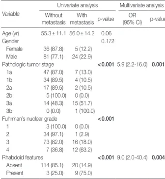
The presence of metastasis showed a significantly strong as- sociation with the presence of rhabdoid features (p<0.001). Me- tastasis was also significantly associated with higher nuclear grade (p<0.001) and pathologic tumor stage (p<0.001) in the univariate analysis but showed no significant correlation with age or gender (Table 3).
In the multivariate analysis, nuclear grade and pathologic tu- mor stage were each recategorized into two groups (low vs high) (for nuclear grade, 1 and 2 vs 3 and 4; for pathologic tumor stage, 1 and 2 vs 3). After adjusting for the presence of rhab-
doid features, nuclear grade, and pathologic tumor stage, the multivariate analysis showed that the presence of rhabdoid fea- tures (p=0.004) and pathologic tumor stage (p=0.001) were significantly associated with metastasis. When the presence of rhabdoid features was used for predicting metastasis, the sensi- tivity, specificity, positive predictive value, and negative predic- tive value were 31%, 97%, 75%, and 85%, respectively.
Correlation between diseaserelated mortality and rhabdoid features
In the Kaplan-Meier analysis (Fig. 2), disease-related mortal- ity was correlated with gender (p=0.047), nuclear grade (p<
0.001), pathologic tumor stage (p<0.001), and presence of rha- bdoid features (p<0.001).
The multivariate model for disease-related mortality includ- ed gender, nuclear grade, pathologic tumor stage, and the pres- ence of rhabdoid features. Nuclear grade and pathologic tumor stage were each recategorized into two groups (low vs high).
The outcome was independently associated with the presence of rhabdoid features (hazard ratio
[HR
], 5.1; 95% confidence in- terval
[CI
], 1.8 to 14.5; p=0.002) and recategorized pathologic
Table 2. Relationship between rhabdoid features and clinicopatho- logic parameters of clear cell-type renal cell carcinoma (RCC)
Clinicpathologic parameters
RCC without rhabdoid features
RCC with rhabdoid features p-value
No. of pateints 134 12
Age (yr) 56.2±12 (19-82) 56.4±10 (45-71) 0.959
Male/Female ratio 94/40 11/1 0.18
Pathologic tumor stage 0.001
1a 54 (40.3) 0 (0.0)
1b 34 (25.4) 4 (33.3)
2a 18 (13.4) 1 (8.3)
2b 5 (3.7) 0 (0.0)
3a 23 (17.2) 6 (50)
3b 0 (0.0) 1 (8.3)
Tumor size (cm) 5.4±2.5 (1.0-13.8) 7.8±2.3 (5.3-12.5) 0.002
Fuhrman’s nuclear grade <0.001
1 3 (2.2) 0 (0.0)
2 35 (26.1) 0 (0.0)
3 87 (64.9) 2 (16.7)
4 9 (6.7) 10 (83.3)
Metastasis <0.001
Absent 114 (85.1) 3 (25.0)
Present 20 (14.9) 9 (75.0)
The data represent the mean±standard deviation (range) or the number of patients (%). The bold values indicate a statistically significant difference (p<
0.05) by independent t-test (for age and tumor size), chi-square test with linear-by-linear association (for pathologic tumor stage and nuclear grade), and Fisher’s exact test (for gender and metastasis).
Table 3. Univariate and multivariate analyses of the clinicopatho- logic parameters of clear cell-type renal cell carcinoma (RCC) relat- ed to metastasis
Variable
Univariate analysis Multivariate analysis Without
metastasis With
metastasis p-value OR
(95% CI) p-value Age (yr) 55.3±11.1 56.0±14.2 0.06
Gender 0.172
Female 36 (87.8) 5 (12.2) Male 81 (77.1) 24 (22.9)
Pathologic tumor stage <0.001 5.9 (2.2-16.0) 0.001
1a 47 (87.0) 7 (13.0)
1b 34 (89.5) 4 (10.5)
2a 17 (89.5) 2 (10.5)
2b 5 (100.0) 0 (0.0)
3a 14 (48.3) 15 (51.7)
3b 0 (0.0) 1 (100.0)
Fuhrman’s nuclear grade <0.001
1 3 (100.0) 0 (0.0)
2 34 (97.1) 1 (2.9)
3 73 (82.0) 16 (18.0)
4 7 (36.8) 12 (63.2)
Rhabdoid features <0.001 9.0 (2.0-40.4) 0.004 Absent 114 (85.1) 20 (14.9)
Present 3 (25.0) 9 (75.0)
Under univariate analysis, the data represent the mean±standard deviation or the number of patients (%). The bold values indicate a statistically signifi- cant difference (p<0.05).
OR, odds ratio; CI, confidence interval.
tumor stage (HR, 4.7; 95% CI, 1.9 to 12.0; p=0.001).
Immunohistochemical findings
The results of the immunohistochemical studies are summa- rized in Table 4. The rhabdoid component showed diffuse or globular cytoplasmic positivity for vimentin (100%) in all 12 cases (Fig. 3). No case showed loss of INI1 (0%). Five were pos- itive for pan-CK (42%), six were positive for EMA (50%), and nine were positive for CD10 (75%). Myogenic markers includ- ing SMA and desmin were negative in all cases (0%). CK7, E- cadherin, and c-Kit were also negative in all cases (0%). p53 pos- itivity was observed in one (8%) case, and the Ki-67 labeling
index ranged from <1 to 25% (mean, 5.5 ± 2.3%). IMP3 posi- tivity was noted in five (42%) cases. IMP3 immunostaining re- sults of the non-rhabdoid areas were available for seven cases, and all of them were positive for IMP3. Interestingly, the rhab- doid areas were negative for IMP3 in three cases with IMP3- positive non-rhabdoid areas.
DISCUSSION
Malignant neoplasms with rhabdoid features have been re- ported in various organs including the kidney, urinary bladder, and prostate;
3-6however, the presence of rhabdoid features in
Fig. 2. Kaplan-Meier curves depicting the impact of gender (A), Fuhrman’s nuclear grade (B), pT stage (C), and presence of rhabdoid features (D) on the disease-associated mortality of patients (n=146) with clear renal cell carcinoma.
1.0
0.8
0.6
0.4
0.2
0.0
Cummulative survival rate
Time (day)
0 1,000 2,000 3,000 4,000 5,000 p=0.047
Female Male
1.0
0.8
0.6
0.4
0.2
0.0
Cummulative survival rate
Time (day)
0 1,000 2,000 3,000 4,000 5,000 p<0.001
Nuclear grade I Nuclear grade II Nuclear grade III Nuclear grade IV
A 1.0
0.8
0.6
0.4
0.2
0.0
Cummulative survival rate
Time (day)
0 1,000 2,000 3,000 4,000 5,000 p<0.001
pT stage Ia pT stage Ib pT stage IIa pT stage IIb pT stage IIIa pT stage IIIb
1.0
0.8
0.6
0.4
0.2
0.0
Cummulative survival rate
Time (day)
0 1,000 2,000 3,000 4,000 5,000 p<0.001
Rhabdoid feature () Rhabdoid feature (+)
C D
B
conventional neoplasms is best characterized in the kidney. Since Gokden et al.
3systematically analyzed RCC with rhabdoid fea- tures, several reports have emphasized the poor prognosis asso- ciated with the rhabdoid component.
3-6In neoplasms of other organs such as the stomach, thyroid, meninges, and urinary bladder,
7-10the rhabdoid component has also been associated with aggressive behavior. In the kidney, rhabdoid features are predo minantly associated with clear cell-type RCCs, but there are reports of associations with papillary- and chromophobe- type RCCs, collecting duct carcinoma, renal medullary carcino- ma, urothelial carcinoma, synovial sarcoma, and mixed stromal and epithelial tumor.
2-6,11-15We found a rhabdoid component in 6.9% of the 174 cases in this study, which is comparable to the results of previous studies.
3-6The reported range of frequency (3.2-7.4%) may be attributed to the selection criteria; one study with the lowest frequency included tumors with rhabdoid fea- tures of more than 10% only.
3-6In our study, the rhabdoid fea- tures were associated with clear cell-type RCCs only, and collect- ing duct carcinoma and renal medullary carcinoma were not in-
Fig. 3. Specimens showing positive staining for vimentin (A) and integrase interactor-1 (B), and negative staining for p53 (C). The Ki-67 label- ing index varied from 1% (left in D) to 25% (right in D).
A B
C D
Table 4. Comparison of immunostaining results of rhabdoid cells in renal cell carcinoma between this study and previous studies
Staining Present study Kuroiwa et al.4 Leroy et al.6 Vimentin 12/12 (100) 8/8 (100) 14/14 (100)
SMA 0/12 (0) 0/8 (0) 0/14 (0)
Desmin 0/12 (0) 0/8 (0) 0/14 (0)
EMA 6/12 (50) 6/8 (75) 11/14 (78)
Pan-cytokeratin 5/12 (42) 9/14 (65)
CK7 0/12 (0)
E-cadherin 0/12 (0)
CD10 9/12 (75)
p53 1/12 (8) 10/14 (71)
c-Kit 0/12 (0) 1/14 (7)
IMP3 5/12 (42)
INI1 12/12 (100)
AE1/AE3 6/8 (75)
CAM 5.2 4/8 (50)
HHF-35 0/8 (0)
Chromogranin 0/8 (0)
The data represent the positive cases/total cases (%).
SMA, smooth muscle actin; EMA, epithelial membrane antigen; CK7, cyto- keratin 7; IMP3, insulin-like growth factor mRNA binding protein 3; INI1, in- tegrase interactor-1.
cluded. Their incidence among clear cell-type RCCs was 8.2%.
When we investigated the prognostic significance of rhab- doid features in RCC, the presence of rhabdoid cells was associ- ated with poor prognostic factors such as a higher nuclear grade, higher pathologic stage, and accompanying tumor necrosis. In the multivariate analysis, the presence of rhabdoid features was one of the independent risk factors for metastasis. Additionally, the presence of rhabdoid features was independently associated with disease-related mortality. Therefore, the presence of a rhab- doid component should receive special attention and should be included in pathologic reports, as in the case of meningioma.
8,16Notably, most rhabdoid components were closely associated with necrosis in our study. Leroy et al.
6and Kuroiwa et al.
4re- ported areas of necrosis adjacent to the rhabdoid component in 13 of 14 (93%) and all eight cases (100%), respectively. There- fore, it is advisable to include necrotic tissue during gross ex- amination to obtain additional prognostic information.
The nature and histogenesis of rhabdoid cells in conventional renal neoplasms have not been clearly defined. Rhabdoid cells generally express vimentin and variably express pan-CK and EMA. Myogenic markers are negative. Our immunostaining results are in agreement with these findings. The protein ex- pression patterns somewhat overlap with those seen in rhabdoid tumors, and, therefore, a possibility of a composite tumor con- sisting of conventional neoplasm and a rhabdoid tumor can be considered. However, the conserved nuclear expression of INI1 in the present study as well as in other studies supports the view that the development of rhabdoid features in conventional neo- plasms is unrelated to the histogenesis of rhabdoid tumor.
17,18Leroy et al.
6reported that p53 expression is higher in rhabdoid areas than in non-rhabdoid areas, implicating p53 in tumor de- differentiation; however, we observed p53 expression in only one case. Miyagi et al.
19also reported the lack of p53 expression or mutation in lung adenocarcinoma with rhabdoid features. This contradictory finding in the kidney needs further clarification.
Higher proliferation activity of the rhabdoid component has been reported in the kidney, lung, and meninges.
4,16,19Kuroiwa et al.
4estimated the proliferation activity of the rhabdoid com- ponent as 9.89 ± 8.35%, which is higher than that of the con- ventional RCC component. In our study, the Ki-67 labeling in- dex was 5.5 ± 2.3%, which was not statistically different from the Ki-67 labeling index of the non-rhabdoid component (data not shown). This disagreement may be attributed to the stain- ing fields (whole slide vs microarray) or organ differences and needs further investigation. Our immunostaining results sug- gest that the development of rhabdoid features is not related to
the histogenesis of rhabdoid tumor and may not be associated with a p53 mutation or higher proliferation activity.
Factors other than high proliferation activity may contribute to the aggressive behavior of the rhabdoid component, includ- ing loss of cell junctions, cell-cell interactions or cell-extracellu- lar matrix (ECM) interaction, and changes associated with isch- emia or necrosis. Most rhabdoid cells are scattered individually and are associated with necrosis. The loss of cell junctions or cell-cell and cell-ECM interaction has been regarded as an im- portant step in the process of tumor metastasis.
20,21We did not study the expression patterns of variable adhesion molecules ex- cept E-cadherin; thus, this possibility could not be demonstrat- ed. Necrosis in clear cell-type RCC is considered to be a poor prognostic factor, but the underlying mechanisms have not been clarified.
22,23IMP3 is an oncofetal protein known to be a good marker for RCC metastasis.
24,25Interestingly, the frequency of IMP3 expression in the rhabdoid area was lower than in the non- rhabdoid area.
In the kidney, neoplasms with rhabdoid features should be distinguished from rhabdoid tumors and rhabdomyosarcoma.
Rhabdoid tumor, a highly aggressive neoplasm of childhood, is predominantly composed of rhabdoid cells. On ultrastructural study, the characteristic cytoplasmic hyaline inclusion is a whorl of intermediate filaments. This tumor has a molecular hallmark:
biallelic inactivation of the hSNF5/INI1 tumor suppressor gene.
The loss of INI1 can be demonstrated by immunohistochemi- cal staining
18and observed in renal and extrarenal rhabdoid tu- mors, renomedullary carcinomas, and, variably, in epithelioid sarcomas. Rhabdomyosarcomas are very rarely documented in the kidney. They show characteristic striated muscle differentia- tion and myoglobin and desmin positivity.
In conclusion, the rhabdoid component of RCC was an inde- pendent prognostic factor of metastasis; therefore, its identifica- tion is critical. The underlying mechanism of the aggressive be- havior needs further investigation.
REFERENCES
1. Weeks DA, Beckwith JB, Mierau GW, Luckey DW. Rhabdoid tumor of kidney: a report of 111 cases from the National Wilms’ Tumor Study Pathology Center. Am J Surg Pathol 1989; 13: 439-58.
2. Weeks DA, Beckwith JB, Mierau GW, Zuppan CW. Renal neoplasms mimicking rhabdoid tumor of kidney: a report from the National Wilms’ Tumor Study Pathology Center. Am J Surg Pathol 1991; 15:
1042-54.
3. Gökden N, Nappi O, Swanson PE, et al. Renal cell carcinoma with rhabdoid features. Am J Surg Pathol 2000; 24: 1329-38.
4. Kuroiwa K, Kinoshita Y, Shiratsuchi H, et al. Renal cell carcinoma with rhabdoid features: an aggressive neoplasm. Histopathology 2002; 41: 538-48.
5. Shannon B, Stan Wisniewski Z, Bentel J, Cohen RJ. Adult rhabdoid renal cell carcinoma. Arch Pathol Lab Med 2002; 126: 1506-10.
6. Leroy X, Zini L, Buob D, Ballereau C, Villers A, Aubert S. Renal cell carcinoma with rhabdoid features: an aggressive neoplasm with overexpression of p53. Arch Pathol Lab Med 2007; 131: 102-6.
7. Ueyama T, Nagai E, Yao T, Tsuneyoshi M. Vimentin-positive gastric carcinomas with rhabdoid features: a clinicopathologic and immu- nohistochemical study. Am J Surg Pathol 1993; 17: 813-9.
8. Perry A, Scheithauer BW, Stafford SL, Abell-Aleff PC, Meyer FB.
“Rhabdoid” meningioma: an aggressive variant. Am J Surg Pathol 1998; 22: 1482-90.
9. Parwani AV, Herawi M, Volmar K, Tsay SH, Epstein JI. Urothelial carcinoma with rhabdoid features: report of 6 cases. Hum Pathol 2006; 37: 168-72.
10. Albores-Saavedra J, Hernandez M, Sanchez-Sosa S, Simpson K, An- geles A, Henson DE. Histologic variants of papillary and follicular carcinomas associated with anaplastic spindle and giant cell carci- nomas of the thyroid: an analysis of rhabdoid and thyroglobulin inclusions. Am J Surg Pathol 2007; 31: 729-36.
11. Shannon BA, Cohen RJ. Rhabdoid differentiation of chromophobe renal cell carcinoma. Pathology 2003; 35: 228-30.
12. Kuroda N, Satake H, Miyazaki E, Hayashi Y, Hiroi M, Enzan H. Col- lecting duct carcinoma exhibiting diastase-resistant PAS-positive globular cytoplasmic inclusions and rhabdoid features arising in adult polycystic kidney disease: a case report. Int J Surg Pathol 2004;
12: 171-7.
13. Sukov WR, Cheville JC, Lager DJ, Lewin JR, Sebo TJ, Lewin M. Ma- lignant mixed epithelial and stromal tumor of the kidney with rhab- doid features: report of a case including immunohistochemical, mol- ecular genetic studies and comparison to morphologically similar renal tumors. Hum Pathol 2007; 38: 1432-7.
14. Jun SY, Choi J, Kang GH, Park SH, Ayala AG, Ro JY. Synovial sar- coma of the kidney with rhabdoid features: report of three cases.
Am J Surg Pathol 2004; 28: 634-7.
15. Cheng JX, Tretiakova M, Gong C, Mandal S, Krausz T, Taxy JB. Re- nal medullary carcinoma: rhabdoid features and the absence of INI1 expression as markers of aggressive behavior. Mod Pathol 2008; 21:
647-52.
16. Kepes JJ, Moral LA, Wilkinson SB, Abdullah A, Llena JF. Rhabdoid transformation of tumor cells in meningiomas: a histologic indica- tion of increased proliferative activity: report of four cases. Am J Surg Pathol 1998; 22: 231-8.
17. Biegel JA, Kalpana G, Knudsen ES, et al. The role of INI1 and the SWI/SNF complex in the development of rhabdoid tumors: meet- ing summary from the workshop on childhood atypical teratoid/
rhabdoid tumors. Cancer Res 2002; 62: 323-8.
18. Hoot AC, Russo P, Judkins AR, Perlman EJ, Biegel JA. Immunohis- tochemical analysis of hSNF5/INI1 distinguishes renal and extra- renal malignant rhabdoid tumors from other pediatric soft tissue tumors. Am J Surg Pathol 2004; 28: 1485-91.
19. Miyagi J, Tsuhako K, Kinjo T, Iwamasa T, Hashimoto H, Ishikawa S.
Rhabdoid tumour of the lung is a dedifferentiated phenotype of pulmonary adenocarcinoma. Histopathology 2000; 37: 37-44.
20. Gutwein P, Schramme A, Voss B, et al. Downregulation of junction- al adhesion molecule-A is involved in the progression of clear cell renal cell carcinoma. Biochem Biophys Res Commun 2009; 380: 387- 91.
21. Bissell MJ, Radisky D. Putting tumours in context. Nat Rev Cancer 2001; 1: 46-54.
22. Lee SE, Byun SS, Oh JK, et al. Significance of macroscopic tumor necrosis as a prognostic indicator for renal cell carcinoma. J Urol 2006; 176(4 Pt 1): 1332-7.
23. Tollefson MK, Thompson RH, Sheinin Y, et al. Ki-67 and coagula- tive tumor necrosis are independent predictors of poor outcome for patients with clear cell renal cell carcinoma and not surrogates for each other. Cancer 2007; 110: 783-90.
24. Jiang Z, Chu PG, Woda BA, et al. Analysis of RNA-binding protein IMP3 to predict metastasis and prognosis of renal-cell carcinoma: a retrospective study. Lancet Oncol 2006; 7: 556-64.
25. Jiang Z, Chu PG, Woda BA, et al. Combination of quantitative IMP3 and tumor stage: a new system to predict metastasis for patients with localized renal cell carcinomas. Clin Cancer Res 2008; 14: 5579- 84.