INTRODUCTION
Laryngeal cancer is one of the most common head and neck human cancers (excluding skin cancer) and continues to be a major unsolved health problem.1 Laryngeal squamous cell car- cinoma (LSCC) is the most common histological type of laryn-
geal malignancy.2 It composed the most part of laryngeal can- cers. Most patients of LSCC were diagnosised firstly between 50 to 75 years of age. The male-to-female sex ratio of laryngeal cancer was from 6:1 to 10:1 in most cases.3 Current methods for the diagnosis of early-stage LSCC were quite unsatisfacto- ry.4 Nowdays, only less than 50% of patients with LSCC were in early stage at diagnosis, due to the lack of effective early-diag- nosis methods.5,6 As effective biomarker is the key of early diag- nosis of cancer and therefore the identification of new bio- markers for LSCC is in severe need.
MicroRNAs (miRNAs), an abundant class of 17–25 nucleo- tides that comprise small noncoding RNAs, post-transcription- ally regulate gene expression by directly binding to the 3’ un- translational region (3’ UTR) of target miRNAs.7 Until now, over 1000 miRNA genes have been identified in mammals; howev- er, revealing their roles in physiology and pathology remains an ongoing process. Recently, it has been suggested that miRNAs
The Expression of MicroRNA-155 in Plasma and Tissue Is Matched in Human Laryngeal Squamous Cell Carcinoma
Jian-ling Wang1, Xin Wang1, Dong Yang1, and Wen-jie Shi2
1Department of Otolaryngol Head Neck Surgery, General Hospital of Tianjin Medical University, Tianjin;
2Department of Otolaryngol Head Neck Surgery, Tianjin First Central Hospital, Tianjin, China.
Purpose: Tumor-associated microRNAs have been detected in cancer, though whether plasma microRNA-155 (miR-155) could be a potential biomarker for laryngeal squamous cell carcinoma (LSCC) prognosis is unclear. We aimed to determine how miR- 155 can be used to predict the clinical characteristics of patients with LSCC and correctly diagnose them.
Materials and Methods: We collected tissue samples and peripheral blood samples before and after treatment from 280 LSCC cases and 560 controls. Real-time quantitative reverse transcription PCR was employed in this study to compare the relative ex- pression of miR-155.
Results: A total of 280 LSCC patients and 560 age- and sex-matched controls were included in the study. The miR-155 level was more up-regulated in LSCC tissue than in the non-tumor tissues (13.6±2.4 vs. 3.1±0.80, p<0.001). Additionally, a significantly higher miR-155 level in plasma samples from LSCC patients than in those of the controls (8.9±1.25 vs. 1.8±0.8, p<0.001) was reported.
Tissue miR-155 showed an area under the curve (AUC) of 0.933, with a sensitivity of 82.6% and a specificity of 89.2%. The AUC for plasma miR-155 was 0.757, with a sensitivity of 58.4% and a specificity of 69.5%. When early LSCC in TNM I stage was considered, tissue miR-155 showed an area under the curve of 0.804, with a sensitivity of 85.2% and a specificity of 87.3%.
Conclusion: The expression of tissue and plasma miR-155 were significantly up-regulated in patients with LSCC. Our work will serve as a basis for further investigation, preferably large-scale validation in clinical trials.
Key Words: MiR-155, laryngeal squamous cell carcinoma, diagnosis, biomarker Yonsei Med J 2016 Mar;57(2):298-305
http://dx.doi.org/10.3349/ymj.2016.57.2.298 pISSN: 0513-5796 · eISSN: 1976-2437
Received: January 8, 2015 Revised: March 24, 2015 Accepted: April 22, 2015
Corresponding author: Dr. Wen-jie Shi, Department of Otolaryngol Head Neck Surgery, Tianjin First Central Hospital, No. 24 Fukang road, Nankai Area, Tianjin 300192, China.
Tel: 86-22-60362255, Fax: 86-22-23626199, E-mail: [email protected]
•The authors have no financial conflicts of interest.
© Copyright: Yonsei University College of Medicine 2016
This is an Open Access article distributed under the terms of the Creative Com- mons Attribution Non-Commercial License (http://creativecommons.org/licenses/
by-nc/3.0) which permits unrestricted non-commercial use, distribution, and repro- duction in any medium, provided the original work is properly cited.
participate in the regulation of diverse biological processes.
Bioinformatics data have indicated that a single miRNA can bind to hundreds of miRNA targets, and these targets could theoretically regulate every biological process.8 Each miRNA, though powerful, has also been characterized as being margin- al (a given gene can be targeted by several miRNAs, and a giv- en miRNA typically exerts a modest repression).9 One miRNA, microRNA-155 (miR-155) is known to regulate multiple aspects of types of cancer progression in addition to demonstrating a correlation with poor prognosis.10,11 Located within the noncod- ing B cell integration cluster (BIC) gene, miR-155 was first dis- covered through retroviral integrations in B cell lymphomas.12 Currently miR-155 is found to be amplified in many cancers, irrespective of retroviral integration. As one of the more highly evaluated miRNAs, miR-155 is an established oncogenic miR- NA, and many of its targets are known.13-15 MiR-155 is known to regulate cellular proliferation and survival through multiple mechanisms, including the mitogen activated protein kinases (MAPKs) signaling pathways. MiR-155 is a positive multi-fac- eted regulator of MAPK signaling, intervening at heteroge- neous points along the signaling cascade to enhance signaling.
Through the targeting of inositol polyphosphate-5-phospha- tase (SHIP1), miR-155 enhances extracellular signal-regulated kinases (ERK) activation. In a cancer cell line, miR-155 expres- sion enhanced ERK phosphorylation and cellular proliferation;
conversely, the loss of miR-155 expression led to decreased ERK activation in B-cells.16 Downstream MAPK effectors, such as AP-1 complex member c-Jun, are enhanced through miR- 155 targeting of DET1. Targeting of DET1 stabilizes c-Jun tran- scripts and subsequently increases AP-1 activity.17
Recently, it has been reported that plasma contains suffi- ciently stable miRNA species that might be useful as non-in- vasive biomarkers for several cancers, including LSCC.18,19 Thus, the aim of the present study was to evaluate the expression of miR-155 as a potential biomarker for LSCC. However, the as- sociations of plasma with tissue miR-155 levels are not yet clear.
In this study, we investigated plasma and tissue miR-155 ex- pression in a group of patients with LSCC and controls matched according to age and sex.
MATERIALS AND METHODS
Study designs and participants
We collected tissue samples and peripheral blood samples be- fore and after treatment from LSCC cases and controls. In total, 280 LSCC patients and 560 controls were included in this study.
The clinicopathological characteristics (age, gender, smoking status, tumor size, differentiation status, lymph node metasta- sis, stage classification) of study subjects were recorded by trained investigators. Primary LSCC and adjacent noncancer- ous tissues were procured from patients undergoing surgical resections. All tissues were snap-frozen immediately after sur-
gery and stored at -80°C until use. Three milliliters of whole blood were collected in an Ethylenediaminetetraacetic Acid Vacutainer (BD Company, San Diego, CA, USA) for plasma.
Sample collections were centrifuged at 2500 rpm at 4°C for 10 min. The supernatant fluids were then stored at -80°C until to- tal RNA extraction. This study was approved by the Institution- al Review Board (IRB) of the General Hospital of Tianjin Medi- cal University, Tianjin, China.
Plasma and tissue sampling and RNA isolation
Cell-free plasma was isolated via a two-step protocol (2500 rpm at room temperature for 10 min and 14000×g at 4°C for 10 min) within 2 h after collection to prevent contamination of the cel- lular nucleic acids. The resulting plasma was transferred to new tubes and stored at -80°C. The plasma was transferred to RNase/DNase-free tubes and stored at -80°C until RNA isola- tion. The total RNA was isolated from the plasma using a mir- Vana PARIS isolation kit (Ambion, Austin, TX, USA) according to the manufacturer’s instructions for plasma samples without enrichment for small RNAs. Briefly, 400 μL of plasma was used to extract the total RNA. Each sample was eluted in 40 μL of RNAse-free water. Cancer and non-cancer tissues from LSCC cases were immediately flash frozen in liquid nitrogen, and stored at -80°C until RNA isolation using a mirVana PARIS iso- lation kit (Ambion, Austin, TX, USA) according to the manu- facturer’s instructions, which were similar to those described above. The average levels of miR-155 expression in tissues and sera were normalized relative to the average amounts of U6 snRNAm, using the 2-ΔΔCT method. RNA quality was measured using a denaturing 15% polyacrylamide gel.
Quantitative reverse transcription-polymerase chain reaction (qRT-PCR)
The reverse transcription reaction was carried out using the TaqMan MicroRNA Reverse Transcription Kit (Applied Biosys- tems, America) according to the manufacturer’s instructions in a total reaction volume of 7.5 mL. Quantitative polymerase chain reaction (qPCR) was performed in triplicate using Taq- Man Universal PCR Master Mix (Applied Biosystems) in an ABI 7500 Real-Time PCR system (Applied Biosystems) with the fol- lowing conditions: 95°C for 10 min, followed by 40 cycles of 95°C for 15 s and 60°C for 1 min. The cycle threshold (Ct) values were calculated using SDS 2.0.1 software (Applied Biosystems).
Template-free controls for both RT and PCR were included in each experiment to ensure target-specific amplification. In ad- Table 1. Primers Used in qPCR
MiRNAs Primer sequence (5'-3')
MiR-155 Forward TGCCTCCAACTGACTCCTAC Reverse GCGAGCACAGAATAATACGTA
U6 Forward GCTTCGGCAGCACATATACTAAAAT
Reverse CGCTTCACGAATTTGCGTGTCAT qPCR, quantitative polymerase chain reaction; miR-155, microRNA-155.
dition, if CtU6 did not occur within 32 cycles, the assay was re- peated. Samples with low U6 snRNA levels were not excluded from data analyses. Primers used in qPCR are presented in Ta- ble 1.
Statistical analysis
We used the paired Wilcoxon nonparametric test to analyze pairs of non-tumor tissue and cancer samples or between cases and controls. The Mann-Whitney U test was used for the anal- yses of the expression of different miRNAs. Receiver operating characteristic (ROC) curves were drawn, and the areas under the ROC curves (AUCs) were measured to assess the specifici- ty and sensitivity of circulating miR-155 as potential diagnos- tic biomarkers for LSCC. All statistical analyses were performed using SPSS 16.0 software (SPSS Inc., Chicago, IL, USA). Differ- ences were considered significant if p<0.05. All experiments were performed in triplicate and repeated at least three times.
RESULTS
Clinical characteristics of patients and controls A total of 280 LSCC patients and 560 age, sex-matched con- trols were included in the study. The median age of the patients was 64.8±12.1 years, while the median age of the controls was 65.1±9.8 years. No differences were detected between the Table 2. Clinicopathological Features of 280 LSCC Patients and 560 Con-
trols
Clinical features Cases Controls p value
Mean age (yrs) 64.8±12.1 65.1±9.8 0.162
<60 194 326 0.341
≥60 86 234
Gender 0.541
Male 245 488
Female 35 72
Smoking <0.001
Yes 212 311
No 68 249
Tumor size
≤3 cm 243
>3 cm 37
Differentiation
Moderate–well 197
Poor 83
Lymph node metastasis
Negative 167
Positive 113
Stage classification
Stage I 69
Stages II, III, and IV 211 LSCC, laryngeal squamous cell carcinoma.
Table 3. Associations of Aberrant MiR-155 Expression in Tissue and Plasma with Clinicopathological Data of LSCC Patients
Clinical features n Tissue miR-155
p value Plasma miR-155
p value
High Low Low High
Mean age (yrs) 64.8±12.1 64.2±11.4 65.2±10.7 0.492 65.4±9.8 63.9±13.6 0.421
<60 194 101 93 0.365 95 99 0.693
≥60 86 39 47 45 41
Gender 0.673 0.492
Male 245 123 122 119 126
Female 35 17 18 21 14
Smoking 0.676 <0.001
Smoker 212 108 104 120 92
Never-smoker 68 32 36 20 48
Tumor size 0.004 0.033
≤3 cm 243 113 130 115 128
>3 cm 37 27 10 25 12
Differentiation status <0.001 0.515
Moderate–well 197 79 118 103 94
Poor 83 61 22 47 36
Lymph node metastasis <0.001 0.007
Negative 167 60 107 64 103
Positive 113 80 33 76 37
Stage classification <0.001 0.001
Stage I 69 19 50 22 47
Stages II, III, and IV 211 121 90 118 93
miR-155, microRNA-155; LSCC, laryngeal squamous cell carcinoma.
groups (p=0.162). Of the 280 patients, 245 were female, and the others were male. Among the 560 controls, 245 were female, and the others were male (p=0.541). However, when tobacco smoking status was considered, the rate of smoking was high- er in patients with LSCC (p<0.001). The details regarding clini- copathologic features were only collected for the patients, who also came for follow-up treatment. Pathological features were obtained for all cases, among which 243 were in the 0–3- cm group while 37 were in the >3-cm group. Most patients had a tumor of more than stage I (211 of 288, 75.36%). A moder- ately or well differentiated tumor was seen in 197 cases, while poor differentiated tumors comprised 24.64% of the patient population. Of the patients with lymph node metastasis infor-
mation, 167 had negative lymph node metastasis, and 116 had positive lymph node metastasis (Table 2).
Tissues and plasma miRNA-155 levels in cases and controls
Total RNA was isolated from the cancer and control tissues of all LSCC cancer patients. Additionally, plasma miR-155 levels were detected in both cases and controls. In general, the miR- 155 levels were quantified using a quantitative reverse tran- scription PCR assay with U6 as the internal control. In our re- sults, we found that the miR-155 level was more up-regulated in LSCC tissue than in the non-tumor tissues (13.6±2.4 vs. 3.1±
0.80; p<0.001). Moreover, we observed a significantly higher
Fig. 1. Relative expression of tissue and plasma miR-155 in LSCC pa- tients. miR-155, microRNA-155; LSCC, laryngeal squamous cell carcino- ma.
20
15
10
5
0
Tissue Plasma
Relative expression of miR-155
Control Stage I Stage II Stage III Stage IV
* * *
* *
*
Fig. 2. The association between tissue and plasma miR-155 in LSCC cases. miR-155, microRNA-155; LSCC, laryngeal squamous cell carcino- ma.
8.0 7.5 7.0 6.5 6.0 5.5 5.0
Tissue
5 10 15 20 25 30 35 40
Plasma
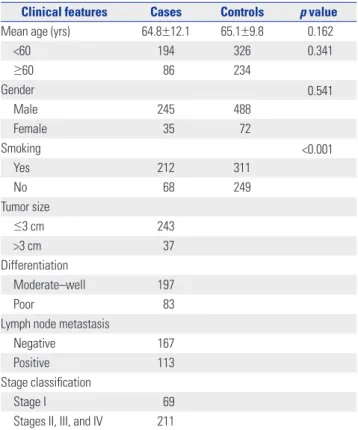
Fig. 3. Receiver operating characteristic curves of tissue and plasma miR-155 for the diagnosis of LSCC. (A) Tissue miR-155. (B) Plasma miR-155. AUC, area under the curve; miR-155, microRNA-155; LSCC, laryngeal squamous cell carcinoma.
1.0
0.8
0.6
0.4
0.2
0.0
1.0
0.8
0.6
0.4
0.2
0.0
AUC=0.933 AUC=0.757
Sensitivity Sensitivity
1-specificity 1-specificity
0.0 0.2 0.4 0.6 0.8 1.0 0.0 0.2 0.4 0.6 0.8 1.0
A B
miR-155 level in plasma samples from LSCC patients than in those from the controls (8.9±1.25 vs. 1.8±0.8; p<0.001). We also reported the miR-155 levels of the LSCC cases for each differ- ent stage. It was found that both tissue and plasma miR-155 levels were higher in stage I LSCC cases. Additionally, the lev- els of miR-155 in stage II, III, and IV were higher than that of stage I (Table 3). However, no increasing trend was observed as the stage increased. The expression data for miR-155 in tissue and plasma are presented in Fig. 1.
We also conducted advanced analyses to detect a possible relationship between the tissue and plasma levels of miR-155.
Both tissue and plasma samples were used in the detection of the miR-155 level. On Pearson correlation analyses, a signifi- cant association was detected (p<0.001; r=0.725) (Fig. 2).
Diagnostic performance of tissue and plasma miR-155 for LSCC
The diagnostic performances of tissue and plasma miR-155 for LSCC were detected. Cutoff points were determined using the highest sum of sensitivity and specificity. The cutoff points for tissue and plasma miR-155 were 7.34 and 3.12, respective- ly. Tissue miR-155 showed an AUC of 0.933, with a sensitivity of 82.6% and a specificity of 89.2% (Fig. 3A). However, the AUC for plasma miR-155 was 0.757, with a sensitivity of 58.4% and a specificity of 69.5% (Fig. 3B).
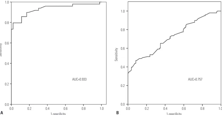
We also conducted advanced analyses on the diagnostic per- formances for early TNM I stage LSCC. As well, the cutoff points were determined using the highest sum of sensitivity and speci- ficity. The cutoff points for tissue and plasma miR-155 were 7.34 and 3.12, respectively. Tissue miR-155 showed an AUC of 0.804,
with a sensitivity of 85.2% and a specificity of 87.3% (Fig. 4A).
However, the AUC for plasma miR-155 was 0.824, with a sensi- tivity of 72.4% and a specificity of 76.9% (Fig. 4B).
The expression of tissue and plasma miR-155 after surgery
A total of 280 LSCC cases were obtained to detect the change of plasma miR-155. Compared to preoperative expression, plasma miR-155 demonstrated significant depression. As shown in Fig.
5, the expression of tissue miR-155 changed from 8.9 to 4.3 (p=0.002). In the smoking group, the expression of plasma miR- 155 was 11.5±1.5, while the postoperative level was 5.2±0.9 (p=0.001). The level of miR-155 in the non-smoking group changed from 8.1±0.9 to 4.0±1.3, and a significant difference
Fig. 4. Receiver operating characteristic curves of tissue and plasma miR-155 for the diagnosis of early-stage LSCC. (A) Tissue miR-155. (B) Plasma miR-155. AUC, area under the curve; miR-155, microRNA-155; LSCC, laryngeal squamous cell carcinoma.
1.0
0.8
0.6
0.4
0.2
0.0
1.0
0.8
0.6
0.4
0.2
0.0
AUC=0.804 AUC=0.824
Sensitivity Sensitivity
1-specificity 1-specificity
0.0 0.2 0.4 0.6 0.8 1.0 0.0 0.2 0.4 0.6 0.8 1.0
A B
Fig. 5. Preoperative and postoperative plasma miR-155 levels of LSCC patients. miR-155, microRNA-155; LSCC, laryngeal squamous cell carci- noma.
14 12 10 8 6 4 2 0
Total Smoker Never-smoker
The expression of miR-155
Pre-operation Post-operation
was detected (p=0.012).
DISCUSSION
Sensitive and specific biomarkers were capital in developing preventive screening kinds of cancers, including LSCC. How- ever, current methods were insufficient in detecting LSCC in the early stage. Recent achievements in imaging technology have greatly improved the diagnosis of LSCC; however, the high cost maked it unavailable in developing countries. In re- cent years, miRNAs have been regarded as new diagnostic tools for the detection of kinds of cancers. As miRNA expression was tissue-specific and quite stable, miRNAs may be used as a mark- er to resolve tumor tissue from normal tissue or as a diagnos- tic tool. Although several groups have investigated the poten- tial application of miRNA as a biomarker for LSCC,20,21 most of the reports have used histological specimens and thus have limited clinical significance. Given that miRNAs are stable in circulation, we propose that they may serve as ideal noninva- sive biomarkers for the diagnosis of LSCC. Therefore, we ana- lyzed both tissue and plasma levels of miR-155 in patients with LSCC and controls and evaluated their diagnostic performance as diagnostic markers of LSCC.
To our knowledge, this is the first study to explore the diag- nostic impact of miR-155 in a case-control study with 500 par- ticipants. Previous reports indicated that miR-155 was up-reg- ulated in several malignancies, such as Hodgkin lymphoma,22 breast cancer,23 and lung carcinoma,24 and perceived an onco- gene, although the diagnostic impact was not addressed in these studies. Similarly, in LSCC, miR-155 has been interpret- ed as an oncogene. In a global miRNA profiling study with 51 formalin-fixed archival LSCC samples using a quantitative re- verse transcription-PCR approach, miRNA expression was cor- related with patients’ clinical parameters. Thirty-eight of 117 (33%) consistently detected miRNAs were significantly differ- entially expressed in malignant tissues when compared to nor- mal tissues, and overexpression of miR-155 in LSCC patients was detected.25
As in most previous studies, the diagnostic effects of miR- 155 on different cancers were analyzed in the tissues.26,27 It has been reported that serum miRNAs were quite stable and could hardly degraded by enzymes. It has also been demonstrated that miRNAs are resistant to cancers. However, as the impor- tance of circling miRNAs is considered in the diagnosis of can- cer, the application of plasma or serum miR-155 in the diagno- sis of breast cancer,28 glioma,29 lung adenocarcinoma,24 and Ho- dgkin lymphoma.22 In a previous study, the levels of miR-155 in cancer tissues or cell lines were significantly higher than in benign tissue or non-cancer cells.30 However, miR-155 levels in plasma were not reported in previous studies. In our study, we conducted a case-control study to identify the expression and association of both tissue and plasma miR-155 in LSCC cases.
In general, our data showed that the expressions of miR-155 in both tissue and plasma were significantly higher. Moreover, we found that the expression levels of miR-155 in tissue and plasma were significantly associated. This result was accor- dant with several previous studies, such as microRNA-375 in colorectal cancer31 and miR-106b~25 expressions in patients with gastric cancers.32 MiR-155 in tissue and plasma has been used as a diagnostic marker for LSCC. According to the require- ments for biomarkers in diseases, we speculated that both tis- sue and plasma miR-155 could be used as diagnostic markers for LSCC. Therefore, all of the data in our study were sufficient for describing diagnostic markers for the diagnosis of LSCC in all stages. Our study was the first to detect both tissue and plas- ma miR-155 in LSCC patients by using qPCR. This technology could be used to discover new biomarkers for LSCC from 280 LSCC cases and 560 controls with reasonable reliability. In the current study, we analyzed the expression of miR-155 in LSCC tissues and plasma samples in order to determine their po- tential value in the diagnosis of LSCC patients and early-stage LSCC. Our data showed that levels of tissue and plasma miR- 155 expression were more up-regulated in LSCC than in the corresponding non-cancerous laryngeal tissues or the con- trols, and the ROC data showed that the AUC for tissue and plasma miR-155 was 0.933 and 0.757, respectively. In stage I LSCC patients, the ROC data indicated that the AUC for tissue and plasma miR-155 was 0.804 and 0824, respectively. Based on these ROC analyses, we found that the tissue and plasma miR-155 levels were potential important diagnostic markers for LSCC. A total of 280 LSCC cases were obtained to detect the change of plasma miR-155. Compared to the preoperative ex- pression, plasma miR-155 demonstrated significant depres- sion. The changes in plasma miR-155 demonstrated the po- tential application of miR-155 for the prognosis of LSCC. In this paper, we found that only plasma miR-155 was differently ex- pressed in the LSCC cases. It is an interesting result that sug- gests that miR-155 level was more related with the pathologi- cal characteristics. When the plasma miR-155 level was consi- dered, the expression of circulating miR-155 was modified after tobacco smoke exposure. In a study conducted by Herberth, et al.,33 it was reported that blood miRNAs are sensitive to envi- ronmental stressors, including tobacco smoke. In conclusion, we found that plasma miRNA was sensitive to different clini- cal and environmental factors.
The tumor promoter roles of miR-155 in different cancers are determined by both in vitro and in vivo study, and an up- regulated level of miR-155 expression would increase cell via- bility, prevent cell apoptosis, and increase tumorigenicity. As an important regulatory molecule, miR-155 played a key role in kidns of oncogenic processes. It could down-regulate B-cell lymphoma 6 (BCL6) protein, which could modulate the inter- leukin 4 responses of B cells.34 Additionally, miR-155 could act to enhance transcription and contribute to the pathogenesis of leukemia. MiR-155 could reduce the expression of BCL6 and
then leads to up-regulation of certain BCL6 targets, including inhibitor of differentiation (Id2), interleukin-6 (IL6), cMyc, and Mip1α/Ccl3, all of which promote cell survival and prolif- eration.35 In an in vitro and in vivo study, it was found that the expression levels of miR-155 in LSCC were significantly higher than those in the control mucosa tissues.36 Down-regulation of SOCS1 expression and elevated expression of STAT3 were also observed in LSCC. The relevance of the three factors was sta- tistically significant. Moreover, knockdown of miR-155 elevat- ed SOCS1expression level, suppressed STAT3 expression, and inhibited hep-2 cell growth, migration, and invasion, whereas overexpression of miR-155 resulted in the opposite effects.
STAT3 protein level in poor or moderate cell differentiation was significantly higher than that in a higher degree of cell differ- entiation. These data demonstrated the aberrant expression and function of miR-155 and its downstream targets in LSCC.
These findings suggest that miR-155 plays a promoting role during the development of LSCC, and it may be a useful marker for the prognosis and assessment of therapeutic effects.30
In conclusion, the expressions of tissue and plasma miR-155 were significantly up-regulated in patients with LSCC. Due to its reasonable sensitivity and specificity for LSCC, tissue and plasma miR-155 could serve as potential biomarkers for deter- mining both LSCC stage and early cases. Our work will serve as a basis for further investigation, preferably large-scale vali- dation in clinical trials, before plasma miR-155 can be used as a routine screening tool for LSCC.
REFERENCES
1. Gourin CG, Starmer HM, Herbert RJ, Frick KD, Forastiere AA, Eisele DW, et al. Short- and long-term outcomes of laryngeal can- cer care in the elderly. Laryngoscope 2015;125:924-33.
2. Landry D, Glastonbury CM. Squamous cell carcinoma of the upper aerodigestive tract: a review. Radiol Clin North Am 2015;53:81-97.
3. Rutt AL, Hawkshaw MJ, Sataloff RT. Laryngeal cancer in patients younger than 30 years: a review of 99 cases. Ear Nose Throat J 2010;89:189-92.
4. Park JM, Jung CK, Choi YJ, Lee KY, Kang JH, Kim MS, et al. The use of an immunohistochemical diagnostic panel to determine the primary site of cervical lymph node metastases of occult squamous cell carcinoma. Hum Pathol 2010;41:431-7.
5. Qiu G, Li Y, Liu Z, Wang M, Ge J, Bai X. Clinical value of serum HMGB1 in diagnosis and prognosis of laryngeal squamous cell carcinoma. Med Oncol 2014;31:316.
6. Vasca V, Vasca E, Freiman P, Marian D, Luce A, Mesolella M, et al.
Keratin 5 expression in squamocellular carcinoma of the head and neck. Oncol Lett 2014;8:2501-4.
7. Cheng CJ, Bahal R, Babar IA, Pincus Z, Barrera F, Liu C, et al. Mi- croRNA silencing for cancer therapy targeted to the tumour mi- croenvironment. Nature 2015;518:107-10.
8. Loh YH, Yi SV, Streelman JT. Evolution of microRNAs and the di- versification of species. Genome Biol Evol 2011;3:55-65.
9. Ceppi P, Peter ME. MicroRNAs regulate both epithelial-to-mesen- chymal transition and cancer stem cells. Oncogene 2014;33:269-78.
10. Xu TP, Zhu CH, Zhang J, Xia R, Wu FL, Han L, et al. MicroRNA-155 expression has prognostic value in patients with non-small cell
lung cancer and digestive system carcinomas. Asian Pac J Cancer Prev 2013;14:7085-90.
11. Lind EF, Ohashi PS. Mir-155, a central modulator of T-cell re- sponses. Eur J Immunol 2014;44:11-5.
12. Zheng Y, Xiong S, Jiang P, Liu R, Liu X, Qian J, et al. Glucocorti- coids inhibit lipopolysaccharide-mediated inflammatory re- sponse by downregulating microRNA-155: a novel anti-inflam- mation mechanism. Free Radic Biol Med 2012;52:1307-17.
13. Bera A, VenkataSubbaRao K, Manoharan MS, Hill P, Freeman JW.
A miRNA signature of chemoresistant mesenchymal phenotype identifies novel molecular targets associated with advanced pan- creatic cancer. PLoS One 2014;9:e106343.
14. Chen L, Jiang K, Jiang H, Wei P. miR-155 mediates drug resistance in osteosarcoma cells via inducing autophagy. Exp Ther Med 2014;8:527-32.
15. Pareek S, Roy S, Kumari B, Jain P, Banerjee A, Vrati S. MiR-155 in- duction in microglial cells suppresses Japanese encephalitis virus replication and negatively modulates innate immune responses. J Neuroinflammation 2014;11:97.
16. Zhu J, Chen T, Yang L, Li Z, Wong MM, Zheng X, et al. Regulation of microRNA-155 in atherosclerotic inflammatory responses by targeting MAP3K10. PLoS One 2012;7:e46551.
17. Anzi S, Finkin S, Shaulian E. Transcriptional repression of c-Jun’s E3 ubiquitin ligases contributes to c-Jun induction by UV. Cell Signal 2008;20:862-71.
18. Tutar Y. miRNA and cancer; computational and experimental ap- proaches. Curr Pharm Biotechnol 2014;15:429.
19. Ayaz L, Görür A, Yarog˘lu HY, Ozcan C, Tamer L. Differential ex- pression of microRNAs in plasma of patients with laryngeal squa- mous cell carcinoma: potential early-detection markers for laryn- geal squamous cell carcinoma. J Cancer Res Clin Oncol 2013;139:
1499-506.
20. Sun X, Song Y, Tai X, Liu B, Ji W. MicroRNA expression and its de- tection in human supraglottic laryngeal squamous cell carcino- ma. Biomed Rep 2013;1:743-6.
21. Wu TY, Zhang TH, Qu LM, Feng JP, Tian LL, Zhang BH, et al. MiR- 19a is correlated with prognosis and apoptosis of laryngeal squa- mous cell carcinoma by regulating TIMP-2 expression. Int J Clin Exp Pathol 2013;7:56-63.
22. Jones K, Nourse JP, Keane C, Bhatnagar A, Gandhi MK. Plasma microRNA are disease response biomarkers in classical Hodgkin lymphoma. Clin Cancer Res 2014;20:253-64.
23. Vimalraj S, Miranda PJ, Ramyakrishna B, Selvamurugan N. Regu- lation of breast cancer and bone metastasis by microRNAs. Dis Markers 2013;35:369-87.
24. Gao F, Chang J, Wang H, Zhang G. Potential diagnostic value of miR-155 in serum from lung adenocarcinoma patients. Oncol Rep 2014;31:351-7.
25. Hui AB, Lenarduzzi M, Krushel T, Waldron L, Pintilie M, Shi W, et al. Comprehensive MicroRNA profiling for head and neck squa- mous cell carcinomas. Clin Cancer Res 2010;16:1129-39.
26. Gasparini P, Cascione L, Fassan M, Lovat F, Guler G, Balci S, et al.
microRNA expression profiling identifies a four microRNA signa- ture as a novel diagnostic and prognostic biomarker in triple neg- ative breast cancers. Oncotarget 2014;5:1174-84.
27. Kopp KL, Ralfkiaer U, Nielsen BS, Gniadecki R, Woetmann A, Ødum N, et al. Expression of miR-155 and miR-126 in situ in cu- taneous T-cell lymphoma. APMIS 2013;121:1020-4.
28. Sochor M, Basova P, Pesta M, Dusilkova N, Bartos J, Burda P, et al.
Oncogenic microRNAs: miR-155, miR-19a, miR-181b, and miR- 24 enable monitoring of early breast cancer in serum. BMC Can- cer 2014;14:448.
29. Sun J, Shi H, Lai N, Liao K, Zhang S, Lu X. Overexpression of mi-
croRNA-155 predicts poor prognosis in glioma patients. Med On- col 2014;31:911.
30. Zhao XD, Zhang W, Liang HJ, Ji WY. Overexpression of miR-155 promotes proliferation and invasion of human laryngeal squa- mous cell carcinoma via targeting SOCS1 and STAT3. PLoS One 2013;8:e56395.
31. Xu L, Li M, Wang M, Yan D, Feng G, An G. The expression of mi- croRNA-375 in plasma and tissue is matched in human colorectal cancer. BMC Cancer 2014;14:714.
32. Zhang R, Wang W, Li F, Zhang H, Liu J. MicroRNA-106b~25 ex- pressions in tumor tissues and plasma of patients with gastric cancers. Med Oncol 2014;31:243.
33. Herberth G, Bauer M, Gasch M, Hinz D, Röder S, Olek S, et al. Ma- ternal and cord blood miR-223 expression associates with prena-
tal tobacco smoke exposure and low regulatory T-cell numbers. J Allergy Clin Immunol 2014;133:543-50.
34. Basso K, Schneider C, Shen Q, Holmes AB, Setty M, Leslie C, et al.
BCL6 positively regulates AID and germinal center gene expres- sion via repression of miR-155. J Exp Med 2012;209:2455-65.
35. Sandhu SK, Volinia S, Costinean S, Galasso M, Neinast R, Santha- nam R, et al. miR-155 targets histone deacetylase 4 (HDAC4) and impairs transcriptional activity of B-cell lymphoma 6 (BCL6) in the Eμ-miR-155 transgenic mouse model. Proc Natl Acad Sci U S A 2012;109:20047-52.
36. Eis PS, Tam W, Sun L, Chadburn A, Li Z, Gomez MF, et al. Accu- mulation of miR-155 and BIC RNA in human B cell lymphomas.
Proc Natl Acad Sci U S A 2005;102:3627-32.