Original Article
Altered expression of microRNA miR-21, miR-155, and let-7a and their roles in pulmonary neuroendocrine tumors
pin_2845583..591Hyoun Wook Lee,1Eun Hee Lee,1Seung Yeon Ha,2Chang Hun Lee,3Hee Kyung Chang,4Sunhee Chang,5 Kun Young Kwon,6Il Seon Hwang,6Mee Sook Roh7and Jeong Wook Seo8
Departments of Pathology,
1Samsung Changwon Hospital, Sungkyunkwan University School of Medicine, Changwon,
2
Gachon University of Medicine and Science, Incheon,
3Busan National University School of Medicine, Busan,
4Kosin University College of Medicine, Busan,
5Inje University Ilsan Paik Hospital, Ilsan,
6Keimyung University College of Medicine, Daegu,
7Dong-A University College of Medicine, Busan,
8Prevention and Management Center, Regional Cardiocerebrovascular Center, Dong-A University Medical Center, Busan, Korea
MicroRNA (miRNA) has a critical effect on tumorigenesis through post-transcriptional modification and is considered to be potential biomarkers for cancer diagnosis and treat- ment monitoring. We evaluated the expression pattern of three selected miRNAs (miR-21, miR-155, and let-7a) to evaluate their potential roles by quantitative reverse transcription-polymerase chain reaction using formalin- fixed and paraffin-embedded tissues of 63 surgically resected pulmonary neuroendocrine (NE) tumors (19 typical carcinoids (TCs), 6 atypical carcinoids (ACs), 19 large cell NE carcinomas (LCNECs), and 19 small cell lung carcino- mas (SCLCs). Control amplification for U6 small nuclear RNA (U6) was performed in all samples. Normalized Ct values were calculated (CtExperimental miRNA-CtU6) for each case and recorded. The expression levels of miR-21 and miR-155 were significantly higher in high-grade NE carcinomas (LCNECs and SCLCs) than in carcinoid tumors (TCs and ACs) (each P < 0.001). The expression level of miR-21 in carcinoid tumors with lymph node metastasis was signifi- cantly higher than in carcinoid tumors without lymph node metastasis (P= 0.010). To the best of our knowledge, the present study is the first to examine the expression patterns of miR-21 and miR-155 as an adjunctive diagnostic tool or clinically relevant biomarkers for pulmonary NE tumors.
Key words: let-7a, lung, microRNA, miR-155, miR-21, neuroen- docrine tumor
Pulmonary neuroendocrine (NE) tumors form a distinct group of neoplasms that share characteristic morphologic, immu- nohistochemical, ultrastructural, and molecular features.
They span a clinical spectrum, from low-grade typical carci- noid (TC) and intermediate-grade atypical carcinoid (AC) to high-grade large cell NE carcinoma (LCNEC) and small cell lung carcinomas (SCLC).1Currently, the 2004 World Health Organization (WHO) classification of pulmonary NE tumors is based on combined architectural patterns with the two most relevant parameters, the mitotic index and presence of necrosis, observed by hematoxylin and eosin (H&E) staining, for the purpose of recognizing the four different subtypes.2 However, they represent a wide spectrum of phenotypically distinct entities, from which pulmonary NE tumors can some- times be difficult to differentiate, even for an expert patholo- gist.3,4 Moreover, reproducible and objective pathologic criteria with clinical and prognostic value must be established when comparing the various grades of pulmonary NE tumors.
With respect to the difficult issues surrounding the differential diagnosis of pulmonary NE tumors, we think that the molecu- lar data has pathological and clinical relevance.
Since only a few institutions have frozen tissue banks, most molecular studies of these tumors have been retrospec- tive studies performed on formalin-fixed and paraffin- embedded (FFPE) tissue samples. Therefore, the type of molecular studies has been limited. Recently, FFPE tissue using reverse transcription (RT)-polymerase chain reaction (PCR) has become an accurate and robust method for microRNA (miRNA) analysis.5
MicroRNA molecules are evolutionarily-conserved, small non-coding RNA molecules (19–24 nucleotides in size) and, unlike mRNA, do not encode amino-acid sequences.6,7 The miRNAs can function as endogenous negative gene regulators at the posttranscriptional level by cleavage and/or Correspondence: Mee Sook Roh, MD, PhD, Department of Pathol-
ogy, Dong-A University College of Medicine 1,3-ga, Dongdaeshin- dong, Seo-gu, Busan 602-715, South Korea. Email: msroh@
dau.ac.kr
Received 17 March 2012. Accepted for publication 18 June 2012.
© 2012 The Authors
Pathology International © 2012 Japanese Society of Pathology and Blackwell Publishing Asia Pty Ltd
Pathology International 2012; 62: 583–591 doi:10.1111/j.1440-1827.2012.02845.x
translational repression of their mRNA targets, thereby con- trolling a wide range of biological functions such as cellular proliferation, differentiation, and apoptosis.7,8 Dysregulated expression of miRNAs has been identified in a variety of human malignancies, which suggests that miRNAs can func- tion as potential oncogenes or tumor suppressor genes, depending on the cellular context and the target genes.7–11 These observed changes in miRNA also imply that miRNA could potentially be useful diagnostic and prognostic markers in cancer.
Recent studies have documented a relationship between the aberrant expression of a class of miRNA and the patho- genesis of many human cancers, including lung cancer.12–14 However, little is known about the expression levels or func- tions of miRNAs in pulmonary NE tumors. We hypothesized that specific miRNA biomarkers may exist that could accu- rately and reliably distinguish subtypes of pulmonary NE tumors. In the present study, we selected 3 miRNAs (miR-21, miR-155, and let-7a) from published human miRNA data that are more closely related to tumor development and progression.15–17 It is known that miR-21 and miR-155 are overexpressed in a variety of cancers,15,16whereas let-7a is underexpressed in malignancies.17
Here we report on the expression patterns of miR-21, miR-155, and let-7a to evaluate their potential as diagnostic tools or relevant biomarkers by quantitative RT-PCR using FFPE tissues of 63 surgically resected pulmonary NE tumors including all four subtypes.
MATERIALS AND METHODS Patients and tissue samples
Formalin-fixed and paraffin-embedded specimens obtained from 63 patients with a histologic diagnosis of pulmonary NE tumor were retrieved from the files at a number of institutions in South Korea. They were registered at the Departments of Pathology of Samsung Changwon Hospital, Gachon Univer- sity Gil Hospital, Busan National University Hospital, Kosin
University Hospital, Inje University Ilsan Paik Hospital, Keimyung University Hospital, and Dong-A University Medical Center, between 2001 and 2008. The institutional review board of each institute approved our study, and written informed consent was obtained from all the patients before surgery to permit the use of their resected samples for research. To ensure that there would be enough specimens for pathologic examination, only surgically resected cases were considered. To determine an accurate histologic diag- nosis of the pulmonary NE tumors, H&E-stained slides from each case were reviewed by three experienced pulmonary pathologists (S.Y.H, C.H.L., M.S.R.)18,19based on the revised WHO classification of 2004,2 and only unequivocal cases were used. Combined SCLC with LCNEC, or with other non- small cell lung carcinoma (NSCLC), and combined LCNEC with other NSCLC were not included in this study to deter- mine the precise difference of miRNA expression patterns among four subtypes of pulmonary NE tumors. To demon- strate the NE phenotype, immunohistochemical staining for such general NE markers as chromogranin-A, synaptophysin and CD56 was performed, if that was necessary. Immuno- histochemically, the tumor was considered as positive for NE markers if the tumor cells exhibited focal, patchy, or diffuse staining. Finally, the 63 cases enrolled in this study consisted of 19 TCs, 6 ACs, 19 LCNECs, and 19 SCLCs. Representa- tive histologic features for each subtype of pulmonary NE tumor are shown in Figure 1.The clinicopathological charac- teristics of the 63 pulmonary NE tumors are summarized in Table 1.
RNA extraction
Tumor areas were confirmed and marked on H&E-stained slides under the microscope. A representative FFPE tissue block from each case and four 10mm unstained sections were used for RNA extraction. The non-tumor areas on the paraffin slides were manually removed with surgical blades, and the remaining tissue on the slide was scraped into an Eppendorf tube for RNA extraction. Deparaffinization was
Table 1 The clinicopathological characteristics of the 63 pulmonary neuroendocrine tumors
Characteristics TC (n= 19) (%) AC (n= 6) (%) LCNEC (n= 19) (%) SCLC (n= 19) (%)
Age (years)
Mean 49.6 47.4 65.5 64.2
Range 24–67 36–66 50–77 54–78
Gender (M : F) 12:7 3:3 18:1 14:5
Size (cm)
Mean 2.9 3.7 4.7 4.5
Range 0.7–5.5 1.4–7.0 1.7–7.3 1.2–10.0
Lymphovascular invasion 3 (15.8) 2 (33.3) 10 (52.6) 12 (63.2)
Lymph node metastasis 2 (10.5) 2 (33.3) 8 (42.1) 10 (52.6)
AC, atypical carcinoid; F, female; LCNEC, large cell neuroendocrine carcinoma; M, male; SCLC, small cell lung cancer; TC, typical carcinoid.
© 2012 The Authors
performed by incubating the sections in xylene three times for 10 min, and then the sections were placed in absolute ethanol for three washes of 10 min each. The samples were then air dried for 30 min at room temperature. Total RNA was extracted from the dried sections using the Ambion Recov- erAll Total Nucleic Acid Isolation Kit (Applied Biosystems, Foster City, CA, USA). This procedure involves DNase treat- ment, purification, and RNA elution. The quality and quantity of isolated total RNA were measured using a NanoDrop ND-1000 spectrophotometer (Thermo Fisher Scientific, Rockford, IL, USA). Purified total RNA samples were stored at-80°C until used for analysis.
Quantitative RT-PCR
Complementary DNA was generated using the TaqMan microRNA RT kit with specifically designed stem-loop RT primers for each mature miRNA (miR-21, miR-155, and let- 7a) according to the manufacturer’s instructions. We also quantified transcripts of U6 small nuclear RNA (U6) as an endogenous control for normalizing the levels of target miRNA. The RT reactions contained 10 ng of total RNA as the template, 5ml of gene-specific stem-loop RT primer, 1.5ml of 10 RT buffer, 0.15 ml of 100 mM dNTPs, 1 ml of MultiScribe RTase, and 4.16ml of nuclease-free water.
a
c d
Figure 1 Representative histologic fea-
b
tures for each subtype of pulmonary neu- roendocrine tumor. (a) Typical carcinoid showed an organoid nesting with fibrovascu- lar septa. The tumor cells were uniform and showed a finely granular nuclear chromatin pattern and a moderate amount of eosino- philic cytoplasm. Neither mitosis nor necro- sis was found. (b) Atypical carcinoid showed a punctuate focus of necrosis within tumor nest composed of tumor cells with carcinoid morphology. (c) Large cell neuroendocrine carcinoma consisted of organoid nesting with palisading and rosette-like pattern. The tumor cells were large, polygonal with abun- dant eosinophilic cytoplasm, coarsely gra- nular nuclear chromatin, and prominent nucleoli (inset). (d) Small cell lung carci- noma consisted of a sheet-like growth pattern with extensive necrosis. The tumor cells were smaller than three lymphocytes (arrow) and had scant cytoplasm, finely granular nuclear chromatin, and inconspicu- ous nucleoli (inset).
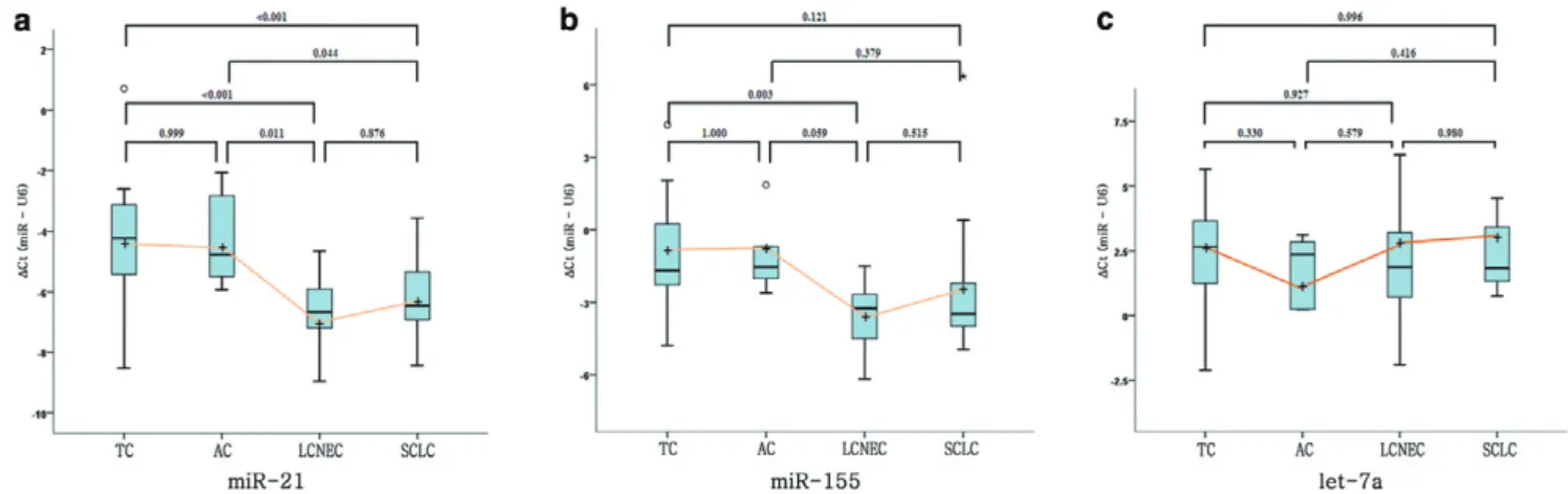
Figure 2 Box-and-whisker plots for comparison of mean normalized Ct (DCt) values of miR-21 (a), miR-155 (b), and let-7a (c) with histologic subtypes of 63 pulmonary neuroendocrine tumors. High-grade neuroendocrine carcinomas (large cell neuroendocrine carcinomas (LCNEC) and small cell lung carcinoma (SCLC)) showed statistically higher expression of miR-21 and miR-155 than carcinoid tumors (typical carcinoid (TC) and atypical carcinoid (AC)), whereas let-7a showed no significant difference.
© 2012 The Authors
The15ml reactions were incubated for 30 min at 16°C, 30 min at 42°C, 5 min at 85°C, and then held at 4°C. Quan- titative real time-PCR was performed using an ABI PRISM 7000 Sequence Detection System (Applied Biosystems, Foster City, CA, USA). The PCR reaction was performed in 10ml aliquots with 5 ml TaqMan Universal MixII, 0.5 ml TaqMan miRNA for each 2ml of miRNA. Reactions were incubated at 95°C for 10 min, followed by 40 cycles of ampli- fication at 95°C for 15 s and at 60°C for 1 min. All experi- ments were performed in duplicate to confirm reproducibility.
Data normalization
The threshold cycle data (Ct) and baseline were determined using auto settings. The Ct value was defined as the frac- tional cycle number at which the fluorescence passed the fixed threshold. U6 was used as the endogenous control to normalize expression levels. Following the amplification, the same threshold was set for analyzing all experiments to compare Ct values derived from different experiments.
The mean Ct values of each sample were determined from duplicate reactions and then normalized against the corresponding U6 Ct values, calculated as delta Ct (DCt = CtExperimental miRNA- CtU6). All data presented were nor- malized Ct (DCt) values.
Statistical analysis
In the first part of the experiments, one way analysis of variance was used to determine the association between the expression levels of three candidate miRNAs and his- tologic subtypes of pulmonary NE tumors. Turkey’s test was further used to compare the expression levels of a marker across multiple groups pairwisely. Contingency table and Student’s t-test were applied to determine the associations between expression levels of miRNAs and clinicopathologic characteristics. A P-value < 0.05 was considered statisti- cally significant.
In addition, receiver operating characteristic (ROC) curve analysis was undertaken using theDCt value of each miRNA to evaluate their ability in diagnosing the histologic subtype of pulmonary NE tumors. Using this approach, areas under the ROC curve (AUC) identified optimal sensitivity and specificity levels in distinguishing histologic subtypes of pulmonary NE tumors based on theDCt value. The AUCs with 95% confi- dence interval that did not include the 0.5-value signified that the miRNA had some ability to distinguish between the sub- types. To measure the agreement between diagnosis based on the cut-off value of miRNA expression level and histologic diagnosis on H&E section, the generalized kappa value was calculated.
All calculations were performed with SPSS version 18.0 for Windows (SPSS Inc., Chicago, IL, USA).
RESULTS
Differential expression of miRNAs according to the histologic subtype of pulmonary NE tumors
RNA was successfully extracted from all FFPE samples. All cases showed uniformly high miRNA quality, with Ct values for control U6 control RNA RT-PCR clustering within less than three amplification cycles for all samples. The Ct range for U6 control RNA varied from 25.1 to 27.9 cycles.
The results for the expression level of miR-21, miR-155, and let-7a according to the histologic subtype of the pulmo- nary NE tumors are shown in Table 2 and Figure 2. Results showed miR-21 to be the most consistently overexpressed in all subtypes of pulmonary NE tumors. The meanDCt values for miR-21 were-4.23 (95% confidence interval (CI): -4.93 to -3.53) in TC, -4.31 (95% CI: -5.55 to -3.07) in AC, -6.59 (95% CI: -7.28 to -5.89) in LCNEC, and -6.22 (95% CI:
-6.91 to -5.52) in SCLC. The expression level of miR-21 was clearly different between the histologic subtypes of pulmo- nary NE tumors (P < 0.001). In pairwise comparison, the expression level of miR-21 was significantly higher in high- Table 2 Correlation of normalized Ct values of miR-21, miR-155, and let-7a with histologic subtypes of 63 pulmonary neuroendocrine tumors
Diagnosis
Normalized Ct value of miRNA (95% confidence interval)
miR-21 miR-155 let-7a
TC -4.23 (-4.93 to -3.53) -1.13 (-2.08 to -0.18) 2.51 (1.61 to 3.41)
AC -4.31 (-5.55 to -3.07) -1.08 (-2.78 to -0.61) 0.95 (0.66 to 2.55)
LCNEC -6.59 (-7.28 to -5.89) -3.58 (-4.53 to -2.63) 2.12 (1.22 to 3.02)
SCLC -6.22 (-6.91 to -5.52) -2.65 (-3.60 to -1.70) 2.37 (1.47 to 3.27)
P value† <0.001 0.003 0.384
Carcinoid tumors -4.25 (-5.00 to -3.50) -1.12 (-1.99 to -0.25) 2.14 (1.17 to 3.10)
High-grade NE carcinomas -6.40 (-6.81 to -5.99) -3.11 (-3.79 to -2.44) 2.24 (1.68 to 2.81)
P value‡ <0.001 <0.001 0.832
†Difference in the mean normalized Ct values of miRNA among the four subtypes of pulmonary neuroendocrine tumors.
‡Difference in the mean normalized Ct values of miRNA between carcinoid tumors and high-grade neuroendocrine carcinomas.
AC, atypical carcinoid; LCNEC, large cell neuroendocrine carcinoma; NE, neuroendocrine; SCLC, small cell lung cancer, TC, typical carcinoid.
© 2012 The Authors
grade NE carcinomas (LCNECs and SCLCs) than in carcinoid tumors (TCs and ACs) (P< 0.001) (Table 2). Fur- thermore, the expression level of miR-21 was significantly different between TC and LCNEC (P< 0.001), between TC and SCLC (P< 0.001), between AC and LCNEC (P = 0.011) and between AC and SCLC (P= 0.044). However, no differ- ence was found between TC and AC (P = 0.999) and between LCNEC and SCLC (P= 0.876) (Fig. 2a).
For miR-155, the meanDCt values for miR-155 were -1.13 (95% CI: -2.08 to -0.18) in TC, -1.08 (95% CI: -2.78 to -0.61) in AC, -3.58 (95% CI: -4.53 to -2.63) in LCNEC, and -2.65 (95% CI: -3.60 to -1.70) in SCLC. The expression level of miR-155 was different between the histologic sub- types of pulmonary NE tumors (P= 0.003). In particular, the expression level of miR-155 was significantly higher in high- grade NE carcinomas than in carcinoid tumors (P< 0.001) (Table 1). Although a significant difference was found between TC and LCNEC (P = 0.003), no difference was found between TC and AC (P= 1.0), between TC and SCLC (P= 0.121), between AC and LCNEC (P = 0.059), between AC and SCLC (P= 0.379), and between LCNEC and SCLC (P= 0.515) (Fig. 2b).
On the other hand, substantial overlaps were seen between the let-7a distribution ranges among the histologic subtypes. The meanDCt values for let-7a were 2.51 (95% CI:
1.61 to 3.41) in TC, 0.95 (0.66 to 2.55) in AC, 2.12 (1.22 to 3.02) in LCNEC, and 2.37 (1.47 to 3.27) in SCLC (Table 1).
The expression level of let-7a was not statistically different between the histologic subtypes of pulmonary NE tumors (P= 0.384) (Fig. 2c).
Predicting value based on miRNAs expression in differential diagnosis of pulmonary NE tumors
Since we found that two miRNAs, miR-21 and miR-155, can differentiate carcinoid tumors from high-grade NE carcino- mas, the cut-off DCt values of miR21 and miR-155 were chosen from the ROC curve analysis in order to maximize sensitivity and specificity. The ROC curves with correspond- ing AUCs for miR-21 and miR-155 expression level in pul- monary NE tumors are shown in Figure 3.
The ROC curve analysis indicated that the DCt value of miR-21 for carcinoid tumors yielded an AUC of 0.846 (95%
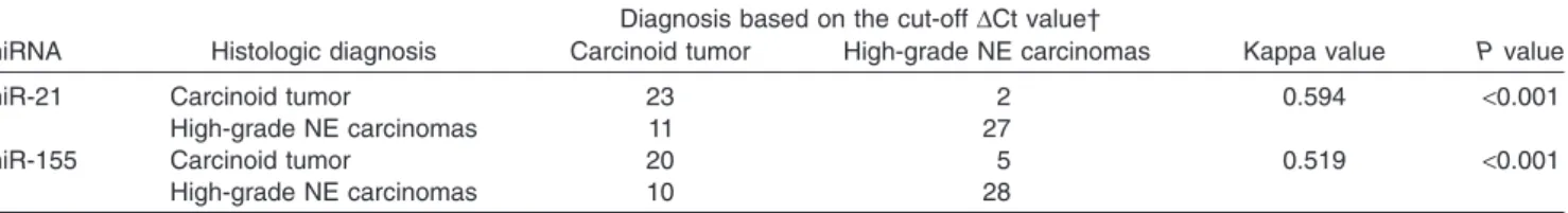
CI: -0.744 to -0.948). At the cut-off DCt value at -5.93 (carcinoid tumor3–5.93, high-grade NE carcinoma <-5.93), the expression level of miR-21 had 92.0% sensitivity and 71.1% specificity in discriminating carcinoid tumors from high-grade NE carcinomas. The kappa value for comparing the diagnosis based on the cut-offDCt value of miR-21 and histologic diagnosis on H&E section was 0.594 (Table 3).
For miR-155, the DCt value of miR-155 for carcinoid tumors yielded an AUC of 0.811 (95% CI:-0.696 to -0.925).
At the cut-off DCt value at -2.32 (carcinoid tumor 3-2.32, Figure 3 Receiver operating characteristic (ROC) curve analysis using normalized Ct (DCt) values of miRNA expression for discriminating carcinoid tumors and high-grade neuroendocrine (NE) carcinomas. (a)DCt value of miR-21 for carcinoid tumors yielded areas under the ROC (AUC) of 0.846 (95% CI:-0.744 to -0.948). At the cut-off DCt value at -5.93 (carcinoid tumor3-5.93, high-grade NE carcinoma <-5.93), the expression level of miR-21 had 92.0% sensitivity and 71.1% specificity in discriminating carcinoid tumors from high-grade NE carcinomas. (b) DCt value of miR-155 for carcinoid tumors yielded an AUC of 0.811 (95% CI: -0.696 to -0.925). At the cut-off DCt value at -2.32 (carcinoid tumor3-2.32, high-grade NE carcinoma <-2.32), the expression level of miR-155 had 80.0% sensitivity and 73.7% specificity in discriminating carcinoid tumors from high-grade NE carcinomas.
© 2012 The Authors
high-grade NE carcinoma <-2.32), the expression level of miR-155 had 80.0% sensitivity and 73.7% specificity in dis- criminating carcinoid tumors from high-grade NE carcino- mas. The kappa value for comparing the diagnosis based on the cut-offDCt value of miR-155 expression and histologic diagnosis on H&E section was 0.519 (Table 3).
Correlation of miRNAs expression with
clinicopathologic characteristics in carcinoid tumors and high-grade NE carcinomas
To further determine the clinical significance of dysregulated expressions of miRNAs, we evaluated expression levels of miR-21, miR-155, and let-7a in relation to various clinico- pathologic characteristics in carcinoid tumors and high-grade NE carcinomas, respectively. We found that the meanDCt value for miR-21 in carcinoid tumors with lymph node metasta- sis (–6.291 1.52) was quite different from that in carcinoid tumors without lymph node metastasis (–3.86 1 1.61) (P =
0.010). Other clinicopathologic characteristics were not corre- lated with the miR-21 expression level. On the other hand, the expression levels of miR-155 and let-7a showed no significant differences according to clinicopathologic characteristics in carcinoid tumors (Table 4). In addition, the expression levels of miR-21, miR-155 and let-7a showed no significant differ- ences according to further histopathological prognostic features beyond necrosis and mitoses, including nuclear pleo- morphism, aerogeneous spreading, palisading, papillary for- mation, and pseudoglandular patterns suggested by the 2004 WHO classificaition2in carcinoid tumors. In high-grade NE carcinomas, no significant correlations between the expres- sion levels of miR-21, miR-155, and let-7a and clinicopatho- logic characteristics were found in high-grade NE carcinomas (Table 5).
DISCUSSION
Since pulmonary NE tumors represent a distinct neoplastic entity that shares a wide spectrum of morphologic character- Table 3 Comparison of diagnosis based on the cut-offDCt value of miRNA expression and histologic diagnosis on hematoxylin and eosin section in 63 pulmonary neuroendocrine tumors
miRNA Histologic diagnosis
Diagnosis based on the cut-offDCt value†
Kappa value P value Carcinoid tumor High-grade NE carcinomas
miR-21 Carcinoid tumor 23 2 0.594 <0.001
High-grade NE carcinomas 11 27
miR-155 Carcinoid tumor 20 5 0.519 <0.001
High-grade NE carcinomas 10 28
†Cut-offDCt value of miR-21 = -5.93, cut-off DCt value of miR-155 = -2.32.
NE, neuroendocrine;DCt, normalized Ct.
Table 4 Correlation between miRNA expressions and clinicopathologic characteristics in 25 carcinoid tumors Clinicopathologic
characteristics
No. of patients (n= 25)
Normalized Ct value of miRNA (mean1 standard deviation)
miR-21 miR-155 let-7a
Sex
Female 10 -3.711 1.48 -0.301 2.79 1.591 1.81
Male 15 -4.611 1.96 -1.671 1.31 2.501 2.62
P value 0.229 0.172 0.353
Age (year)
250 14 -3.731 1.76 -0.921 1.78 1.651 2.79
>50 11 -4.911 1.73 -1.371 2.51 2.761 1.49
P value 0.108 0.606 0.247
Tumor size (cm)
22 8 -4.291 2.65 -1.391 2.58 1.851 3.25
2< and 23 15 -4.321 1.31 -0.971 2.01 2.181 1.70
>3 2 -3.551 2.09 -1.141 1.22 2.961 3.83
P value 0.858 0.908 0.841
Lymphovascular invasion
No 19 -4.101 1.96 -1.091 2.35 1.791 2.54
Yes 6 -4.721 1.24 -1.241 1.10 3.221 1.02
P value 0.475 0.883 0.197
Lymph node metastasis
No 21 -3.861 1.61 -0.861 2.12 1.881 2.44
Yes 4 -6.291 1.52 -2.491 1.55 3.461 1.08
P value 0.010 0.160 0.225
© 2012 The Authors
istics, these tumors have been a difficult challenge for patho- logists to diagnose. Additionally, there are no specific immunohistochemical or molecular markers that allow for separation of these tumors. In this study, miR-21 showed the best separation between high-grade NE carcinomas and carcinoid tumors in pairwise comparison. In addition, the expression level of miR-155 was significantly higher in high- grade NE carcinomas than in carcinoid tumors. However, most of the difficulties in diagnosing pulmonary NE tumors come from problems in distinguishing LCNEC from SCLC and TC from AC.3,4 Unfortunately, no difference was found between TC and AC and between LCNEC and SCLC in the expression levels of miR-21 and miR-155. Moreover, the kappa values for comparing the diagnosis based on the cut-off DCt values of miR-21 and miR-155 expression and histologic diagnosis on H&E section were 0.594 and 0.519, respectively.
These kappa values just made it into the moderate agreement category, which is not yet efficient for routine clinical applica- tion. Hence, it cannot be used as a substitute of accurate morphologic characterization of tumors, and careful correla- tion with routine morphology is essential in all cases. However, miRNAs are well preserved in formalin-fixed tissue, making them ideal candidates for molecular markers used in routinely processed material. Therefore, miR-21 and miR-155 could be employed as an adjunctive maker for the differential diagnosis of pulmonary NE tumors in surgical pathology specimens.
The availability of a reliable marker will be of value, not only for a more reliable and robust subclassification of cases, but also in identifying patients to determine an individual’s risk- benefit ratio in future clinical trials. Pulmonary NE tumors do not constitute a single, uniform entity but build up a variable
clinical behavior. In particular, confusion frequently arises regarding the biologic behavior of carcinoid tumors. Although carcinoid tumors are considered to have a low metastatic potential, lymph node metastases are present in 4–14% of TC cases and 35–64% in AC cases.20The 5- and 10-year disease- free survival rates of patients who had regional lymph node metastasis were 74% and 53%, respectively, compared with 96% and 84%, respectively, in those without lymph node metastasis.21,22Interestingly, in this study, four carcinoid tumor patients diagnosed with lymph node metastasis had high expression levels of miR-21, which would suggest the poten- tial practical use of miR-21 in future clinical management of patients with carcinoid tumors to predict lymph node metasta- sis. Knowledge of a specific molecular alteration associated with metastasis would enable assessment of the risk of tumor dissemination by investigating biopsy material prior to surgery.
Due to the relatively small number of events, however, further confirmation of these results is required.
In addition to diagnostic and clinical considerations, the biological implications of our findings in tumorigenesis of pulmonary NE tumors should also be discussed. Both miR-21 and miR-155 are well known as ‘oncomirs’.11In this study, miR-21 and miR-155 levels were high in almost all the samples, although with a moderate degree of variability. In this regard, it is intriguing that miR-21 and miR-155 overex- pression appears to be a common event in both carcinoid tumors and high-grade NE carcinomas. miR-21, located in chromosome 17q23.1, is the most commonly overexpressed oncogenic miRNA among human cancers from various organs, including the lung.23Although the detailed molecu- lar mechanisms underlying such oncogenic functions of Table 5 Correlation between miRNA expression and clinicopathologic characteristics in 38 high-grade neuroendocrine carcinomas Clinicopathologic
characteristics
No. of patients (n= 38)
Normalized Ct value of miRNA (Mean1 Standard deviation)
miR-21 miR-155 let-7a
Sex
Female 6 -5.671 1.63 -3.131 2.02 2.731 1.39
Male 32 -6.571 1.12 -3.111 2.09 2.141 1.78
P value 0.087 0.986 0.415
Age (year)
250 1 -4.87 -0.4 0.97
>50 37 -6.441 1.25 -3.211 2.00 2.281 1.72
P value · · ·
Tumor size (cm)
22 3 -6.381 0.72 -2.491 3.13 1.551 0.52
2< and 23 25 -6.581 1.26 -3.631 1.33 2.451 1.60
>3 10 -5.821 1.35 -2.731 1.19 1.811 2.29
P value 0.334 0.201 0.508
Lymphovascular invasion
No 16 -6.301 1.35 -3.141 1.34 2.611 1.21
Yes 22 -6.501 1.19 -3.091 2.57 2.821 1.43
P value 0.636 0.945 0.067
Lymph node metastasis
No 20 -6.351 1.18 -3.171 2.65 2.341 1.76
Yes 18 -6.461 1.36 -3.051 1.14 2.141 1.70
P value 0.802 0.854 0.730
© 2012 The Authors
the overexpressed miR-21 remain to be clarified, previous studies have identified several tumor suppressors as molecu- lar targets of miR-21, including tropomyosin 1, programmed cell death 4, PTEN, tissue inhibitor of metalloproteinase 3, and Maspin.24–26It is notable that a high miR-21 expression was associated with a poorer prognosis of pancreatic ductal adenocarcinoma patients.26In this study, we found a signifi- cant correlation between the overexpression of miR-21 and lymph node metastasis of carcinoid tumors. Furthermore, the expression level of miR-21 was significantly higher in high- grade NE carcinomas than in carcinoid tumors. Our finding would suggest that the downstream effects of miR-21 over- expression could be pivotal in establishing a complete malig- nant phenotype. It is known that miR-155 is a downstream effector of transforming growth factor b (TGF-b) induced RhoA expression, contributing to TGF-b-induced epithelial- mesenchymal transition, tight junction dissociation, cell migration, and invasion.27In this context, our study revealed that the expression level of miR-155 was higher in invasive, high-grade NE carcinomas than in carcinoid tumors. Further functional analysis of the target molecules regulated by miR-21 or miR-155 could also be of help to understand the tumorigenesis of pulmonary NE tumors.
In contrast, let-7 is considered to be a tumor suppressor gene that reduces cancer cell growth, and it is underex- pressed in malignant tumors.17 Enhanced let-7 expression leads to decreased high-mobility group A2 expression, a chromatin-remodeling protein that activates proinvasive and prometastatic genes.28Loss or reduction of let-7 also leads to Ras overexpression, thus, promoting cellular growth and contributing to tumorigenesis.29Unfortunately, the expression level of let-7a was not statistically different between the his- tologic subtypes of pulmonary NE tumors and showed no significant differences according to clinicopathologic factors both in carcinoid tumors and high-grade NE carcinomas in this study. These results can be explained by a previous study, in which the same miRNAs had oncogenic or tumor suppressor activity, depending on the tissue or cell type in which they were expressed.10,11
In conclusion, the miR-21 and miR-155 were differentially expressed according to the histologic subtypes of pulmonary NE tumors, and the expression level of miR-21 was signifi- cantly higher in carcinoid tumors with lymph node metastasis than in carcinoid tumors without lymph node metastasis. To the best of our knowledge, the present study is the first to examine the expression patterns of miRNAs as an adjunctive diagnostic tool or clinically relevant biomarkers for pulmonary NE tumors. Taking into account the great importance of miRNAs in tumors, we believe that our results will be of importance for both clinical researchers and those who design miRNA-based novel therapeutics. However, further studies with a large number of cases, especially ones includ- ing more ACs, and identification of additional miRNAs whose
aberrations can be specific to pulmonary NE tumors are needed to validate our findings.
ACKNOWLEDGMENTS
This work was supported by the Dong-A University research fund.
REFERENCES
1 Travis WD. Lung tumours with neuroendocrine differentiation.
Eur J Cancer 2009; 45 (Suppl 1): 251–66.
2 Travis WD, Brambilla E, Muller-Hermelink HK, Harris CC. World Health Organization International Histological Classification of Tumours. Pathology and Genetics of Tumors of the Lung, Pleura, Thymus and Heart. Lyon: IARC press, 2004.
3 den Bakker MA, Willemsen S, Grunberg K et al. Small cell carcinoma of the lung and large cell neuroendocrine carcinoma interobserver variability. Histopathology 2010; 56: 356–63.
4 Travis WD, Gal AA, Colby TV, Klimstra DS, Falk R, Koss MN.
Reproducibility of neuroendocrine lung tumor classification.
Hum Pathol 1998; 29: 272–9.
5 Xi Y, Nakajima G, Gavin E et al. Systemic analysis of microRNA expression of RNA extracted from fresh frozen and formalin- fixed, paraffin-embedded samples. RNA 2007; 13: 1668–74.
6 Bartel DP. MicroRNAs: Genomics, biogenesis, mechanism, and function. Cell 2004; 116: 281–97.
7 Caldas C, Brenton JD. Sizing up miRNAs as cancer genes. Nat Med 2005; 11: 712–4.
8 Lagos-Quintana M, Rauhurt R, Lendeckel W, Tuschl T. Identi- fication of novel genes coding for small expressed RNAs.
Science 2001; 294: 853–8.
9 Bandres E, Agirre X, Ramire N, Zarate R, Garcia-Foncillas J.
MicroRNAs as cancer players: Potential clinical and biological effects. DNA Cell Biol 2007; 26: 273–82.
10 Sassen S, Miska EA, Caldas C. MicroRNA: Implications for cancer. Virchows Arch 2008; 452: 1–10.
11 Esquela-Kerscher A, Slack FJ. Oncomirs-microRNAs with a role in cancer. Nat Rev Cancer 2006; 6: 259–69.
12 Yanaihara N, Caplen N, Bowman E et al. Unique microRNA molecular profiles in lung cancer diagnosis and prognosis.
Cancer Cells 2006; 9: 189–98.
13 Lee JH, Voortmam J, Dingemans AM et al. MicroRNA expres- sion and clinical outcome of small cell lung cancer. PLoS ONE 2011; 6: e21300.
14 Yu SL, Chen HY, Chang GC et al. MicroRNA signature predicts survival and relapse in lung cancer. Cancer Cells 2008; 13:
48–57.
15 Markou A, Tsaroucha EG, Kaklamanis L, Fotinou M, Georgoul- ias V, Lianidou ES. Prognostic value of mature microRNA-21 and microRNA-205 overexpression in non-small cell lung cancer by quantitative real-time RT-PCR. Clin Chem 2008; 54:
1696–704.
16 Volinia S, Calin GA, Liu CG et al. A microRNA expression sig- nature of human solid tumors defines cancer gene targets. Proc Natl Acad Sci USA 2006; 103: 2257–61.
17 Takamizawa J, Konishi H, Yanagisawa K et al. Reduced expres- sion of the let-7 microRNAs in human lung cancers in associa- tion with shortened postoperative survival. Cancer Res 2004;
64: 3753–6.
© 2012 The Authors
18 Lee CH, Chang HK, Lee HW, Shin DH, Roh MS. The interob- server variability for diagnosing pulmonary carcinoid tumor.
Korean J Pathol 2010; 44: 267–71.
19 Ha SY, Han J, Kim WS, Suh BS, Roh MS. Interobserver vari- ability in diagnosing high-grade neuroendocrine carcinoma of the lung and comparing it with the morphometric analysis.
Korean J Pathol 2012; 46: 42–7.
20 Beasley MB, Thunnissen FB, Brambila E et al. Pulmonary atypi- cal carcinoid: Predictors of survival in 106 cases. Hum Pathol 2000; 31: 1255–65.
21 Travis WD, Rush W, Flieder DB et al. Survival analysis of 200 pulmonary neuroendocrine tumors with clarification of criteria for atypical carcinoid and its separation from typical carcinoid.
Am J Surg Pathol 1998; 22: 934–44.
22 Bagheri R, Mashhadi MR, Haghi SZ, Sadrizadh A, Rezaeetalab F. Tracheobronchopulmonary carcinoid tumors: Analysis of 40 patients. Ann Thorac Cardiovasc Surg 2011; 17: 7–12.
23 Chan JA, Krichevsky AM, Kosik KS. MicroRNA-21 is an anti- apoptotic factor in human glioblastoma cells. Cancer Res 2005;
65: 6029–33.
24 Pezzolesi MG, Platzer P, Waite KA, Eng C. Differential expres- sion of PTEN-targeting microRNAs miR-19a and miR-21 in cowden syndrome. Am J Hum Genet 2008; 82: 1141–9.
25 Zhu S, Si ML, Wu H, Mo YY. MicroRNA-21 targets the tumor suppressor gene tropomyosin I (TPM1). J Biol Chem 2007; 282:
14328–36.
26 Nagao Y, Hisaoka M, Matsuyama A et al. Association of microRNA-21 expression with its targets, PDCD4 and TIMP3, in pancreatic ductal adenocarcinoma. Mod Pathol 2012; 25: 112–
21.
27 Kong W, Yang H, He L et al. McroRNA-155 is regulated by the transforming growth beta/Smad pathway and contributes to epi- thelial cell plasticity by targeting RhoA. Mol Cell Biol 2008; 28:
6773–84.
28 Rahman MM, Qian ZR, Wang EL et al. Frequent overexpres- sion of HMGA1 and 2 in gastroenteropancreatic neuroendocrine tumours and its relationship to let-7 downregulation. Br J Cancer 2009; 100: 501–10.
29 Johnson SM, Grosshans H, Shingara J et al. RAS is regulated by the let-7 microRNA family. Cell 2005; 120: 635–47.
© 2012 The Authors