Overexpression of MicroRNA-31 as a Promising Biomarker for Prognosis and Metastasis in Human Colorectal Cancer
Keun Hur1,2*
1Department of Biochemistry and Cell Biology, Cell and Matrix Research Institute, School of Medicine, Kyungpook National University, Daegu 41944, Korea
2BK21 Plus KNU Biomedical Convergence Program, Department of Biomedical Science, Kyungpook National University, Daegu 41944, Korea
Received May 19, 2016 /Revised June 12, 2016 /Accepted June 12, 2016
Colorectal cancer (CRC) is the third most common cancer and a leading cause of cancer-related death worldwide. Although several diagnostic and therapeutic tools have been available, CRC remains diffi- cult to complete cure because of insufficient understanding of the molecular mechanisms underlying this disease progression. MicroRNAs (miRNAs) are small non-coding RNA molecules that strongly regulate gene expression via transcriptional and translational control mechanisms. Many crucial cel- lular pathways are frequently disrupted in cancer development process due to dysregulation of sev- eral miRNAs. Mir-31 functions as an oncogene that modulate expression of multiple cancer related genes. Thus, we aimed to demonstrate clinical relevance of miR-31 in human CRC. Quantitative RT- PCR analysis of miR-31 expression was performed in 175 CRC tissues and 16 normal colonic mucosa (NM). Next, we investigated clinical significances of miR-31 expression in various clinicopathologic features in CRC patients cohort. Mir-31 was significantly up-regulated in CRC tissues compared to NM. In CRC tissues, miR-31 expression level was significantly elevated in a stage-dependent manner, which was associated with poor survival in patients with CRC. High miR-31 levels in CRC tissues significantly correlated with poor prognosis (hazard ratio [HR]=2.4) as well as distant metastasis (odds ratio [OR]=2.3). In conclusion, we identified clinical significance of miR-31 expression in CRC. High miR-31 expression may be clinically able to use as a biomarker for CRC prognosis and predicting metastasis.
Key words : Biomarker, colorectal cancer, metastasis, MicroRNA-31, prognosis
*Corresponding author
*Tel : +82-53-420-4821, Fax : +82-53-422-1466
*E-mail : [email protected]
This is an Open-Access article distributed under the terms of the Creative Commons Attribution Non-Commercial License (http://creativecommons.org/licenses/by-nc/3.0) which permits unrestricted non-commercial use, distribution, and reproduction in any medium, provided the original work is properly cited.
Journal of Life Science 2016 Vol. 26. No. 6. 705~710 DOI : http://dx.doi.org/10.5352/JLS.2016.26.6.705
서 론
대장암은 세계적으로 3번째로 흔한 암종이며, 암으로 인한 사망의 주요 원인이 되고 있다[21]. 현재 대장암의 진단방법으 로 contrast enhanced computed tomography (CT), positron emission tomography-CT (PET-CT), 그리고 magnetic reso- nance imaging (MRI)와 같은 이미지를 이용한 방법[4] 및 fe- cal occult-blood test, colonoscopy와 같은 방법이[29] 널리 사 용되고 있다. 이렇듯 대장암의 진단과 치료를 위해 다양한 방 법이 이용되고는 있으나 병의 진행에 관여하는 분자메커니즘 이해의 부족 때문에 여전히 완전한 치료는 어려운 실정이다.
MicroRNAs (miRNAs)는 단백질 정보를 코딩하고 있지 않 은 작은 RNA 단편이다. 이러한 miRNAs는 특정 유전자의 전
사과정 또는 번역과정을 조절하는 강력한 유전자 조절자로서 의 기능을 가진다[5, 15]. 암의 발생과정에서는 세포의 분화, 이동과 같은 중요한 세포신호 전달 과정에 손상이 빈번하게 발생 하는데, 다양한 miRNAs의 이상발현이 그 원인이 되고 있다. 암화 과정에서 mir-17-92 cluster의 경우 과발현 그리고 miR-107의 경우 저발현을 보이는데 이러한 이상 발현이 암세 포의 angiogenesis 과정을 조장한다고 알려져 있다[9, 32]. 뿐 만 아니라, miR-21과 miR-31의 과발현은 암세포의 침윤과 이 동에 직접적으로 영향을 준다는 보고가 있으며[1, 7], miR- 200c의 경우 암의 전이에 중요한 EMT 과정의 조절자로서의 역할을 한다는 것이 알려져 있다[13].
MiRNA-31은 발암과정에 관여하는 다양한 유전자를 조절 하여 암을 유발시키는 암유전자의 역할을 한다고 알려져 있다 [14, 16, 33]. 재미있게도 miR-31은 다양한 암종 별로 그 발현 양상이 다르다고 알려져 있다. 방광암[28, 30], 유방암[2, 24, 26], 식도암[19], 난소암[8], 전립선암[3, 10, 27] 등에서는 miR-31의 발현이 저해되어 있다고 보고 되어있는 반면에 대장 암[6, 20, 23, 25, 31], 폐암[17, 18], 구강암[22] 등에서는 miR-31 의 과발현이 보고 되어 있다.
따라서, 본 연구에서는 대량의 대장암 임상 조직 샘플에서 miR-31 발현 양상을 확인하고 그것의 임상적의의를 규명하고
Table 1. Clinicopathological characteristics of the colorectal can- cer patients
Characteristics CRC Tissue cohort
Patients n=175 Age (Years)
≤65 >65
74 101 Sex
Male Female
102 73 Adenocarcinoma histology
Adenocarcinoma*
Mucinous
162 10 Tumor location
Proximal Distal Rectum
58 49 68 TNM stage
I II III IV
38 53 44 40
*Adenocarcinoma includes well, moderately, and poorly differ- entiated.
자 하였다. 이를 위해 175례의 대장암 조직과 16례의 정상 대 장조직에서 miR-31의 발현을 분석하고, 나아가 임상병리적 요 인들과의 상관관계를 밝혀내고자 하였다. 뿐만 아니라, miR-31 발현이 실제 대장암 환자의 예후예측 및 전이예측이 가능한 바이오마커로서의 활용 가능성이 있는지 검증 하고자 하였다.
재료 및 방법
임상시료
본 연구는 2005년부터 2012년까지 일본 미에대학교병원에 서 대장암 수술을 받았던 175명의 환자를 대상으로 하였다.
분석은 환자로부터 얻은 대장암 조직 175례 및 정상 대장조직 16례를 대상으로 시행하였다. 이 연구는 일본 미에대학교병원 의 기관생명윤리위원회(IRB)에서 승인을 얻어 연구대상자의 동의하에 실시하였다. 총 175례의 대장암 환자군 연령의 중앙 값은 65세였으며, 남자가 102명 여자가 73명 이었다. T-stage, N-stage, M-stage를 이용한 임상적 병기는 미국 암협회 분류 (AJCC classification)에 따라 분류하였다. TNM 병기 분류에 따라, 1기 대장암 환자는 38명, 2기 대장암 환자는 53명, 3기 대장암 환자는 44명, 4기 대장암 환자는 40명 이었다. 대상 환자의 임상 및 병리학적 특성은 Table 1과 같다.
MicroRNA (miRNA)의 분리 및 정량분석
대장암 환자의 Formalin-fixed and paraffin- embedded (FFPE) 조직으로부터 miRNA를 분리하기 위해 RecoverAll Total Nucleic Acid Isolation Kits (Ambion Inc, Austin, TX)를 제조사의 프로토콜에 따라 사용하였다[13]. 추출된 miRNA는 Nanodrop (Nanodrop, USA)을 이용하여 정확한 농도를 측정 하였고, 260/280nm의 ratio를 확인한 후 ratio 1.8 이상인 순도 의 RNA만을 실험에 사용하였다.
MicroRNA (miRNA)의 발현분석
대장암 조직에서 miRNA 발현양을 분석하기 위해 Taqman real-time reverse transcription (RT)–PCR assays (Applied Biosystems, Foster city, CA)를 제조사의 프로토콜에 따라 사 용하였다[11, 12]. 20 ng의 total RNA를 역전사반응을 시켜 complementary DNA를 생성하였다. 그런 후, Taqman probe (Applied Biosystems)를 이용한 real-time RT-PCR을 시행하 여 miRNA의 증폭이 시작되는 Ct 값을 구하였다. 정량적 분석 을 위해 miR-16 Taqman probe를 이용한 real-time RT-PCR을 함께 시행하였으며, 2–ΔCt 분석방법을 이용해 실제 miRNA의 발현양을 계산하였다.
통계학적 분석
실험 결과의 통계적 처리는 student t-test, Kruskal-Wallis test 및 analysis of variance (ANOVA)로 하였으며, p<0.05를 유의한 차이의 한계로 하였다. 생존분석을 위해 Kaplan–
Meier analysis 와 log-rank test가 사용되었다. MiRNA의 발현 이 높은 군과 낮은 군으로 나누기 위해 receiver operating characteristic (ROC) curves를 이용하였다. 대장암 환자의 생 존율 분석은 Kaplan-Meier 생존분석법, 예후 분석은 Cox 비례 위험 모형(Cox proportional hazard model), 암전이 예측 분석 에는 로지스틱 회귀모형(Logistic regression model)을 각각 사용하였다. 모든 데이터는 평균 ± 표준 편차로 기술하였으며, 통계 프로그램은 Medcalc version 12.3 (Broekstraat, Belgium) 와 GraphPad Prism version 5.0 (GraphPad Software, San Diego, CA, USA)을 이용하였다.
결과 및 고찰
인체 대장암 조직에서 miR-31의 발현 양상
인체 대장암 조직에서 miR-31의 발현 양상을 확인하기 위 해서 16례의 정상 대장조직과 32례의 대장암 조직을 무작위로 선택하였다. 일반적으로 대장 조직에서 비교적 발현양이 일정 하다고 알려진 miR-16을 기준으로 miR-31의 상대적인 발현값 을 계산한 결과, Fig. 1에서 보여지는 바와 같이 정상 대장 조직 에 비해 대장암 조직에서 miR-31의 발현이 약 9배 정도 증가되 어 있었다. 뿐만 아니라 이러한 대장암 조직에서의 miR-31 발
Fig. 1. Expression status of miRNA-31 in normal colonic mucosa and colorectal cancer tissues. Quantitative analysis of miR-31 expression in normal colonic mucosa (NM) com- pared to colorectal cancer (CRC) tissues. The gray hori- zontal bar represents mean expression levels; ***p<
0.0001, student t-test; FC, fold change.
현의 증가는 student t-test 결과 통계적으로 매우 유의함 (p<0.0001)을 알 수 있었다.
대장암의 임상병리학적 예후인자와 miR-31 발현의 상관관 계
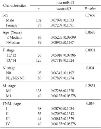
대장암 조직과 정상 대장조직 간의 비교에서 대장암 조직내 miR-31의 과발현 양상을 관찰할 수 있었다. 따라서 그 다음단 계로 175명 전체 대장암 환자의 대장암 조직을 분석하여 miR-31의 발현 정도와 임상병리학적 예후인자의 상관관계를 확인해 보았다(Table 2). 총 175명 대장암 환자의 성별, 나이, 종양의 장벽 침윤 정도(T-stage), 림프절 전이 유무(N-stage), 다른 장기로의 전이 유무(M-stage), 대장암의 병기(TNM stage) 와 같은 임상병리학적 인자의 차이에 따른 miR-31의 발현정도 를 분석해 보았다.
종양의 T-stage에 따른 miR-31의 발현은, T1/T2 (50례)에 비해 T3/T4 (125례)에서 유의하게 증가함을 알 수 있었다(p=
0.0001).
림프절 전이를 나타내는 N-stage에 따른 발현은 림프절 전 이가 없는 N0 환자군(95례)에 비해 림프절 전이가 있는 N1/
N2/N3 환자군(80례)에서 miR-31의 과발현을 관찰할 수 있었 다(p=0.004).
대장암의 병기에 따른 miR-31의 발현은, 1기(38례), 2기(53 례), 3기(44례), 4기(40례)로 병기가 증가할수록 그 발현의 정도 가 유의하게 증가 되는 양상을 보였다(p=0.016, Fig. 2).
따라서 이상의 결과를 종합해 볼 때, 대장암 조직에서 miR-31의 과발현은 종양의 진행 정도 및 악성도와 밀접한 연 관이 있을 것으로 사료된다.
Table 2. Comparison of clinicopathologic parameters
Characteristics hsa-miR-31
n mean±SD P value
Sex Male Female
102 73
0.07078±0.1333 0.07209±0.1091
0.7436
Age (Years) ≤Median >Median
86 89
0.05255±0.09099 0.08945±0.1467
0.0685 T stage
T1/T2 T3/T4
50 125
0.05018±0.09386 0.07718±0.1324
0.0001 N stage
N0 N1/N2/N3
95 80
0.06342±0.1197 0.07829±0.1274
0.004 M stage
M0 M1
135 40
0.07286±0.1328 0.06135±0.08278
0.2831 TNM stage
I II III IV
38 53 44 40
0.05780±0.1034 0.07067±0.1343 0.08812±0.1529 0.06135±0.08278
0.016
Fig. 2. Expression of miR-31 associates with the TNM stages. Gray dots illustrating tissue miR-31 levels in different TNM stages (I, II, III, and IV) of CRCs (n=175). Statistically significant difference was determined using Anova test.
MiR-31 과발현이 대장암 환자의 생존 및 예후에 미치는 영향
앞선 분석결과 miR-31의 과발현이 대장암의 발병과 밀접한 연관이 있었기에, miR-31의 발현 양상이 실제 대장암 환자의 생존률에 미치는 영향을 조사해 보았다. 우선 175명 대장암 환자를 대상으로 하여 그들의 생존기간을 바탕으로 Receiver Operating Characteristic (ROC) curve를 그리고 민감도(sen-
Fig. 3. Kaplan-Meier Survival Analysis. Overall survival analysis based on miR-31 expression. The p values were de- termined by log-rank test. All statistical tests were two- sided.
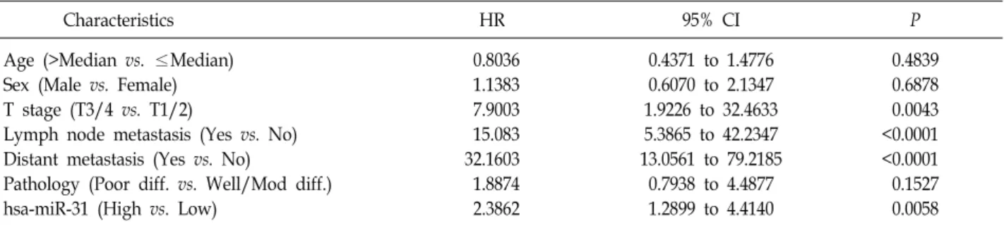
Table 3. Association between miRNA expression and CRC prognosis (Cox proportional hazards model)
Characteristics HR 95% CI P
Age (>Median vs. ≤Median) Sex (Male vs. Female) T stage (T3/4 vs. T1/2)
Lymph node metastasis (Yes vs. No) Distant metastasis (Yes vs. No)
Pathology (Poor diff. vs. Well/Mod diff.) hsa-miR-31 (High vs. Low)
0.8036 1.1383 7.9003 15.083 32.1603 1.8874 2.3862
0.4371 to 1.4776 0.6070 to 2.1347 1.9226 to 32.4633 5.3865 to 42.2347 13.0561 to 79.2185
0.7938 to 4.4877 1.2899 to 4.4140
0.4839 0.6878 0.0043
<0.0001
<0.0001 0.1527 0.0058 HR, hazard ratio; CI, confidence interval
Table 4. Association between miRNA expression and CRC distant metastasis (Logistic regression model)
Characteristics OR 95% CI P
Age (>Median vs. ≤Median) Sex (Male vs. Female) T stage (T3/4 vs. T1/2) N stage (N1/2/3 vs. N0) hsa-miR-31 (High vs. Low)
0.5137 1.0136 6.00E+00
25.1163 2.31E+00
0.2476 to 1.0659 0.4907 to 2.0937 1.7500 to 20.5717 7.3223 to 86.1511 1.1166 to 4.7843
0.0703 0.9708 0.0006
< 0.0001 0.0226 OR, odds ratio; CI, confidence interval
sitivity)와 특이도(specificity)가 최대가 되는 지점을 기준으로 miR-31 과발현 환자 그룹(75례)과 저발현 환자 그룹(98례)으 로 나누었다. 그런 후 Kaplan-Meier 생존분석을 시행해 보았 다(Fig. 3). 그 결과 log rank test상 p=0.0045으로 통계적으로 유의하게 miR-31 발현이 높은 대장암 환자 그룹이 miR-31 발 현이 상대적으로 낮은 환자 그룹에 비해 5년 생존율이 나쁜 것으로 나타났다.
실제 miR-31의 과발현이 대장암 환자의 생존율에 영향을 미치는 예후 인자가 될 수 있는지 알아보기 위해 Cox 비례위 험 모형(Cox proportional hazard model)을 이용해 임상병리 학적 예후인자(나이, 성별, T-stage, N-stage, M-stage, Patho- logic differentiation)와 miR-31 발현을 분석해 보았다(Table
3). 대장암 환자의 나이(p=0.4839), 성별(p=0.6878), Pathologic differentiation (p=0.1527)는 환자의 예후와 통계적으로 유의 성은 없었다. 하지만, T-stage (p=0.0043), N-stage (p<0.0001), M- stage (p<0.0001), 그리고 miR-31 과발현(p=0.0058)은 통계 적으로 유의하게 대장암 환자의 예후와 밀접한 연관성이 있는 것으로 판명되었다.
MiR-31 발현 양상을 이용한 대장암의 전이 예측 암전이는 대장암을 비롯한 대부분의 암환자의 생존율과 밀 접한 연관이 있는 것으로 알려져 있다. 따라서 본 연구진은 로지스틱 회귀모형(Logistic regression model)을 이용하여 대 장암 환자의 생존 및 예후와 유의한 연관성을 보인 miR-31의 과발현이 대장암의 전이를 예측할 수 있는지 분석해 보았다 (Table 4). 임상병리학적 인자들 중에서 대장암 환자의 나이 (p=0.0703)와 성별(p=0.9708)의 차이는 대장암 환자의 전이와 통계적으로 유의한 연관성을 보이지 않았다. 하지만, T-stage (p=0.0006), N-stage (p<0.0001), 그리고 miR-31 과발현(p=
0.0226)은 통계적으로 유의하게 대장암 환자의 원격전이와 연 관성이 있는 것으로 나타났다.
결론적으로, 대장암 조직에서 특이하게 과발현 되는 miR-31은 환자의 예후예측 및 전이예측에 도움이 되는 유용한 바이오마커로 활용될 수 있는 가능성이 높다고 사료된다. 따 라서 향후 대량의 대장암 조직뿐만 아니라 혈액과 같은 보다 다양한 임상샘플을 이용한 후속 검증연구가 이루어져야 할 것이다.
감사의 글
이 논문은 2014학년도 경북대학교 신임교수정착연구비에 의하여 연구되었음.
References
1. Asangani, I. A., Rasheed, S. A., Nikolova, D. A., Leupold, J. H., Colburn, N. H., Post, S. and Allgayer, H. 2008.
MicroRNA-21 (miR-21) post-transcriptionally downregulates tumor suppressor Pdcd4 and stimulates invasion, intra- vasation and metastasis in colorectal cancer. Oncogene 27, 2128-2136.
2. Augoff, K., McCue, B., Plow, E. F. and Sossey-Alaoui, K.
2012. miR-31 and its host gene lncRNA LOC554202 are regu- lated by promoter hypermethylation in triple-negative breast cancer. Mol. Cancer 11, 5.
3. Bhatnagar, N., Li, X., Padi, S. K., Zhang, Q., Tang, M. S.
and Guo, B. 2010. Downregulation of miR-205 and miR-31 confers resistance to chemotherapy-induced apoptosis in prostate cancer cells. Cell Death Dis. 1, e105.
4. Bipat, S., van Leeuwen, M. S., Ijzermans, J. N., Comans, E.
F., Planting, A. S., Bossuyt, P. M., Greve, J. W. and Stoker, J. 2007. Evidence-base guideline on management of color- ectal liver metastases in the Netherlands. Neth. J. Med. 65, 5-14.
5. Calin, G. A. and Croce, C. M. 2006. MicroRNA signatures in human cancers. Nat. Rev. Cancer 6, 857-866.
6. Cekaite, L., Rantala, J. K., Bruun, J., Guriby, M., Agesen, T. H., Danielsen, S. A., Lind, G. E., Nesbakken, A., Kallionie- mi, O., Lothe, R. A. and Skotheim, R. I. 2012. MiR-9, -31, and -182 deregulation promote proliferation and tumor cell survival in colon cancer. Neoplasia 14, 868-879.
7. Cottonham, C. L., Kaneko, S. and Xu, L. 2010. miR-21 and miR-31 converge on TIAM1 to regulate migration and in- vasion of colon carcinoma cells. J. Biol. Chem. 285, 35293- 35302.
8. Creighton, C. J., Fountain, M. D., Yu, Z., Nagaraja, A. K., Zhu, H., Khan, M., Olokpa, E., Zariff, A., Gunaratne, P. H., Matzuk, M. M. and Anderson, M. L. 2010. Molecular profil- ing uncovers a p53-associated role for microRNA-31 in in- hibiting the proliferation of serous ovarian carcinomas and other cancers. Cancer Res. 70, 1906-1915.
9. Dews, M., Homayouni, A., Yu, D., Murphy, D., Sevignani, C., Wentzel, E., Furth, E. E., Lee, W. M., Enders, G. H., Mendell, J. T. and Thomas-Tikhonenko, A. 2006. Augmenta- tion of tumor angiogenesis by a Myc-activated microRNA cluster. Nat. Genet. 38, 1060-1065.
10. Fuse, M., Kojima, S., Enokida, H., Chiyomaru, T., Yoshino, H., Nohata, N., Kinoshita, T., Sakamoto, S., Naya, Y., Nakagawa, M., Ichikawa, T. and Seki, N. 2012. Tumor sup- pressive microRNAs (miR-222 and miR-31) regulate molec- ular pathways based on microRNA expression signature in prostate cancer. J. Hum. Genet. 57, 691-699.
11. Hur, K., Toiyama, Y., Okugawa, Y., Ide, S., Imaoka, H.,
Boland, C. R. and Goel, A. 2015. Circulating microRNA-203 predicts prognosis and metastasis in human colorectal cancer. Gut. doi: 10.1136/gutjnl-2014-308737. [Epub ahead of print]
12. Hur, K., Toiyama, Y., Schetter, A. J., Okugawa, Y., Harris, C. C., Boland, C. R. and Goel, A. 2015. Identification of a metastasis-specific MicroRNA signature in human colorectal cancer. J. Natl. Cancer Inst. 107, 1-11.
13. Hur, K., Toiyama, Y., Takahashi, M., Balaguer, F., Nagasaka, T., Koike, J., Hemmi, H., Koi, M., Boland, C. R. and Goel, A. 2013. MicroRNA-200c modulates epithelial-to-mesen- chymal transition (EMT) in human colorectal cancer metastasis. Gut 62, 1315-1326.
14. Laurila, E. M. and Kallioniemi, A. 2013. The diverse role of miR-31 in regulating cancer associated phenotypes. Genes Chromosomes Cancer 52, 1103-1113.
15. Lewis, B. P., Burge, C. B. and Bartel, D. P. 2005. Conserved seed pairing, often flanked by adenosines, indicates that thousands of human genes are microRNA targets. Cell 120, 15-20.
16. Li, T., Luo, W., Liu, K., Lv, X. and Xi, T. 2015. miR-31 pro- motes proliferation of colon cancer cells by targeting E2F2.
Biotechnol. Lett. 37, 523-532.
17. Liu, C. J., Kao, S. Y., Tu, H. F., Tsai, M. M., Chang, K. W.
and Lin, S. C. 2010. Increase of microRNA miR-31 level in plasma could be a potential marker of oral cancer. Oral Dis.
16, 360-364.
18. Liu, C. J., Tsai, M. M., Hung, P. S., Kao, S. Y., Liu, T. Y., Wu, K. J., Chiou, S. H., Lin, S. C. and Chang, K. W. 2010.
miR-31 ablates expression of the HIF regulatory factor FIH to activate the HIF pathway in head and neck carcinoma.
Cancer Res. 70, 1635-1644.
19. Lynam-Lennon, N., Reynolds, J. V., Marignol, L., Sheils, O.
M., Pidgeon, G. P. and Maher, S. G. 2012. MicroRNA-31 modulates tumour sensitivity to radiation in oesophageal adenocarcinoma. J. Mol. Med (Berl). 90, 1449-1458.
20. Schee, K., Boye, K., Abrahamsen, T. W., Fodstad, O. and Flatmark, K. 2012. Clinical relevance of microRNA miR-21, miR-31, miR-92a, miR-101, miR-106a and miR-145 in color- ectal cancer. BMC Cancer 12, 505.
21. Siegel, R. L., Miller, K. D. and Jemal, A. 2016. Cancer sta- tistics, 2016. CA. Cancer J. Clin. 66, 7-30.
22. Siow, M. Y., Ng, L. P., Vincent-Chong, V. K., Jamaludin, M., Abraham, M. T., Abdul Rahman, Z. A., Kallarakkal, T.
G., Yang, Y. H., Cheong, S. C. and Zain, R. B. 2014.
Dysregulation of miR-31 and miR-375 expression is asso- ciated with clinical outcomes in oral carcinoma. Oral Dis.
20, 345-351.
23. Slaby, O., Svoboda, M., Fabian, P., Smerdova, T., Knoflick- ova, D., Bednarikova, M., Nenutil, R. and Vyzula, R. 2007.
Altered expression of miR-21, miR-31, miR-143 and miR-145 is related to clinicopathologic features of colorectal cancer.
Oncology 72, 397-402.
24. Sossey-Alaoui, K., Downs-Kelly, E., Das, M., Izem, L., Tubbs, R. and Plow, E. F. 2011. WAVE3, an actin remodeling pro- tein, is regulated by the metastasis suppressor microRNA,
초록:MicroRNA-31 과발현을 이용한 대장암의 예후예측 및 전이예측 바이오마커 발굴
허 근*
(경북대학교 의과대학 생화학세포생물학교실)
대장암은 세계적으로 3번째로 흔한 암종이며, 암으로 인한 사망의 주요 원인이 되고 있다. 비록 다양한 진단 방법이나 치료 방법이 이용되고는 있으나 병의 진행에 관여하는 분자메커니즘 이해의 부족 때문에 여전히 완전한 치료는 어려운 실정이다. 마이크로알엔에이는 단백질 정보를 코딩하고 있지 않은 작은 알엔에이 단편이다. 이러한 마이크로알엔에이는 특정 유전자의 전사과정 또는 번역과정을 조절하는 강력한 유전자 조절자로서의 기능을 가 진다. 암의 발생과정에서 중요한 세포신호 전달 과정의 손상이 빈번하게 발생 하는데, 다양한 마이크로알엔에이의 이상발현이 그 원인이 되고 있다. 마이크로알엔에이-31은 암유전자의 역할을 하며 발암과정에 관여하는 다양한 유전자를 조절한다고 알려져 있다. 따라서, 본 연구에서는 대장암에서 마이크로알엔에이-31 발현의 임상적의의를 규명하고자 하였다. 175례의 대장암 조직과 16례의 정상 대장조직에서 실시간 유전자 증폭장치를 이용하여 마이 크로알엔에이-31의 발현을 분석하고, 임상병리적 요인들과의 상관관계를 분석하고 임상적 유용성을 연구해 보았 다. 마이크로알엔에이-31은 정상조직에 비해 대장암 조직에서 과발현이 되어 있었다. 175례 대장암 조직을 이용한 분석에서 마이크로알엔에이-31의 발현은 병기의 진행 정도에 따라 발현이 증가 되고 있었으며, 실제 마이크로알 엔에이-31의 발현이 높은 대장암 환자군의 생존률이 그렇지 않은 환자군에 비해 통계적으로 유의하게 나쁜 것으 로 확인 되었다. Cox 비례위험 모형과 로지스틱 회귀 모형을 이용한 분석에서 마이크로알엔에이-31의 과발현이 직접적으로 대장암 환자의 예후 및 원발전이와 연관성이 있는 것이 확인 되었다. 따라서, 이상의 연구결과를 종합 해볼 때 대장암에서 과발현 된 마이크로알엔에이-31은 대장암 환자의 예후예측 및 전이예측 바이오마커로서의 활용 가능성이 높다고 볼 수 있다.
miR-31, during the invasion-metastasis cascade. Int. J.
Cancer 129, 1331-1343.
25. Sun, F., Wang, J., Pan, Q., Yu, Y., Zhang, Y., Wan, Y., Wang, J., Li, X. and Hong, A. 2009. Characterization of function and regulation of miR-24-1 and miR-31. Biochem. Biophys.
Res. Commun. 380, 660-665.
26. Valastyan, S., Reinhardt, F., Benaich, N., Calogrias, D., Szasz, A. M., Wang, Z. C., Brock, J. E., Richardson, A. L. and Weinberg, R. A. 2009. A pleiotropically acting microRNA, miR-31, inhibits breast cancer metastasis. Cell 137, 1032-1046.
27. Veerla, S., Panagopoulos, I., Jin, Y., Lindgren, D. and Hoglund, M. 2008. Promoter analysis of epigenetically con- trolled genes in bladder cancer. Genes Chromosomes Cancer 47, 368-378.
28. Wang, S., Li, Q., Wang, K., Dai, Y., Yang, J., Xue, S., Han, F., Zhang, Q., Liu, J. and Wu, W. 2013. Decreased expression of microRNA-31 associates with aggressive tumor pro- gression and poor prognosis in patients with bladder cancer.
Clin. Transl. Oncol. 15, 849-854.
29. Weizman, A. V. and Nguyen, G. C. 2010. Colon cancer screening in 2010: an up-date. Minerva. Gastroenterol. Dietol.
56, 181-188.
30. Wszolek, M. F., Rieger-Christ, K. M., Kenney, P. A., Gould, J. J., Silva Neto, B., Lavoie, A. K., Logvinenko, T., Libertino, J. A. and Summerhayes, I. C. 2011. A MicroRNA expression profile defining the invasive bladder tumor phenotype.
Urol. Oncol. 29, 794-801.
31. Xu, X. M., Qian, J. C., Deng, Z. L., Cai, Z., Tang, T., Wang, P., Zhang, K. H. and Cai, J. P. 2012. Expression of miR-21, miR-31, miR-96 and miR-135b is correlated with the clinical parameters of colorectal cancer. Oncol. Lett. 4, 339-345.
32. Yamakuchi, M., Lotterman, C. D., Bao, C., Hruban, R. H., Karim, B., Mendell, J. T., Huso, D. and Lowenstein, C. J.
2010. P53-induced microRNA-107 inhibits HIF-1 and tumor angiogenesis. Proc. Natl. Acad. Sci. USA 107, 6334-6339.
33. Zheng, W., Liu, Z., Zhang, W. and Hu, X. 2015. miR-31 func- tions as an oncogene in cervical cancer. Arch. Gynecol. Obstet.
292, 1083-1089.