Vol. 24, No. 4, 2012 491
Letter to the Editor
Received August 9, 2012, Revised September 3, 2012, Accepted for publication September 9, 2012
Corresponding author: Moon-Kyun Cho, M.D., Department of Dermatology, Soonchunhyang University Seoul Hospital, 59 Daesagwan-ro, Yongsan-gu, Seoul 140-743, Korea. Tel: 82-2-709-9368, Fax: 82-2-709-9374, E-mail: [email protected]
This is an Open Access article distributed under the terms of the Creative Commons Attribution Non-Commercial License (http://
creativecommons.org/licenses/by-nc/3.0) which permits unrestricted non-commercial use, distribution, and reproduction in any medium, provided the original work is properly cited.
ing the medication.
REM, first described by Steigleder, is a rare cutaneous disease, characterized by persistent erythema in a reticular pattern and/or confluent erythematous papules and pla- ques, with a predilection of location on the central area of the chest or on the upper back; and rarely on the arms, face and abdominal region3-5. It predominantly afflicts middle-aged women3-5. The typical histopathology is interstitial deposits of mucin in the dermis. The patho- genesis remains unclear3,4.
Antimalarial drugs are the treatment of choice for REM3-5. Hydroxychloroquine, a derivative of chloroquine, is fre- quently used as the first-line treatment, and it always results in rapid improvement within about a month of starting the treatment; however, recurrence is also common5. The present patient responded well to hydroxy- chloroquine. Unfortunately, he had hypomnesis after administrated the drug. Based on the dosage-adverse effects relationship, we considered that hydroxychloro- quine is responsible for the hypomnesis, but the mechani- sm is unclear.
Although such a condition is very rare and the present
patient had finally recovered, practitioners should be aware of it as it may have a negative impact on patient's quality of life, and even lead severe problem.
REFERENCES
1. Kuhn A, Ruland V, Bonsmann G. Cutaneous lupus erythema- tosus: update of therapeutic options part I. J Am Acad Derma- tol 2011;65:e179-193.
2. Gül U, Cakmak SK, Kiliç A, Gönül M, Bilgili S. A case of hydroxychloroquine induced pruritus. Eur J Dermatol 2006;
16:586-587.
3. Kreuter A, Scola N, Tigges C, Altmeyer P, Gambichler T. Cli- nical features and efficacy of antimalarial treatment for reticul- ar erythematous mucinosis: a case series of 11 patients. Arch Dermatol 2011;147:710-715.
4. Karadag AS, Simsek GG. Reticular erythematous mucinosis syndrome with telangiectasias. Indian J Dermatol Venereol Leprol 2010;76:86.
5. Suárez-Amor O, Pérez-Bustillo A, González-Morán MA, Ra- mírez-Santos A, Rodríguez-Prieto MA. Reticular erythematous mucinosis: partial response to treatment with topical tacroli- mus. Actas Dermosifiliogr 2010;101:105-106.
http://dx.doi.org/10.5021/ad.2012.24.4.491
Tripartite Motif-Containing 29 Expression in Squamous Cell Carcinoma
Moon-Kyun Cho, M.D.
Department of Dermatology, College of Medicine, Soonchunhyang University, Seoul, Korea
Dear Editor:
Tripartite motif-containing 29 (TRIM29), also known as the ataxia telangiectasia group D complementing gene, is reduced in certain cancers by some reports, although, others report higher expression of TRIM29 in several cancers.
While the exact functions of TRIM proteins have yet to be
specified, they undergo viral replication, and participate in signal transduction and development in many human diseases, especially cancer.
A few members of the TRIM family are associated with cellular growth and processes, such as transcriptional regulation, cell growth, apoptosis, development and onco- genesis1. Several TRIM proteins play many roles in the
492 Ann Dermatol Letter to the Editor
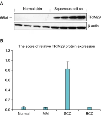
Fig. 1. (A) TRIM29 protein was expressed on squamous cell carci- nomal, normal skin tissue by western blot analysis. (B) The score of relative TRIM29 expression amount in western blotting.
TRIM29: tripartite motif-containing 29, MM: malignant mela- noma, SCC: squamous cell carcinoma, BCC: basal cell carci- noma.
cellular processes, including the development and growth of diseases, but the physiological and pathological functions of TRIM29 have not been confirmed; while its presence in the cell membrane has been observed2.
TRIM29 is typically expressed in the placenta, lung, thymus, prostate, testis, and colon, but not in the heart, brain, skeletal muscle, pancreas, spleen, ovary, or the small intestine, as measured by Northern blot analysis3. There have not been many studies on TRIM29, but it is assessed that TRIM29 expression is suppressed in breast and prostate cancer4-6.
In a study by Schlomm et al.7, cDNA microarray analysis on prostatic cancer cells and noncancerous prostatic cells conducted using laser microdissection, resulted in 216 different genes, with TRIM29 as one of the downregulated genes.
Similarly, Ernst et al.4 reported that TRIM29 was shown to be downregulated.
Liu et al.6 reported that TRIM29 functions as a tumor suppressor in nontumorigenic breast cells and invasive estrogen receptor positive breast cancer using cDNA microarray analysis.
TRIM29's functions may differ depending on the cell type, but there have not been any studies on the role of TRIM29
on tumorigenesis.
TRIM29 over-expresses in lung, bladder, colorectal, ovarian, gastric, pancreatic and endometrial cancers and in multiple myeloma, and recent studies show that TRIM29 is associated with tumor invasion and lymph node metastasis.
A recent report correlated TRIM29 expression in gastric cancer and poor histological grade, large tumor size, extent of tumor invasion, and lymph node metastasis8. The Institutional Review Board of Seoul Soonchunhyang University Hospital reviewed and approved this research protocol, which involved the use of tissue samples.
We analyzed a total of 6 normal skin tissues and 18 malignant skin tumor tissues, including 6 malignant mela- noma (MM), 6 squamous cell carcinoma (SCC), and 6 basal cell carcinoma (BCC), using Western blot and immunohistochemistry.
The human MM cell line G361 served as a positive control for the TRIM29 expression.
The human MM cell line G361 was not expressed for TRIM29 protein.
In this study, TRIM29, which indicates the metastatic potentiality in gastric cancer, was measured in BCC, SCC and MM cancers, using Western blot and immuno- histologic method. The results show that the expression of TRIM29 was not expressed in 6 cases of BCC with slow growth and poor metastasis. In 6 cases of SCC, which is aggressive and locally invasive, but not metastatic cancer, the expression of TRIM29 was strongly positive (Fig. 1A).
In the 6 cases of MM, which is invasive and has distant sites metastatic potential through hematogenous and lymphagenous paths, TRIM29 protein was not expressed.
In all six MM and BCC, TRIM29 was not expressed (data not shown). The amount of expression in Western blotting is displayed graphically (Fig. 1B).
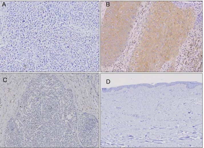
Immunohistochemical study showed that the staining pattern of TRIM29 in normal human skin tissues mirrored those of a Western blot analysis (Fig. 2).
The exact function and mechanism of TRIM29 during tumor genesis have not been confirmed.
The possible roles and mechanisms of TRIM29 protein expression in malignant skin cancers will be examined in additional studies.
REFERENCES
1. Quaderi NA, Schweiger S, Gaudenz K, Franco B, Rugarli EI, Berger W, et al. Opitz G/BBB syndrome, a defect of midline development, is due to mutations in a new RING finger gene on Xp22. Nat Genet 1997;17:285-291.
2. Reymond A, Meroni G, Fantozzi A, Merla G, Cairo S, Luzi L,
Vol. 24, No. 4, 2012 493
Letter to the Editor
Fig. 2. Immunohistochemical analysis of tripartite motif-containing 29 (TRIM29) expression in (A) malignant melanoma, (B) squamous cell carcinoma, (C) basal cell carcinoma, and (D) normal skin tissue. Only squamous cell carcinoma displayed TRIM29 expression.
(A, B, C: ×200, D: ×100).
et al. The tripartite motif family identifies cell compartments.
EMBO J 2001;20:2140-2151.
3. Hosoi Y, Kapp LN. Expression of a candidate ataxia- telangiectasia group D gene in cultured fibroblast cell lines and human tissues. Int J Radiat Biol 1994;66(6 Suppl):
S71-76.
4. Ernst T, Hergenhahn M, Kenzelmann M, Cohen CD, Bonrouhi M, Weninger A, et al. Decrease and gain of gene expression are equally discriminatory markers for prostate carcinoma: a gene expression analysis on total and micro- dissected prostate tissue. Am J Pathol 2002;160:2169-2180.
5. Nacht M, Ferguson AT, Zhang W, Petroziello JM, Cook BP, Gao YH, et al. Combining serial analysis of gene expression and array technologies to identify genes differentially
expressed in breast cancer. Cancer Res 1999;59:5464-5470.
6. Liu J, Welm B, Boucher KM, Ebbert MT, Bernard PS. TRIM29 functions as a tumor suppressor in nontumorigenic breast cells and invasive ER+ breast cancer. Am J Pathol 2012;
180:839-847.
7. Schlomm T, Luebke AM, Sültmann H, Hellwinkel OJ, Sauer U, Poustka A, et al. Extraction and processing of high quality RNA from impalpable and macroscopically invisible prostate cancer for microarray gene expression analysis. Int J Oncol 2005;27:713-720.
8. Kosaka Y, Inoue H, Ohmachi T, Yokoe T, Matsumoto T, Mimori K, et al. Tripartite motif-containing 29 (TRIM29) is a novel marker for lymph node metastasis in gastric cancer.
Ann Surg Oncol 2007;14:2543-2549.