• 검색 결과가 없습니다.

Squamous Cell Carcinoma of the Gallbladder Presenting with a Cholecystoduodenal Fistula

N/A
N/A
Protected

Academic year: 2021

Share "Squamous Cell Carcinoma of the Gallbladder Presenting with a Cholecystoduodenal Fistula"

Copied!
5
0
0

로드 중.... (전체 텍스트 보기)

전체 글

(1)

© Copyright

Keimyung University School of Medicine 2016

Gallbladder cancer (GBC) is the most common primary hepatobiliary carcinoma and the sixth most common gastrointestinal malignancy.

Adenocarcinoma accounts for the vast majority of GBCs (80–95%), whereas squamous cell carcinoma constitutes only 0–3.3% of GBCs. A 69-year-old man was suspected to have GBC with a cholecystoduodenal fistula on an abdominal computed tomography scan. He underwent esophagogastroduodenoscopy, which revealed that the duodenum was obstructed by the mass. Duodenal and biliary stents were successfully placed using endoscopic retrograde cholangiopancreatography. Pathology obtained from the duodenum revealed the mass to be a squamous cell carcinoma.

Key Words: Cholecystoduodenal fistula, Endoscopic retrograde cholan- giopancreatography, Gallbladder, Squamous cell carcinoma

Introduction

Gallbladder cancer (GBC) is the most common malignancy of the biliary tract and accounts for 1–2% of all malignancies in Korea [1].

Although it is a rare malignancy, most GBCs are incurable due to the anatomical position of the gallbladder, rapid progression and early metastasis [2]. Adenocarcinoma accounts for the vast majority of GBCs (80–95%), whereas squamous cell carcinoma constitutes only 0–3.3% of GBCs [3]. Herein, we report a case of squamous cell carcinoma of the gallbladder presenting with a cholecystoduodenal fistula.

Received: February 11, 2016 Revised: May 19, 2016 Accepted: June 01, 2016

Corresponding Author: Kyong Joo Lee, M.D., Department of Internal Medicine,

Yonsei University Wonju College of Medicine, 20 Ilsan-ro, Wonju 26426, Korea

Tel: +82-33-741-0507 E-mail: [email protected]

• The authors report no conflict of interest in this work.

Department of Internal Medicine, Pathology

1

, Yonsei University Wonju College of Medicine, Wonju, Korea

Seung Kook Cho, M.D., Young Bean Ko, M.D., Soon Chang Park, M.D., Sang Jun Lee, M.D., Jae Hyun Kim, M.D., Mee Yon Cho

1

, M.D., Jae Woo Kim, M.D., Kyong Joo Lee, M.D.

Squamous Cell Carcinoma of the Gallbladder Presenting with a

Cholecystoduodenal Fistula

(2)

Case Report

A 69-year-old poultry worker presented to a local hospital with anorexia, abdominal discomfort, and weight loss of 15 kg over the past month. Partial obstruction of the duodenum was found using esophagogastroduodenoscopy (EGD), and he was transferred to our department for further evaluation.

There was no family history of cancer, but he was a chronic alcoholic who drank daily and had a 50-pack- year smoking history. He had no medical history, except for controlled essential hypertension. He was chronically ill-looking, and his vital signs were stable.

Vague epigastric tenderness was found on the physical examination. Initial laboratory tests were as follows: white blood cell count 15,840/mm 3 , C-reactive protein 16.0 mg/dL, alkaline phosphatase 665 U/L, gamma-glutamyl transpeptidase 102 U/L, total bilirubin 1.32 mg/dL, carcinoembryonic antigen 7.45 ng/mL, and carbohydrate antigen 19-9 67.6 U/mL. Chest and abdomen x-rays were normal except for mild pleural effusion on the left lung.

Abdominal computed tomography and magnetic resonance imaging scans showed a heterogeneous mass (6 × 6 cm) in liver segments V and VI, with an

irregular surface, which arose from the gallbladder (GB) without evidence of gallstones and was invading into the adjacent duodenum, common hepatic duct (CHD) and proximal common bile duct, resulting in associated intra-hepatic duct dilatation and a cholecystoduodenal fistula (Fig. 1). Positron emission tomography showed intense hyper-metabolism in the GB with a maximum standardized uptake value of 9.2, which was invading into the adjacent duodenum and liver (T4), and no evidence of lymph node involvement or distant metastasis (N0M0) was found.

EGD showed an encircling mass at the duodenum that was obstructing 90% of the duodenal lumen, and a biopsy was performed using biopsy forceps (Fig.

2A). Under fluoroscopic guidance, a 20 mm × 6 cm uncovered metal duodenal stent (Taewoong Medical Co., Ltd., Goyang-si, Korea) was successfully placed into the duodenum for symptom palliation (Fig. 2B).

S u b s e q u e n t l y , e n d o s c o p i c r e t r o g r a d e cholangiopancreatography was performed through the duodenal stent with a side-viewing duodenoscope (TJF-260; Olympus, Tokyo, Japan) and a 10 mm × 6 cm uncovered metal biliary stent (Taewoong Medical Co., Ltd., Goyang-si, Korea) was inserted across the obstruction in the CHD (Fig. 2C&D). The histologic

Fig. 1. Abdominal computed tomography scan. (A) Heterogeneous mass at the gallbladder. (B) Cholecystoduodenal fistula.

A B

(3)

examination confirmed the diagnosis of moderately differentiated squamous cell carcinoma of the GB (Fig. 3). The symptoms were alleviated after the placement of duodenal and biliary stents. Afterwards, the patient refused any active treatment including chemotherapy or radiation therapy; he has now been followed up for two months on an outpatient basis and is in fair clinical condition.

Discussion

In this case, the patient presented with anorexia and abdominal discomfort and was diagnosed with

advanced squamous cell carcinoma of the GB. Pure squamous cell carcinoma of gallbladder accounts for 0 - 3.3% of GBCs [3]. Although the patient did not undergo surgery, the specimens obtained from duodenal mass showed pure squamous cell carcinoma.

The GB wall’s innermost surface is lined by a single layer of columnar cells; the lamina propria, a muscular layer, an outer perimuscular layer and serosa lie beneath the epithelia [4,5]. Since a normal GB does not contain squamous epithelium, the origin of squamous cell carcinoma in the GB becomes a question. Three theories of the origin of squamous cell carcinoma in the GB have been suggested: (1) Fig. 2. Endoscopic findings. (A) Biopsy performed on an encircling mass at the duodenum. (B) Duodenal stent placed into the duodenum. (C) Obstructed common hepatic duct. (D) Biliary stent inserted across the obstruction in the common hepatic duct.

A

C

B

D

(4)

from malignant transformation of heterotopic squamous epithelium; (2) from preexisting metaplastic squamous epithelium; and (3) from squamous metaplasia of adenocarcinoma [6].

However, the first two theories have their limitations in that (1) there is no documented report of the presence of congenital heterotopic squamous epithelium, and (2) squamous metaplasia of columnar epithelium does not exist in non-neoplastic conditions including chronic cholecystitis and chole-lithiasis [7]. Therefore, squamous metaplasia of adenocarcinoma is the most plausible theory as of now. According to this theory, an initial adeno-carcinoma undergoes squamous metaplasia to form adenosquamous carcinoma, and the metaplastic component grows rapidly and replaces the adeno-carcinoma component to make pure squamous cell carcinoma [6,8]. This may explain why adeno-squamous carcinoma and pure squamous cell carcinoma have resulted in more advanced stage and a poorer prognosis than adenocarcinoma in several retrospective studies [2,3,9].

According to recent research, CD109 was negative in all normal gallbladder and adenocarcinoma tissues, while CD109 positive cells were found in 86.7% of

pure squamous cell carcinoma and 91.7% of adenosquamous carcinoma [10]. Although this research concluded that CD109 may be involved in the pathogenesis of gallbladder squamous cell carcinomas and present as a novel marker for therapeutic intervention, the molecular mechanisms underlying the genesis and progression of GBCs are still unclear. Also, there are differences in the clinical courses of adenosquamous carcinoma and pure squamous cell carcinoma, in that adenosquamous carcinoma is aggressive and metastasizes widely, while pure squamous cell carcinoma is localized and rarely metastasizes, even when the tumor forms a huge mass [6,9,11]. In this case, the tumor mass formed a huge mass and invaded the duodenum and the bile duct. As a result, cholecystoduodenal fistula was found and there was no evidence of distant metastasis. Although we cannot confirm the presenting tumor is adenosquamous carcinoma or pure squamous cell carcinoma due to the limited nature of punch biopsy, we have assumed that our case is pure squamous cell carcinoma since a huge mass was localized and did not metastasize.

Surgical resection is the primary treatment for Fig. 3. Squamous cell carcinoma with extensive keratinization including pearl formation. (A) H&E stain × 100. (B) H&E stain × 400.

A B

(5)

GBCs, and systemic chemotherapy provides modest benefit in the treatment of advanced GBCs [12,13].

Unfortunately, due to the anatomical position of the gallbladder, rapid progression and early metastasis, most patients present with advanced stage disease, in which GBCs are unresectable or unable to undergo complete resection. Therefore, most treatment plans place emphasis on pain control and palliative care. In our case, endoscopic intervention assisted the patient in coping with cancer. Palliative surgery can also be employed to improve quality of life by easing pain or other symptoms caused by advanced stage cancer, and other various modalities and approach to hospice care should be further studied to pursue better quality of life.

References

1. Jung KW, Won YJ, Kong HJ, Oh CM, Cho H, Lee DH, et al. Cancer statistics in Korea: incidence, mortality, survival, and prevalence in 2012. Cancer Res Treat 2015;47:127-41.

2. Hundal R, Shaffer EA. Gallbladder cancer: epidemiology and outcome. Clin Epidemiol 2014;6:99-109.

3. Song HW, Chen C, Shen HX, Ma L, Zhao YL, Zhang GJ, et al. Squamous/adenosquamous carcinoma of the gallbladder: analysis of 34 cases and comparison of clinicopathologic features and surgical outcomes with adenocarcinoma. J Surg Oncol 2015;112:677-80.

4. Miyazaki K, Tsutsumi N, Kitahara K, Mori M, Sasatomi E, Tokunaga O, et al. Hepatopancreatoduodenectomy for squamous and adenosquamous carcinoma of the gallbladder. Hepatogastroenterology 1995;42:47-50.

5. Saito A, Noguchi Y, Doi C, Mukai K, Fukuzawa K, Yoshikawa T, et al. A case of primary adenosquamous/

squamous cell carcinoma of gallbladder directly invaded duodenum. Hepatogastroenterology 1999;46:204-7.

6. Roppongi T, Takeyoshi I, Ohwada S, Sato Y, Fujii T, Honma M, et al. Minute squamous cell carcinoma of the gallbladder: a case report. Jpn J Clin Oncol 2000;30:43- 5.

7. Kumar A, Singh MK, Kapur BM. Synchronous double malignant tumors of the gall bladder. A case-report of squamous cell carcinoma with an angiosarcoma. Eur J Surg Oncol 1994;20:63-7.

8. Cubertafond P, Gainant A, Cucchiaro G. Surgical treatment of 724 carcinomas of the gallbladder. Results of the French Surgical Association Survey. Ann Surg 1994;219:275-80.

9. Roa I, Ibacache G, Munoz S, de Aretxabala X.

Gallbladder cancer in Chile: pathologic characteristics of survival and prognostic factors: analysis of 1,366 cases.

Am J Clin Pathol 2014;141:675-82.

10. Dong F, Lu C, Chen X, Guo Y, Liu J. CD109 is a novel marker for squamous cell/adenosquamous carcinomas of the gallbladder. Diagn Pathol 2015;10:137.

11. Karasawa T, Itoh K, Komukai M, Ozawa U, Sakurai I, Shikata T. Squamous cell carcinoma of gallbladder- report of two cases and review of literature. Acta Pathol Jpn 1981;31:299-308.

12. Oohashi Y, Shirai Y, Wakai T, Nagakura S, Watanabe H, Hatakeyama K. Adenosquamous carcinoma of the gallbladder warrants resection only if curative resection is feasible. Cancer 2002;94:3000-5.

13. Kalayarasan R, Javed A, Sakhuja P, Agarwal AK.

Squamous variant of gallbladder cancer: is it different

from adenocarcinoma? Am J Surg 2013;206:380-5.

수치

Fig. 1. Abdominal computed tomography scan. (A) Heterogeneous mass at the gallbladder

참조

관련 문서

Distinction of basaloid squamous cell carcinoma from adenoid cystic and small cell undifferentiated carcinoma by immunohistochemistry. Basaloid squamous cell carcinoma of the

(A, B) The follow-up computed tomography scan reveals spontaneous regression of the lacrimal sac squamous cell carcinoma 15 months after the surgical biopsy (A: coronal view, B:

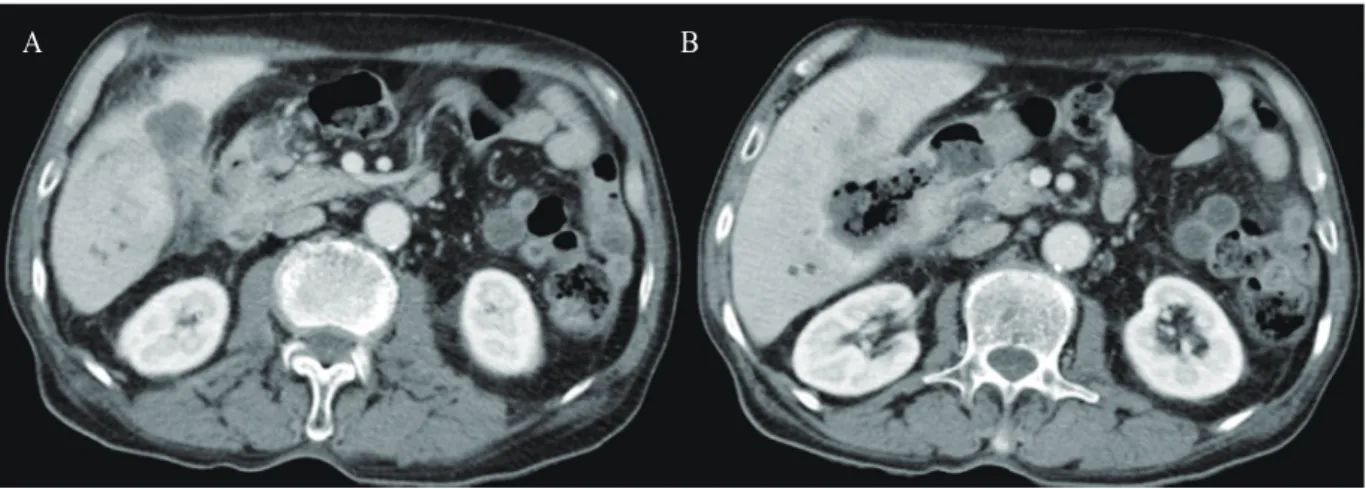
The left renal tumor was misdiagnosed as a renal abscess but finally diagnosed as squamous cell carcinoma metastasized from the intrahepatic bile duct.. The patient expired because

Results: Analyses on each tumor marker indicated that squamous cell carcinoma in the head and neck region had statistically significant correlation with p53, SCC-Ag(TA-4),

Basaloid squamous carcinoma of the uterine cervix is a rare tumor type characterized by an ulcerated, infiltrating growth pattern; nests or cords of small basaloid cells;

We herein report on an interesting case of vulval metastasis from squamous cell carcinoma of the cervix with an unusual clinical manifestation resembling lymphagioma

Acantholytic squamous cell carcinoma is a well-defined variant of squamous cell cancer in which significant portions of the neoplastic proliferation show a pseudoglandular or tubular

Extensive squamous metaplasia simulating squamous cell carcinoma in benign breast papillo- matosis.. Significance of squamous cells in fine nee- dle aspiration