이에 혈액종양을 진단하는데 최근 규명된 유전자 검사를 포함한 검사 지침 개정의 필요성이 대두되었다. 대한진단혈액학회의 표준 화위원회에서는 2016년에 발간된 혈액종양 초진단 검사항목 지침 [2]을 개정하고자 하였다. 초진단 검사항목 지침의 개정을 위해 2017년 3월부터 진단혈액 표준화위원회에서 개정된 WHO의 진단 기준을 검토하고, National Comprehensive Cancer Network (NCCN) Guidelines [3-11], European LeukemiaNet recommenda- tions [12, 13]을 포함한 국제 혈액종양 진단 지침을 바탕으로 혈액 종양 초진단 검사항목 지침 개정안을 2018년 9월 대한 진단혈액학 회 워크숍에서 발표하였다. 그 내용을 바탕으로 지침 개정을 발간 하고자 한다.
혈액종양 초진단 검사항목 지침의 개정은 2017 WHO 혈액종양 의 분류[1] 및 차례에 따라 만성골수성백혈병(chronic myeloid leu- kemia, BCR-ABL1-positive), 골수증식종양(myeloproliferative neoplasms), 골수형성이상/골수증식종양(myelodysplastic/myelo- proliferative neoplasms), 골수형성이상증후군(myelodysplastic 최근 차세대염기서열 분석법 및 유전자 발현 분석 등의 검사법
이 혈액종양의 진단, 새로운 발병기전 및 치료 표적을 규명하는데 이용이 증가하면서, WHO 혈액종양 분류가 2017년 개정되었다[1].
혈액종양 초진단 검사항목 지침 개정
Revision of Laboratory Testing Guidelines for Initial Diagnosis of Hematologic Neoplasms
김인숙1*·이자영2*·공선영3·이승태4·허정원5·남명현6·김명신7·조영욱8·허희진9·송재우4·박효순10 In-Suk Kim, M.D.1*, Ja Young Lee, M.D.2*, Sun-Young Kong, M.D.3, Seung-Tae Lee, M.D.4, Jungwon Huh, M.D.5, Myung-Hyun Nam, M.D.6, Myungshin Kim, M.D.7, Young-Uk Cho, M.D.8, Hee-Jin Huh, M.D.9, Jeawoo Song, M.D.4, Hyosoon Park, M.D.10
부산대학교 의과대학 진단검사의학과1, 인제대학교 의과대학 진단검사의학과2, 국립암센터 진단검사의학과3, 연세대학교 의과대학
진단검사의학과4, 이화여자대학교 의과대학 진단검사의학과5, 고려대학교 의과대학 진단검사의학과6, 가톨릭대학교 의과대학 진단검사의학과7,
울산대학교 의과대학 진단검사의학과8, 동국대학교병원 진단검사의학과9, 성균관대학교 의과대학 강북삼성병원 진단검사의학과10
Department of Laboratory Medicine1*, Pusan National University School of Medicine, Yangsan; Department of Laboratory Medicine2*, Inje University College of Medicine, Busan; Department of Laboratory Medicine3, National Cancer Center, Goyang; Department of Laboratory Medicine4, Yonsei University School of Medicine, Seoul; Department of Laboratory Medicine5, Ewha Womans University College of Medicine, Seoul; Department of Laboratory Medicine6, Korea University School of Medicine, Seoul; Department of Laboratory Medicine7, College of Medicine, The Catholic University of Korea, Seoul; Department of Laboratory Medicine8, University of Ulsan College of Medicine, Seoul;
Department of Laboratory Medicine9, Dongkuk University Hospital, Seoul; Department of Laboratory Medicine10, Kangbuk Samsung Medical Center, Sungkyunkwan University School of Medicine, Seoul, Korea
Vol. 10, No. 1: 10-24, January 2020
https://doi.org/10.3343/lmo.2020.10.1.10 진단혈액학
Corresponding author: Sun-Young Kong, M.D., Ph.D.
https://orcid.org/0000-0003-0620-4058
Department of Laboratory Medicine, Center for Diagnostic Oncology and Translational Epidemiology Research Branch, Hospital and Research Institute, National Cancer Center, 323 Ilsan-ro, Ilsandong-gu, Goyang 10408, Korea
Tel: +82-31-920-1735, Fax: +82-31-920-1339, E-mail: [email protected]
*These authors contributed equally as first authors.
Received: February 20, 2019 Revision received: April 24, 2019 Accepted: May 8, 2019
This article is available from http://www.labmedonline.org 2020, Laboratory Medicine Online
This is an Open Access article distributed under the terms of the Creative Commons Attribution Non-Commercial License (http://creativecommons.org/licenses/by-nc/4.0/) which permits unrestricted non-commercial use, distribution, and reproduction in any medium, provided the original work is properly cited.
The standardization committee of the Korean Society for Laboratory Hematology revised laboratory testing guidelines in order that hematologic neoplasms could be diagnosed according to the revised 4th edition of WHO classification of tumors of haematopoietic and lymphoid tissues. The new guidelines were revised based on an extensive review of international guidelines that included the National Comprehensive Cancer Network Guidelines, and European LeukemiaNet recommendations that are based on the revised WHO classification. We expect that the newly revised guidelines will improve clinical decisions, standardize laboratory tests, and enhance the development of new molecular technologies that are inte- grated into diagnostic algorithms via ongoing consensus initiatives.
Key Words: Hematologic neoplasms, Diagnosis, Laboratory testing, Guidelines
2017-03-16 https://crossmark-cdn.crossref.org/widget/v2.0/logos/CROSSMARK_Color_square.svg
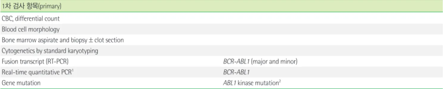
Table 1. 만성골수성백혈병(chronic myeloid leukemia, BCR-ABL1-positive) 1차 검사 항목(primary)
CBC, differential count Blood cell morphology
Bone marrow aspirate and biopsy±clot section Cytogenetics by standard karyotyping
Fusion transcript (RT-PCR) BCR-ABL1 (major and minor)
Real-time quantitative PCR1 BCR-ABL1
Gene mutation ABL1 kinase mutation2
1Major molecular response (MMR)를 확인하기 위해 3개월마다 측정함. MMR (BCR-ABL1 <0.1% internal scale or log reduction 3.0).
2임상적으로 tyrosine kinase inhibitor 내성이 의심될 때 시행함.
Abbreviation: CBC, complete blood cell count.
2차 검사 항목(secondary)
FISH1 BCR-ABL1
Immunohistochemistry (biopsy or clot section) CD342, CD1172, CD613
Cytochemistry Reticulin/collagen4
Myeloperoxidase (MPO) or Sudan black B5 Periodic acid Schiff (PAS)5
Nonspecific esterase5
Immunophenotyping6 - Acute panel
CD2, cCD3, CD5, CD7, CD10, CD13, CD19, CD20, cCD22, CD33, CD34, CD45, CD56, cCD79a, CD117, HLA-DR, MPO, TdT
Gene mutation JAK2 V617F, CALR, MPL W515K/L7
1 염색체 검사에서 정상인 경우나 분열중기세포를 수확하지 못한 경우 및 역전사효소-중합연쇄반응(RT-PCR) 검사에서 BCR-ABL1 유전자 재배열을 확인하지 못했을 때 반드시 시행 함. 골수 검체를 채취하지 못한 경우 말초혈액으로 검사를 시행함.
2Abnormal localization of immature precursors (ALIP)여부를 확인하기 위해 시행함.
3거대핵세포(megakaryocyte) 확인을 위해 시행함.
4섬유화(fibrosis)가 의심될 때 골수생검에서 시행함.
5모세포기(blast phase)일 때 급성백혈병의 cytochemistry와 동일하게 시행함.
6모세포기일 때 급성백혈병의 acute panel과 동일하게 시행함.
7필라델피아 음성인 골수증식종양(myeloproliferative neoplasms)과 감별이 어려울 때 시행함.
Abbreviations: cCD3, cytoplasmic CD3; cCD22, cytoplasmic CD22; cCD79a, cytoplasmic CD79a.
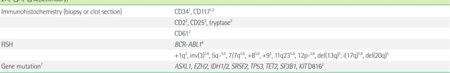
Table 2. 골수증식종양(myeloproliferative neoplasms) 1차 검사 항목(primary)
CBC, differential count Blood cell morphology Serum LDH level Serum erythropoietin
Serum vitamin B12 level1, Serum tryptase1 Bone marrow aspirate and biopsy±clot section
Cytochemistry Reticulin/collagen2
Immunophenotyping3 CD3, CD4, CD8, CD5, CD7, CD27
Cytogenetics by standard karyotyping
Fusion transcript (RT-PCR) BCR-ABL1
FISH FIP1L1-PDGFRA4,5, PDGFRB4, FGFR14, PCM1-JAK24
Gene mutation JAK2V617F6,7, JAK2 exon 127, CALR6, MPL W515K/L6, CSF3R8
1만성호산구백혈병이 의심될 때 시행함.
2섬유화가 의심될 때 골수생검에서 시행함.
3Lymphocyte-variant hypereosinophilia 배제 위해 시행.
4만성호산구백혈병이 의심될 때, FIP1L1-PDGFRA, PDGFRB break apart, FGFR1, PCM1-JAK2 재배열 음성임을 확인하기 위해 시행함.
5RT-PCR로 시행할 수도 있음.
6진성혈소판증가증, 일차골수섬유증, 미분류 골수증식종양이 의심될 때 JAK2, CALR, MPL 돌연변이를 시행함.
7진성적혈구증가증이 의심될 때, JAK2 V617F를 우선적으로 시행하고 JAK2 V617F 돌연변이 음성일 때 JAK2 exon 12 돌연변이 검사를 추가적으로 시행함.
8만성호중구백혈병 의심될 때 CSF3R 돌연변이 검사를 시행함.
Abbreviation: LDH, lactate dehydrogenase. (Continued to the next page)
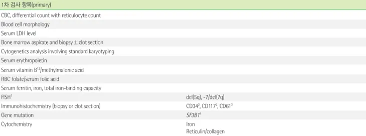
Table 3. 골수형성이상/골수증식종양(myelodysplastic/myeloproliferative neoplasms) 1차 검사 항목(primary)
CBC, differential count with reticulocyte count Blood cell morphology
Bone marrow aspirate and biopsy±clot section Cytogenetics analysis involving standard karyotyping Serum erythropoietin
Serum vitamin B12/methylmalonic acid RBC folate/serum folic acid
Serum ferritin, iron, total iron-binding capacity
Immunohistochemistry (biopsy or clot section) CD341, CD1171, CD612
Cytochemistry Iron
Reticulin/collagen
Fusion transcript BCR-ABL1
FISH FIP1L1-PDGFRA3,4, PDGFRB3,5, FGFR13, PCM-JAK23
inv(3)/t(3q)6, del(5q)6
Gene mutation6 JAK2 V617F, MPL W515K/L, CALR
1ALIP 여부를 확인하기 위해 시행함.
2거대핵세포 확인 및 거핵구형성이상(dysmegakaryopoiesis) 판별을 위해 시행함.
3Eosinophilia 동반될 때 시행함.
4RT-PCR로 시행할 수도 있음.
5만성골수단구백혈병(chronic myelomonocytic leukemia, CMML)에서 tyrosine kinase inhibitors 반응유무 확인을 위해 시행함.
6MPN 병력이 있거나 형태학적으로 MPN 혹은 MDS/MPN with ring sideroblast and thrombocytosis의심될 때 시행함.
2차 검사 항목(secondary)
Immunohistochemistry (biopsy or clot section) CD141, CD641, CD11c1
FISH2 -Y, del(3q), -7/del(7q)1, +8, del(11q), del(12p), i(17q), +19, del(20q), +21 Flow cytometry for dyshematopoietic MDS clone3 CD10, CD11b, CD13, CD15, CD16, CD33, CD45, CD64
Gene mutation PTPN111, NF11, CBL1, JAK31, KRAS1,4,5, NRAS1,4,5, SETBP11,4,5, TET24, DNMT3A4, ASXL14, EZH24, SRSF24, U2AF14, ZRSR24, TP534, STAG24, IDH1/IDH24, RUNX14, ETV64, PHF64, BCOR4, NPM14, CSF3R4,5, ETNK14,5, SF3B14,5,6 Hemoglobin F1
GM-CSF hypersensitivity assay1,7
1소아기골수단구백혈병(juvenile myelomonocytic leukemia, JMML) 의심될 때 시행함.
2염색체검사에서 분열중기세포를 수확하지 못했을 때 시행할 수 있으며 해당 염색체 이상이 의심되거나 복잡핵형(complex karyotype)일 때 시행함.
3본 패널은 조혈세포의 형성이상 판별을 위한 것으로 만성골수단구백혈병(Chronic myelomonocytic leukemia, CMML) subtype 2일 경우 급성백혈병의 acute panel을 적용함.
4CMML 의심되나 myelodysplasia가 관찰되지 않을 경우 ASXL1, TET2, SRSF2, SETBP1을 포함한 유전자돌연변이 검사를 1차 검사로 시행 가능함.
5BCR-ABL1 음성 비정형만성골수성백혈병(atypical chronic myeloid leukemia, BCR-ABL1 negative) 의심될 때 시행함.
6MDS/MPN with ring sideroblast and thrombocytosis의심될 때 시행함.
7국내에 시행기관 없음.
Table 2. Continued 2차 검사 항목(secondary)
Immunohistochemistry (biopsy or clot section) CD341, CD1171,2 CD22, CD252, tryptase2 CD613
FISH BCR-ABL14
+1q5, inv(3)5,6, 5q-5,6, 7/7q5,6, +85,6, +95, 11q235.6, 12p-5,6, del(13q)5, i(17q)5,6, del(20q)5
Gene mutation7 ASXL1, EZH2, IDH1/2, SRSF2, TP53, TET2, SF3B1, KIT D8162
1Abnormal localization of immature precursors (ALIP) 여부를 확인하기 위해 시행함.
2비만세포(mastocytes)가 증가되었을 때 시행함.
3거대핵세포 확인을 위해 시행함.
4RT-PCR 법으로 BCR-ABL1 유전자 재배열을 확인하지 못했거나, 염색체 검사에서 t(9;22)(q34;q11.2)이 관찰되지 않았음에도 불구하고 CML을 완전히 배제할 수 없는 경우에 시행함.
5염색체검사에서 확인하였다면 FISH 추가할 필요 없음.
6MPN의 Dynamic International Prognostic Scoring System (DIPSS)-plus score를 정하기 위해 필요함.
7MPN의 클론성 증명 및 예후와 연관된 분자유전학적 마커 확인을 위해 시행함.
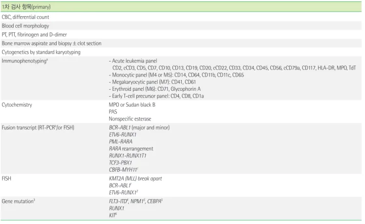
Table 4. 골수형성이상증후군(myelodysplastic syndrome) 1차 검사 항목(primary)
CBC, differential count with reticulocyte count Blood cell morphology
Serum LDH level
Bone marrow aspirate and biopsy±clot section Cytogenetics analysis involving standard karyotyping Serum erythropoietin
Serum vitamin B12/methylmalonic acid RBC folate/serum folic acid
Serum ferritin, iron, total iron-binding capacity
FISH1 del(5q), -7/del(7q)
Immunohistochemistry (biopsy or clot section) CD342, CD1172, CD613
Gene mutation SF3B14
Cytochemistry Iron
Reticulin/collagen
1염색체검사에서 분열중기세포를 수확하지 못했을 때 시행할 수 있으며 해당 염색체 이상이 의심되거나 복잡핵형(complex karyotype)일 때 시행함.
2ALIP 여부를 확인하기 위해 시행함.
3거대핵세포 확인 및 거핵구형성이상(dysmegakaryopoiesis) 판별을 위해 시행함.
4MDS에서 ring sideroblast가 관찰될 때 시행함.
Abbreviation: PNH, paroxysmal nocturnal hemoglobinuria.
2차 검사 항목(secondary)
FISH1 -Y, inv(3)/t(3q)/del(3q), +8, del(11q), del(12p), i(17q), +19, del(20q) - t(8;21)2, t(15;17)2, inv(16)2
Flow cytometry for dyshematopoietic MDS clone3 CD10, CD11b, CD13, CD15, CD16, CD33, CD45, CD64 Flow cytometry for PNH clone4
Gene mutation TET2, DNMT3A, ASXL1, EZH2, SRSF2, U2AF1, ZRSR2, TP53, STAG2, NRAS, CBL, IDH1/IDH2, SETBP1, PHF6, BCOR, PRPF8, SF3B15, JAK2 V617F5, MPL W515K/L5, RUNX16, GATA26, NF16, ETV66, ANKRD266, DDX416, SRP726, CEBPA6, TERT/TERC7, FANC genes7,10, DKC7,ELA2/HAX1/GFI17
Thyroid stimulating hormone Copper, ceruloplasmin8
Viral antigen/antibody assays9 HBV, HCV, CMV, herpes simplex, parvovirus B19, HIV Chromosome breakage analysis10
1염색체검사에서 분열중기세포를 수확하지 못했을 때 시행할 수 있으며 해당 염색체 이상이 의심되거나 복잡핵형(complex karyotype)일 때 시행함.
2급성골수성백혈병(AML)을 배제하기 위해 시행함.
3본 패널은 조혈세포의 형성이상 판별을 위한 것으로 MDS with excess blasts일 경우 급성백혈병의 acute panel을 적용함.
4적혈구, 호중구 및 단구를 대상으로 하고, fluorescent aerolysin (FLAER)과 CD55 또는 CD59를 포함하여 시행할 것을 권장함.
5MDS에서 혈소판증가증이 관찰될 때 시행함.
6Familial MDS 의심될 때 시행함(Supplementary Table 1 참조).
7Inherited BM failure syndrome 의심될 때 임상양상에 따라 시행함.
8위장관 흡수장애, 위 우회로조성술 및 아연보충요법을 받는 환자에서 시행함.
9형성이상(dysplasia) 원인을 배제하기 위해 시행할 수 있음.
10소아 MDS, hypoplastic MDS에서 판코니빈혈과의 감별을 위해 시행함.
Abbreviations: HBV, hepatitis B virus; HCV, hepatitis C virus; CMV, cytomegalovirus.
syndrome), 급성백혈병(acute leukemia), 형질세포종양(plasma cell neoplasm), 림프종의 골수침범(lymphoma with bone marrow involvement), 이식후림프증식질환(post-transplant lymphoprolif- erative disorder), 조직구종양(histiocytic neoplasm) 순으로 정리 하였다(Tables 1-9). 골수증식종양의 경우 진단 시 감별진단이 필 요한 호산구증을 동반한 PDGFRA 등의 유전자 재조합을 포함한 종양(myeloid/lymphoid neoplasms with eosinophilia and gene rearrangement)에 속하는 질환을 포함시켰다. 질환별 진단 시 시행
해야 할 검사 항목들은 필수검사항목을 1차 검사로, 추천검사항 목은 2차 검사로, 연구검사항목은 3차 검사로 Table에 정리하였다.
급성백혈병과 림프종의 골수침범은 2차와 3차 검사는 계열에 따 라 분류하여 설명하였다(Table 5, Table 7). 급성백혈병과 골수형성 이상증후군 등 germline predisposition에 대해서 Supplementary Table 1을 추가하였으며, 림프종의 골수침범에 대해서는 NCCN 지 침에 따른 세포의 크기 및 모양에 따른 분류를 Supplementary Ta- ble 2와 Supplementary Table 3에 추가 기술하였다. 검사 별 보험
Table 5. 급성백혈병(acute leukemia) 1차 검사 항목(primary)
CBC, differential count Blood cell morphology PT, PTT, fibrinogen and D-dimer
Bone marrow aspirate and biopsy±clot section Cytogenetics by standard karyotyping
Immunophenotypinga - Acute leukemia panel
CD2, cCD3, CD5, CD7, CD10, CD13, CD19, CD20, cCD22, CD33, CD34, CD45, CD56, cCD79a, CD117, HLA-DR, MPO, TdT - Monocytic panel (M4 or M5): CD14, CD64, CD11b, CD11c, CD65
- Megakaryocytic panel (M7): CD41, CD61 - Erythroid panel (M6): CD71, Glycophorin A - Early T-cell precursor panel: CD4, CD8, CD1a
Cytochemistry MPO or Sudan black B
PAS
Nonspecific esterase Fusion transcript (RT-PCRb/or FISH) BCR-ABL1 (major and minor)
ETV6-RUNX1 PML-RARA RARA rearrangement RUNX1-RUNX1T1 TCF3-PBX1 CBFB-MYH11c
FISH KMT2A (MLL) break apart
BCR-ABL11 ETV6-RUNX12
Gene mutation3 FLT3-ITD4, NPM15, CEBPA5
RUNX1 KIT6
a보험에서 18종만 인정하고 있음.
b보험에서 7종만인정하고있음.
c현재 RT-PCR보험코드없음.
1ALL, MPAL에서필수적으로시행함.
2소아 ALL에서 iAMP21확인이필요함.
3AML에서필수적으로시행함.
4FLT3-ITD, allelic ratio 반정량측정을권장함.
5정상핵형 AML 진단시 시행함.
6Core binding factor (CBF)- AML 진단 시 시행함.
Abbreviations: PT, prothrombin time; PTT, partial thromboplastin time.
2차 검사 항목(secondary)-급성골수성백혈병(acute myeloid leukemia)
Immunohistochemistry (biopsy or clot section)a CD31, CD341, CD79a1, CD1171, TdT1 CD612, Glycophorin A3
Cytochemistry Reticulin/collagen4
FISH5 inv(3)/t(3;3), del(5q), 7/del(7q), TP53
BCR-ABL1, PML-RARA, RUNX1-RUNX1T1, CBFB
Real-time quantitative PCRb RUNX-RUNX1T1, PML-RARA6, CBFB-MYH11, NPM1, WT1, BAALC
RT-PCRC DEK-NUP214, MLLT3-KMT2A, RBM15-MKL1
Gene mutation7 FLT3-TKD, NRAS, TP53
ABL1 kinase mutation8
DNMT3A, IDH1, IDH2, TET2, MLL-PTD, ASXL1, ETV6, EZH2, CBL, JAK2
a보험은 8종만인정하고있음.
b보험인정항목: RUNX-RUNX1T1, PML-RARA, BCR-ABL1(major/minor), CBFB-MYH11, NPM1, WT1, BAALC.
c현재 보험코드 없음.
1골수흡인검체로 유세포 분석이 불가능할 경우 반드시 시행함.
2거대핵세포 확인을 위해 시행함.
3적백혈병(erythroleukemia)이 의심될 때 시행함.
4섬유화가 의심될 때 골수생검에서 시행함.
5 염색체검사에서 분열중기세포를 수확하지 못했을 때 진단명에 따라 시행함. 그 외 해당 염색체 이상이 의심될 때, 복잡핵형일 때 혹은 해당 이상이 의심되나 G-banded karyotype 또는 RT-PCR에서 관찰하지 못한 경우에 시행.
6급성전골수세포백혈병(acute promyelocytic leukemia)으로 진단되는 경우 잔존질환 확인을 위해 진단 시 필수로 시행함.
7유전자 숫자 등을 고려하여 NGS 패널로 검사하는 것이 효율적일 수 있음.
8BCR-ABL1 양성 AML 환자인 경우 진단 시 시행할 수 있음. (Continued to the next page)
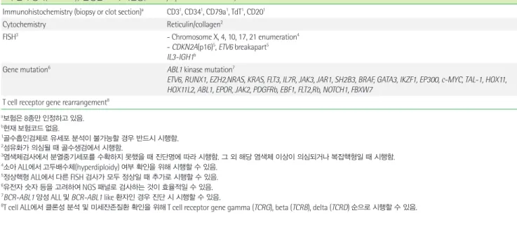
Table 5. Continued
2차 검사 항목(secondary)-급성림프모구백혈병(acute lymphoblastic leukemia) Immunohistochemistry (biopsy or clot section)a CD31, CD341, CD79a1, TdT1, CD201
Cytochemistry Reticulin/collagen2
FISH3 - Chromosome X, 4, 10, 17, 21 enumeration4
- CDKN2A(p16)5, ETV6 breakapart5 IL3-IGH1b
Gene mutation6 ABL1 kinase mutation7
ETV6, RUNX1, EZH2,NRAS, KRAS, FLT3, IL7R, JAK3, JAR1, SH2B3, BRAF, GATA3, IKZF1, EP300, c-MYC, TAL-1, HOX11, HOX11L2, ABL1, EPOR, JAK2, PDGFRb, EBF1, FLT2,Rb, NOTCH1, FBXW7
T cell receptor gene rearrangement8
a보험은 8종만인정하고있음.
b현재보험코드없음.
1골수흡인검체로 유세포 분석이 불가능할 경우 반드시 시행함.
2섬유화가 의심될 때 골수생검에서 시행함.
3염색체검사에서 분열중기세포를 수확하지 못했을 때 진단명에 따라 시행함. 그 외 해당 염색체 이상이 의심되거나 복잡핵형일 때 시행함.
4소아 ALL에서고두배수체(hyperdiploidy) 여부 확인을 위해 시행할 수 있음.
5정상핵형 ALL에서다른 FISH 검사가 모두 정상일 때 추가로 시행할 수 있음.
6유전자 숫자 등을 고려하여 NGS 패널로 검사하는 것이 효율적일 수 있음.
7BCR-ABL1 양성 ALL 및 BCR-ABL1 like 환자인 경우 진단 시 시행할 수 있음.
8T cell ALL에서 클론성 분석 및 미세잔존질환 확인을 위해 T cell receptor gene gamma (TCRG), beta (TCRB), delta (TCRD) 순으로 시행할 수 있음.
3차 검사 항목(tertiary)-급성림프모구백혈병(acute lymphoblastic leukemia)
Gene mutation (germ line)1,2 CEBPA, DDX41, RUNX1, ANKRD26, ETV6, GATA2, SRP72, TERT, TERC, DKC, ELA2, HAX1, GFI1, PTPN11, CBL, KRAS, NF1, BLM, ATG2B/GSKIP
TP53, BRCA1/2
Fanconi anemia and marrow failure syndrome genes Noonan syndrome genes
Down syndrome
1유전자 숫자 등을 고려하여 NGS 패널로 검사하는 것이 효율적일 수 있음.
2 환자의 임상양상, 과거력 및 가족력을 고려할 때 유전성 질환이 의심될 경우, 소아 및 젊은 나이에 발생하였고 dysplastic feature를 동반할 경우, 관련 유전자들은 빈도 및 임상적 연 관성에 따라 유전자를 선정하여 검사할 수 있음 (Supplementary Table 1 참조).
Table 6. 형질세포종양(plasma cell neoplasms) 1차 검사 항목(primary)
CBC, differential count Blood cell morphology BUN/creatinine, albumin Electrolytes with calcium Serum free light chain assay Serum quantitative immunoglobulins
Serum protein electrophoresis, immunofixation electrophoresis
24h urine for total protein, urine protein electrophoresis, urine immunofixation electrophoresis Bone marrow aspirate and biopsy±clot section
Immunohistochemistry (biopsy or clot section) CD138, kappa/lambda
Immunophenotyping CD19, CD38, CD45, CD56, CD138
Cytogenetics by standard karyotyping
FISH IGH-FGFR3, IGH-MAF, del(17p), 1q21 amplification
Serum LDH level β2-microglobulin
Abbreviation: BUN, blood urea nitrogen. (Continued to the next page)
Table 7. 림프종의 골수침범(lymphoma with bone marrow involvement) 1차 검사 항목(primary)
CBC, differential count Blood cell morphology Serum LDH level
Bone marrow aspirate and biopsy±clot section1, 2
Immunohistochemistry (biopsy or clot section) - Non Hodgkin lymphoma: CD3, CD20 - Hodgkin lymphoma: CD3, CD15, CD20, CD30
Immunophenotypinga,3 - Initial panel: CD2, CD3, cCD3, CD5, CD7, CD10, CD19, CD20, cCD22, CD34, CD45, CD56 - B-cells: CD23, CD38, kappa/lambda, FMC7, TdT
- T-cells : CD4, CD8, TCR-ɑβ, TCR-ɣ Cytogenetics by standard karyotyping4
a비호지킨림프종(골수검체)에 대해 다음과 같은 경우에 보험 적용됨.
1) 골수 이외의 부위(장기, 조직 등)에 병변이 없으나, 골수 침범이 의심되는 경우.
2) 골수 이외의 부위에 병변이 의심되나, 조직검사가 불가능한 경우 (2015.12.15. 시행).
1골수생검은 양측 검사를 권장함.
2림프종 의심 세포의 크기 및 표현형에 따라 진단과정을 세분화하여 진행할 수 있음 (Supplementary Table 2 and 3 참조)
3림프종 의심 세포가 25% 이상일 경우 시행할 것을 권장하며 5% 이상이면 2차 검사 항목으로 시행 가능함.
4CLL/SLL인경우, B-cell mitogens 또는 CpG-stimulated metaphase karyotype 추가로 시행 추천함.
2차 검사 항목(secondary)-B세포 림프종(B cell lymphoma)
Immunohistochemistry (biopsy or clot section)1 - High-grade B cell lymphoma (double hit lymphoma, triple hit lymphoma): Ki-67, BCL2, BCL6, MYC - Hairy cell leukemia: Annexin A1, CD103
- Follicular lymphoma, diffuse large B-cell lymphoma (DLBCL), double hit lymphoma, triple hit lymphoma:
BCL2, BCL6, MUM1, MYC, CD10 - Mantle cell lymphoma: CyclinD1 - ALK+DLBCL: ALK
Cytochemistry Tartrate-resistant acid phosphatase (TRAP)2
Fusion transcript (RT-PCR) - Large B cell lymphoma: IRF4 (MUM1) rearrangementa
Gene mutation BRAF V600E2
MYD88 L265P3
FISH4 - Chronic lymphocytic leukemia: del(11q), IGH-CCND1, del(17p), CEP12, del(13q)
- Follicular lymphoma: IGH-BCL2 - Mantle cell lymphoma: IGH-CCND1
- Burkitt lymphoma: IGH-MYC and MYC break apart
- Diffuse large B-cell lymphoma, High-grade B-cell lymphoma (double hit, triple hit): IGH-MYC or MYC break apart, IGH-BCL2 break apart or BCL2 break apart, BCL6 break apart
- MALT lymphoma: API2 (BIRC3)/MALT1
Real-time quantitative PCR EBV quantitative PCR5
Immunoglobulin gene / T-cell receptor gene rearrangement β2-microglobulin
a현재 보험코드 없음.
1골수흡인검체로 유세포 분석이 불가능할 경우 1차 검사 항목에서 선택하여 시행하는 것을 권장하며 CD 항원 검사 항목은 골수흡인 및 골수생검에서 분석 가능함.
2Hairy cell leukemia가 의심될 때 시행함.
3Lymphoplasmacytic lymphoma가 의심될 때 시행함.
4다음에 해당하는 림프종의 골수침범이 의심되는 경우 관련 FISH 검사를 반드시 시행함.
5Burkitt lymphoma, post-transplant lymphoproliferative disorder, other EBV-associated lymphoproliferative disorders가의심될때시행함.
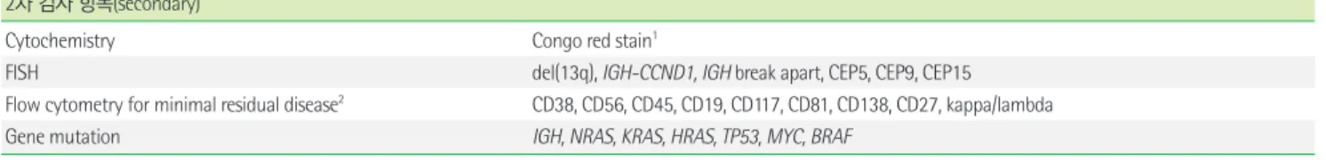
Abbreviations: MALT, mucosa-associated lymphoid tissue; EBV, Epstein-Barr virus. (Continued to the next page) Table 6. Continued
2차 검사 항목(secondary)
Cytochemistry Congo red stain1
FISH del(13q), IGH-CCND1, IGH break apart, CEP5, CEP9, CEP15
Flow cytometry for minimal residual disease2 CD38, CD56, CD45, CD19, CD117, CD81, CD138, CD27, kappa/lambda
Gene mutation IGH, NRAS, KRAS, HRAS, TP53, MYC, BRAF
1아밀로이드증(amyloidosis) 의심될 때 골수생검이나 골수절편에서 시행함.
2클론성 분석 및 미세잔존질환 확인을 위해 시행.
Table 8. 이식후림프증식질환(post-transplant lymphoproliferative disorder) 1차 검사 항목(primary)
CBC, differential count Blood cell morphology
Bone marrow aspirate and biopsy±clot section1
Immunohistochemistry - PH, IM, FFH, polymorphic, monomorphic PTLD2: CD3, CD20, CD56, CD138, kappa/lambda - Classic Hodgkin lymphoma PTLD: CD3, CD15, CD20, CD30
EBV encoded RNA in situ hybridization (EBER-ISH)
Immunophenotypinga,3 - Initial panel: CD2, cCD3, CD5, CD7, CD10, CD19, CD20, cCD22, CD34, CD45, CD56 - B-cells: CD23, CD138, kappa/lambda, FMC7, TdT
- T-cells: CD4, CD8 Cytogenetics by standard karyotyping
a보험에서 백혈병에 한해 18종만 인정하고 있음.
1골수생검은 양측 검사를 권장함.
2Monomorphic PTLD의 경우 2차 검사항목은 림프종의 골수침범에 준하여 실시함.
3림프종 의심 세포가 25% 이상일 경우 시행할 것을 권장하며 5% 이상이면 2차 검사 항목으로 시행 가능함.
Abbreviations: PH, plasmacytic hyperplasia; IM, infectious mononucleosis; FFH, florid follicular hyperplasia; PTLD, posttransplant lymphoproliferative disorders; EBV, Epstein- Barr virus.
Table 7. Continued
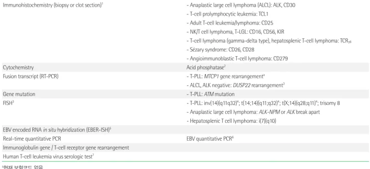
2차 검사 항목(secondary)-T세포 림프종(T cell lymphoma)
Immunohistochemistry (biopsy or clot section)1 - Anaplastic large cell lymphoma (ALCL): ALK, CD30 - T-cell prolymphocytic leukemia: TCL1
- Adult T-cell leukemia/lymphoma: CD25 - NK/T cell lymphoma, T-LGL: CD16, CD56, KIR
- T-cell lymphoma (gamma-delta type), hepatosplenic T-cell lymphoma: TCRγδ
- Sézary syndrome: CD26, CD28
- Angioimmunoblastic T-cell lymphoma: CD279
Cytochemistry Acid phosphatase2
Fusion transcript (RT-PCR) - T-PLL: MTCP1 gene rearrangementa
- ALCL, ALK negative: DUSP22 rearrangement3
Gene mutation - T-PLL: ATM mutation
FISH3 - T-PLL: inv(14)(q11q32)4; t(14;14)(q11;q32)4; t(X;14)(q28;q11)4; trisomy 8
- Anaplastic large cell lymphoma: ALK-NPM or ALK break apart - Hepatosplenic T cell lymphoma: i(7)(q10)
EBV encoded RNA in situ hybridization (EBER-ISH)5
Real-time quantitative PCR EBV quantitative PCR6
Immunoglobulin gene / T-cell receptor gene rearrangement Human T-cell leukemia virus serologic test7
a현재 보험코드 없음.
1골수흡인검체로 유세포 분석이 불가능할 경우 1차 검사 항목에서 선택하여 시행하는 것을 권장하며 CD 항원 검사 항목은 골수흡인 및 골수생검에서 분석 가능함.
2T-cell prolymphocytic leukemia가 의심될 때 시행함.
3다음에 해당하는 림프종의 골수침범이 의심되는 경우 관련 FISH 검사를 추가로 시행할 수 있음.
4해당검사는 TCRAD (T cell receptor A/D) break apart probe로 검사 가능함.
5T-cell large granular lymphocytic leukemia, hepatosplenic T-cell lymphoma, subcutaneous panniculitis-like T-cell lymphoma 등이 의심될 때 시행함.
6Extranodal NK/T cell lymphoma, post-transplant lymphoproliferative disorder, other EBV-associated lymphoproliferative disorders가 의심될 때 시행함.
7Adult T-cell leukemia/lymphoma가 의심될 때 시행함.
Abbreviations: MALT, mucosa-associated lymphoid tissue; EBV, Epstein-Barr virus.
관련 설명은 알파벳 소문자, 검사에 대한 상세 설명은 아라비아 숫 자 순으로 첨자 표기하였으며 지침서 특성상 주석은 한글로 기술 하였다.
빠르게 변화하는 분야이지만 혈액종양 초진단 검사항목 제2차 지침이 혈액종양 진단에 유용하게 활용되고, 발전하는 검사들의 국내 보급이 활발해지며, 검사 표준화에 기여하길 기대한다.
요 약
대한진단혈액학회에서는 혈액종양환자를 대상으로 표준화된 진단 절차에 대한 임상 진료 권장사항을 제공할 수 있는 지침을 개 정하고자 하였다. WHO 개정 4판, National Comprehensive Can- cer Network Guidelines, European LeukemiaNet recommenda- tions을 포함한 국제 지침을 바탕으로 한 혈액종양 초진단 검사항 목 지침을 개정하였다. 본 개정 지침은 국내 혈액종양 진단에 유용 하게 활용되고 검사표준화에 기여하며 지속적인 협의 과정을 통 해 진단 알고리즘에 통합된 새로운 분자 기술 발전을 향상시킬 수 있을 것이다.
이해관계
저자들은 본 연구와 관련되어 어떠한 이해관계도 없음을 밝힙니다.
REFERENCES
1. Swerdlow SH, Campo E, et al. eds. WHO classification of tumours of haematopoietic and lymphoid tissues. Revised 4th ed. Lyon, France:
IARC, 2017.
2. Lee JY, Kim HK, Huh J, Kim M, Kong SY, Cho YW, et al. Test guide- lines for initial diagnosis of hematologic neoplasms. Lab Med Online 2016;6:1-7.
3. Radich JP, Deininger M, Abboud CN, Altman JK, Berman E, Bhatia R, et al. Chronic myeloid leukemia, version 1.2019, NCCN clinical prac- tice guidelines in oncology. J Natl Compr Canc Netw 2018; 16:1108-35.
4. Mesa RA, Jamieson C, Bhatia R, Deininger MW, Fletcher CD, Gerds AT, et al. NCCN guidelines insights: Myeloproliferative neoplasms, ver- sion 2.2018. J Natl Compr Canc Netw 2017; 15:1193-207.
5. Greenberg PL, Stone RM, Al-Kali A, Barta SK, Bejar R, Bennett JM, et al. Myelodysplastic syndromes, version 2.2017, NCCN clinical practice guidelines in oncology. J Natl Compr Canc Netw 2017; 15:60-87.
6. O’Donnell MR, Tallman MS, Abboud CN, Altman JK, Appelbaum FR, Arber DA, et al. Acute myeloid leukemia, version 3.2017, NCCN clinical practice guidelines in oncology. J Natl Compr Canc Netw 2017; 15:926- 57.
7. Brown PA, Shah B, Fathi A, Wieduwilt M, Advani A, Aoun P, et al. NCCN guidelines insights: Acute lymphoblastic leukemia, version 1.2017. J Natl Compr Canc Netw 2017;15:1091-102.
8. Kumar SK, Callander NS, Alsina M, Atanackovic D, Biermann JS, Cas- Table 9. 조직구종양(histiocytic neoplasm)
1차 검사 항목(primary) CBC, differential count Blood cell morphology
Bone marrow aspirate and biopsy±clot section1
Immunohistochemistry - CD1a, S100, Langerin2
Cytogenetics by standard karyotyping
1골수생검은 양측 검사를 권장함. 특히 Langerhans cell histiocytosis의 multisystem disease에서는 반드시 시행해야 함.
2Langerhans cell histiocytosis 진단 시 가장 검출 빈도가 높은 검사 항목임.
2차 검사 항목(secondary)
Immunohistochemistry1 CD3, CD20, CD4, CD21, CD35, CD68, CD123, CD163, Factor XIIIa, Fascin, Lysozyme, TCL1
Gene mutation BRAF V600E2
Gene assay HUMARA (X-linked androgen receptor gene assay)3,4
Immunoglobulin gene/T-cell receptor gene rearrangement
1Langerhans cell histiocytosis와다른 histiocytic neoplasm과의 감별을 위한 종목임.
2Langerhans cell histiocytosis를 비롯한 다양한 종류의 histiocytic neoplasm에서 보고되어 있음.
3Langerhans cell histiocytosis에서 단클론성 패턴을 보임.
4국내 시행기관 없음.
tillo J, et al. NCCN guidelines insights: Multiple myeloma, version 3.2018.
J Natl Compr Canc Netw 2018;16:11-20.
9. Horwitz SM, Zelenetz AD, Gordon LI, Wierda WG, Abramson JS, Ad- vani RH, et al. NCCN guidelines insights: Non-Hodgkin’s lymphomas, version 3.2016. J Natl Compr Canc Netw 2016;14:1067-79.
10. Horwitz SM, Ansell SM, Ai WZ, Barnes J, Barta SK, Choi M, et al. NCCN guidelines insights: T-cell lymphomas, version 2.2018. J Natl Compr Canc Netw 2018;16:123-35.
11. Hoppe RT, Advani RH, Ai WZ, Ambinder RF, Aoun P, Armand P, et al.
NCCN guidelines insights: Hodgkin lymphoma, version 1.2018. J Natl Compr Canc Netw 2018;16:245-54.
12. Döhner H, Estey E, Grimwade D, Amadori S, Appelbaum FR, Büchner T, et al. Diagnosis and management of AML in adults: 2017 ELN recom- mendations from an international expert panel. Blood 2017;129:424- 47.
13. Heidel FH, Gale RP, Hochhaus A. Managing myeloproliferative neo- plasms evidence based on the ELN treatment recommendations 2018.
Leukemia 2018;32:1055-6.