Treatment effects of mandibular total arch distalization using a ramal plate
Objective: The purpose of this study was to evaluate treatment effects after distalization of the mandibular dentition using ramal plates through lateral cephalograms. Methods: Pre- and post-treatment lateral cephalograms and dental casts of 22 adult patients (11 males and 11 females; mean age, 23.9 ± 5.52 years) who received ramal plates for mandibular molar distalization were analyzed. The treatment effects and amount of distalization of the mandibular molars were calculated and tested for statistical significance. The significance level was set at p < 0.001. Results: The mandibular first molar distalization at the crown and root were 2.10 mm (p < 0.001) and 0.81 mm (p = 0.011), respectively. In the evaluation of skeletal variables, there was a significant increase in the Wits appraisal (p < 0.001). In the evaluation of the soft tissue, there was no significant effect on upper lip position, but the lower lips showed a significant retraction of 2.2 mm (p < 0.001). Conclusions: The mandibular molars showed a significant amount of distalization accompanied by limited extrusion and mesiobuccal rotation of the crowns. A ramal plate may be a viable device for mandibular total arch distalization in Class III patients who are reluctant to undergo orthognathic surgery.
[Korean J Orthod 2016;46(4):212-219]
Key words: Distalizing, Miniplate, Class III treatment, Total arch distalization, Cephalometrics
Jonghan Yu
aJae Hyun Park
b,cMohamed Bayome
d,eSungkon Kim
fYoon-Ah Kook
fYoonji Kim
fChang-Hyen Kim
ga
Department of Orthodontics, Graduate School of Clinical Dental Science, The Catholic University of Korea, Seoul, Korea
b
Postgraduate Orthodontic Program, Arizona School of Dentistry & Oral Health, A.T. Still University, Mesa, AZ, USA
c
Graduate School of Dentistry, Kyung Hee University, Seoul, Korea
d
Department of Dentistry, Graduate School, The Catholic University of Korea, Seoul, Korea
e
Department of Postgraduate Studies, the Universidad Autonóma del Paraguay, Asunción, Paraguay
f
Department of Orthodontics, Seoul St.
Mary’s Hospital, College of Medicine, The Catholic University of Korea, Seoul, Korea
g
Department of Oral and Maxillofacial Surgery, Seoul St. Mary’s Hospital, College of Medicine, The Catholic
University of Korea, Seoul, Korea Received August 14, 2015; Revised January 11, 2016; Accepted January 19, 2016.
Corresponding author: Chang-Hyen Kim.
Associate Professor, Department of Oral and Maxillofacial Surgery, Seoul St. Mary’s Hospital, College of Medicine, The Catholic University of Korea, 222 Banpo-daero, Seocho-gu, Seoul 06591, Korea.
Tel +82-2-2258-1776 e-mail [email protected]
*This study was partly supported by funding from the Department of Dentistry and Graduate School of Clinical Dental Science, the Catholic University of Korea.
© 2016 The Korean Association of Orthodontists.
The authors report no commercial, proprietary, or financial interest in the products or companies described in this article.
This is an Open Access article distributed under the terms of the Creative Commons Attribution Non-Commercial License (http://creativecommons.org/licenses/by-nc/4.0) which permits unrestricted non-commercial use, distribution, and reproduction in any medium, provided the original work is properly cited.
pISSN 2234-7518 • eISSN 2005-372X
http://dx.doi.org/10.4041/kjod.2016.46.4.212
INTRODUCTION
The achievement of a pleasing profile with non- surgical treatment of Class III patients has always been challenging. Several modalities have been applied for Class III camouflage treatment.
1-3Several intraoral appliances have been introduced for mandibular molar distalization,
4,5but unfortunately, most require patient compliance. Non-compliance appliances, such as a distal extension lingual arch, Jones jig, or Franzulum appliance, usually produce tipping, anchorage loss, and flaring of the anterior teeth.
6-8Temporary skeletal anchorage devices (TSADs) have been applied to overcome these drawbacks in molar distalization.
9,10However, the placement of miniscrews between the interradicular spaces can result in inter- ference with the distalization process, and the miniscrews might require relocation during distalization. To avoid this disadvantage, Poletti et al.
11installed a miniscrew in the retromolar region for distalization of the lower dentition. However, the placement of a single miniscrew on each side might not be sufficient to with stand the forces required for total arch distalization. Therefore, Sugawara et al.
12,13placed a skeletal ancho rage system behind the second molar at the mandibular body and evaluated the treatment effects of this appliance.
Recently, Kook et al.
14have reported the application of a plate placed at the retromolar fossa of the ramus for mandibular dentition distalization. This appliance might
have a force vector more parallel to the occlusal plane, and is exposed through the retro molar region, which should cause less irritation than buccal miniscrews that are exposed through the movable vestibular mucosa and buccal cheek. However, the treatment effects of a ramal plate have not been evaluated.
The purpose of this study was to evaluate the treat- ment effects after distalization of the mandibular denti- tion using a ramal plate through lateral cephalo grams and three-dimensional (3D) virtual models.
MATERIALS AND METHODS
The sample consisted of pre- and post-treatment lateral cephalograms of 22 adult patients (11 males and 11 females; mean age, 23.9 ± 5.52 years) who had received ramal plates for mandibular molar distalization in the Department of Orthodontics, Seoul St. Mary’s Hospital, The Catholic University of Korea. The sample size required to detect a difference of at least 1.5 units with a standard deviation of 2 units using a power of 0.8 and a probability of type I error (α) = 0.05 was 16 patients.
Approval was obtained from the institutional review board of the Catholic University of Korea (KC14RISE0483) and informed consent was obtained from all subjects according to the Declaration of Helsinki. The inclusion criteria were (1) patients older than 18 years at the start of treatment, (2) with dental Class III malocclusion with more than a one-half cusp discrepancy at the molars, (3)
A
A B B
C
C D D
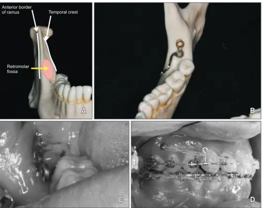
Retromolar fossa Anterior border
of ramus Temporal crest
Figure 1. The placement site
of the ramal plate showing
the retromolar fossa. A, The
placement site of the ramal
plate; B, the ramal plate after
adjustment to fit into the
retromolar fossa; C, the ramal
plate emerging through the
attached gingiva; and D, the
ramal plate connected to an
archwire with a power chain
elastic.
missing or extracted third molars, and (4) no syndrome or systemic disease.
Appliance description
The ramal plates were placed in the retromolar fossa between the anterior border of the mandibular ramus and the temporal crest. After a mucoperiosteal flap opening was created in the retromolar area, an L-plate (Le Forte system; Jeil Medical Corp., Seoul, Korea) was adapted to fit the bone surface. The hook on the plate was located 3 mm lateral to the buccal surface of the second molar, and between the buccal groove and 3 mm anterior to the distal surface anteroposteriorly (Figure 1).
The third molars were extracted during plate installation.
Each plate was fixated with two miniscrews 5 mm in length (with pilot drilling). The flap was sutured over the plate and the hook was extended through the mucosa.
The anterior screw hole of the plate was cut occlusally to convert it into a hook for easier placement of elastics or nickel-titanium closed-coil springs. The elastics or closed-coil springs were connected to hooks that were crimped to the archwire between the lateral incisors and canines after placement of the plate.
The hooks were adjusted to be in line with the facial axis points of the mandibular dentition so that the traction forces were parallel to the occlusal plane. Power chain elastics were connected from the plate hooks to the first molar bracket hooks to deliver a force of 300 g per side and were replaced every 3 weeks.
The plates were placed after the leveling and alignment of the mandibular dental arch was completed. The distalization started with a 0.019 × 0.025 inch stainless steel archwire that was fully engaged, and ended when an acceptable overjet was achieved.
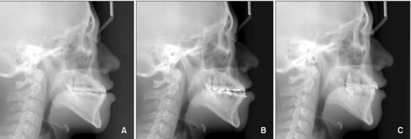
Figure 2 demonstrates the lateral cephalometric records of one of the cases at baseline, after leveling and alignment, and at post-treatment.
Cephalometric analysis
Pre- and post-treatment lateral cephalograms were digitized. For bilateral landmarks, the midpoint between the right and left superimposed landmarks was selected.
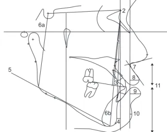
For the evaluation of the total treatment effect, the Frankfort horizontal (FH) line and a perpendicular line (VFH) at the pterygoid point were established as the horizontal and vertical reference lines, respectively. To evaluate the amount of distalization, the mandibular plane (MP) and a perpendicular line (VMP) at the menton were used as the horizontal and vertical reference lines, respectively. The distal surface point of the molar crown and the incisal edge of the incisor were used to identify the crowns. The apex and distal root apex were used to identify the incisor and molar root, respectively. Thirty-one linear and angular cephalometric variables were calculated (Figure 3, 4).
The cephalometric digitization of eight randomly selected cases was repeated 2 weeks later by the same examiner. The intraclass correlation coefficient (ICC) was applied to evaluate intra-examiner reliability. The ICC for all variables ranged between 0.961 and 0.912.
Statistical analysis
Statistical evaluation was performed using SPSS software ver. 16.0 (SPSS Inc., Chicago, IL, USA). The Shapiro-Wilk test was used to assess the normality of the distributions. The paired-sample t-test was applied to evaluate differences between pre- and post-treatment variables, since all these variables were normally distributed. The significance level was set at p < 0.05.
The Bonferroni correction was applied to compensate for multiple comparisons, resulting in a significance level of p < 0.001.
A B C
Figure 2. Cephalometric radiographs are shown. A, Pretreatment; B, after leveling and alignment; and C, post-treatment.
RESULTS
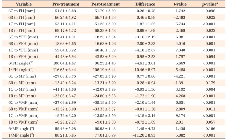
In the evaluation of tooth movement in relation to the FH and VFH as the craniofacial reference lines, there was a significant 3.2 mm (p < 0.001) distalization of the mandibular first molars, while their roots were distalized 2.0 mm with distal tipping of 4.6
o(p < 0.001). The incisors showed a significant retraction of 4.2 mm (p <
0.001) with a lingual inclination of 10.5
o(p < 0.001).
The first molar crowns showed no significant change in vertical position (Table 1).
With respect to the mandibular reference lines, the distalization of the first molars was 2.1 mm (p < 0.001), while their roots were distalized 0.8 mm (p = 0.011).
The amount of distal tipping was not statistically signi- ficant (1.45
o± 4.72
o; p = 0.166). The incisors showed a significant retraction of 4.2 mm (p < 0.001) with a significant lingual inclination of 11.2
o(p < 0.001). The first molar showed 0.77 mm of intrusion, while the incisor demonstrated 0.93 mm of extrusion (Table 1).
In the evaluation of skeletal variables, there was a significant change in the sagittal relationship bet- ween the mandible and maxilla (Wits, 2.4 mm; p <
0.001). However, the A point-nasion-B point (ANB) and
sella-nasion-B point (SNB) angles were not changed significantly. Moreover, the MP angle did not increase significantly (Table 2).
In the evaluation of soft tissue, there was no signi- ficant effect on upper lip position (nasolabial angle, p
= 0.29; true vertical line-upper lip, p = 0.231). However, the lower lip showed a significant retraction of 2.2 mm (p < 0.001). The position of the chin and the ratios between the upper lip and the lower lip and chin showed no significant changes (Table 2).
DISCUSSION
The treatment of Class III patients with moderate mandibular prognathism has always been compli- cated.
15,16The distal movement of mandibular molars is considered a challenging treatment objective. However, the application of TSADs to distalize the molars has increased the range of camouflage treatment. It is difficult to distalize more than 2–3 mm with miniscrews because of the interradicular space,
17-19but with ramal plates, there is no need to consider relocating the screws.
The ramal plate may have an increased stability and can withstand large forces because it is supported by two miniscrews. For mandibular distalization, usually only one miniscrew is placed per side.
10,20Although a
A
B
C
1 D 2
4 3 5
6 7
9 8
11
12 13
14 15
16 16 17 17 18 19 2020
10
Figure 3. Cephalometric reference lines and variables.
A, Frankfort horizontal (FH) plane; B, perpendicular to the FH at the pterygoid; C, mandibular plane (MP); D, perpendicular to MP at menton.
1, Molar crown to B; 2, molar root to B; 3, molar crown to A; 4, molar root to A; 5, molar long axis–A angle; 6, incisor crown to B; 7, incisor root to B; 8, incisor crown to A; 9, incisor root to A; 10, incisor long axis–A angle; 11, molar crown to D; 12, molar root to D; 13, molar crown to C;
14, molar root to C; 15, molar long axis–C angle; 16, incisor crown to D; 17, incisor root to D; 18, incisor crown to C; 19, incisor root to C; and 20, incisor long axis–C angle.
6a
5
6b 1 1 2
3
4 7 8 9
10 11