Effects of bodily retraction of mandibular incisors
versus mandibular setback surgery on pharyngeal
airway space: A comparative study
Objective: The purpose of this study was to compare the changes induced in
the pharyngeal airway space by orthodontic treatment with bodily retraction of the mandibular incisors and mandibular setback surgery without extraction.
Methods: This retrospective study included 63 adult patients (32 men and 31
women). Thirty-three patients who had been treated via four-bicuspid extraction and bodily retraction of the mandibular incisors (incisor retraction, IR group) were compared with 30 patients who had been treated via mandibular setback surgery (MS group) without extraction. Lateral cephalograms were acquired and analyzed before (T1) and after treatment (T2). Results: The superior pharyngeal airway space did not change significantly in either group during treatment. The middle pharyngeal airway space decreased by 1.15 ± 1.17 mm and 1.25 ± 1.35 mm after treatment in the IR and MS groups, respectively, and the decrease was comparable between the two groups. In the MS group, the inferior pharyngeal airway space (E-IPW) decreased by 0.88 ± 1.67 mm after treatment (p < 0.01). The E-IPW was larger in the MS group than in IR group at T1, but it did not differ significantly between the two groups at T2. No significant correlation was observed between changes in the pharyngeal airway space and the skeletal and dental variables in each group. Conclusions: The middle pharyngeal airway space decreased because of the posterior displacement of the mandibular incisors and/or the mandibular body. The E-IPW decreased only in the MS group because of the posterior displacement of only the mandibular body.
[Korean J Orthod 2017;47(6):344-352]
Key words: Bodily retraction, Miniscrew, Mandibular setback surgery, Pharyngeal
airway space Byeong-Tak Keuma
Sung-Hwan Choia,b Yoon Jeong Choia,b Hyoung-Seon Baika Kee-Joon Leea,b
aDepartment of Orthodontics, College
of Dentistry, Yonsei University, Seoul, Korea
bInstitute of Craniofacial Deformity,
College of Dentistry, Yonsei University, Seoul, Korea
Received December 13, 2016; Revised May 30, 2017; Accepted June 1, 2017.
Corresponding author: Kee-Joon Lee.
Professor, Department of Orthodontics, Institute of Craniofacial Deformity, College of Dentistry, Yonsei University, 50-1 Yonsei-ro, Seodaemun-gu, Seoul 03722, Korea.
Tel +82-2-2228-3105 e-mail [email protected]
*This study was supported by a research grant from the College of Dentistry, Yonsei University, 2017 (6-2017-0142).
© 2017 The Korean Association of Orthodontists.
The authors report no commercial, proprietary, or financial interest in the products or companies described in this article.
This is an Open Access article distributed under the terms of the Creative Commons Attribution Non-Commercial License (http://creativecommons.org/licenses/by-nc/4.0) which permits unrestricted non-commercial use, distribution, and reproduction in any medium, provided the original work is properly cited.
pISSN 2234-7518 • eISSN 2005-372X https://doi.org/10.4041/kjod.2017.47.6.344
INTRODUCTION
Narrowing of the pharyngeal airway space (PAS) has been suggested as one of the causes of obstructive sleep apnea.1,2 PAS is associated with the tongue, the
hyoid bone, and their adjacent muscles, and is affe-cted by orthodontic treatment and orthognathic sur-gery. Previous studies have reported that with the inferoposterior movement of the tongue and the hyoid bone immediately after mandibular setback surgery, the PAS showed a corresponding decrease over long-term observation periods in patients with Class III malocclusion.3-7
The effect of orthodontic treatment with incisor re-traction on PAS dimensions arises from changes in intraoral volume. Contemporary orthodontic mechanics incorporating miniscrew-type temporary anchorage devices enable bodily retraction of the maxillary and mandibular incisors in cases of severe protrusion via the effective enforcement of the anchorage segment; however, narrowing of the PAS after treatment has been a concern.8,9 Previous studies have reported that, similar
to mandibular setback surgery, orthodontic treatment with extraction decreases the PAS.10-12
Most studies to date have measured the amount of incisor retraction achieved by incisal tipping movement rather than by bodily movement. The tipping movement of the incisors may have less effect on the tongue and PAS than does the bodily movement. To our knowledge, few studies have investigated the difference between PAS change caused solely by the posterior displacement of the mandibular incisors versus that caused by the simultaneous posterior displacement of both the man-dibular body and incisors.
The aims of this study were to compare changes in the PAS caused by bodily retraction of the mandibular incisors and those caused by mandibular setback surgery without extraction.
MATERIALS AND METHODS
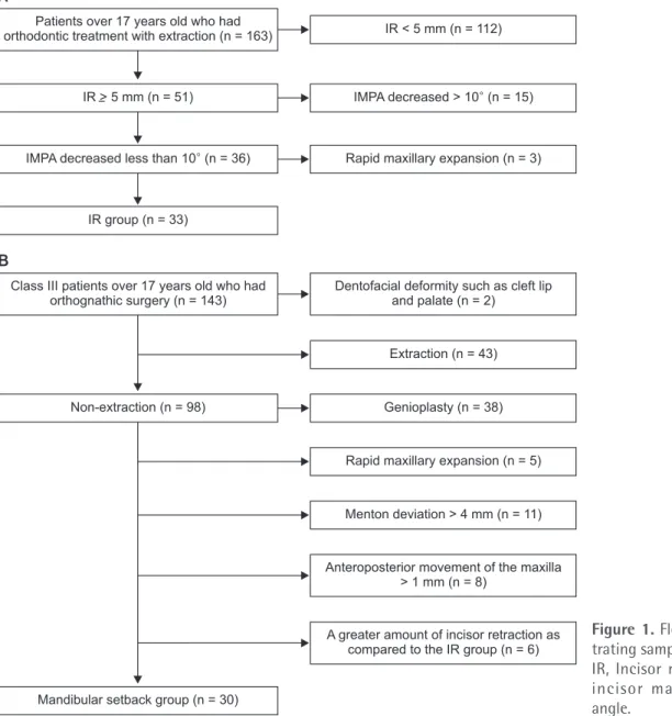
SubjectsThis retrospective study included 163 patients who had undergone orthodontic treatment with extraction at the Department of Orthodontics, Yonsei University Dental Hospital, Seoul, Korea, between 2006 and 2012. Among these patients, 33 (17 men and 16 women; mean age, 24.52 ± 6.15 years) who satisfied the follo-wing inclusion criteria were included in the incisor re-traction (IR) group: over 17 years of age, history of four premolar extractions (a tooth per quadrant), more than 5 mm bodily retraction (reference point: estimated center of resistance located at 0.67 of the root length from the apex of each incisor as measured on a lateral
cephalogram) of an incisor,13,14 no severe dentofacial
deformity such as a cleft lip or palate, no maxillary ex-pansion using rapid maxillary exex-pansion (RME), and less than 10º decrease in the incisor mandibular plane angle (IMPA) (Figure 1A).
All patients in the IR group were treated with pre-adjusted 0.018-inch edgewise brackets with the Roth prescription (Tomy, Tokyo, Japan). After leveling and alignment, tapered miniscrews with 1.8-mm diameter and 7.0-mm threaded length (Orlus No 18107; Ortho-lution, Seoul, Korea) were placed between the maxillary and mandibular second premolars and the first molar under infiltration anesthesia. Thereafter, 0.016 × 0.022-inch stainless steel rectangular archwires with additional labial crown torque (10o) on the incisor segment were
placed in both arches, including the second molars. Short crimpable hooks (TP Orthodontics, LaPorte, IN, USA) were attached distally to the lateral incisor. A retraction force of 150 g was provided by using elastic chains (Ormco, Glendora, CA, USA), and the chains were replaced every 4 weeks. Space closure was performed independently in each arch.
We chose 36 patients who satisfied our inclusion criteria for the mandibular setback (MS) group from a pool of 143 patients who had undergone orthognathic surgery at our hospital between 2006 and 2012. These 36 patients met the following inclusion criteria: over 17 years of age, history of orthognathic surgery without extraction, no severe dentofacial deformity, no RME, no genioplasty, and no severe facial asymmetry over 4 mm of menton deviation from the facial midline. Patients who had over 1 mm anteroposterior movement of the maxilla were excluded, in order to remove the effect on the PAS.15-17 Among the 36 patients chosen, six who had
incisor retraction of over 9 mm were excluded in order to evenly match the amount of posterior displacement of the mandibular incisors between the two groups. The final MS group included in the study comprised 30 patients (15 men and 15 women; n = 3, one-jaw sur-gery; n = 27, two-jaw sursur-gery; mean age, 22.78 ± 4.82 years) (Figure 1B). Patients in the MS group underwent bilateral one-piece Le Fort I osteotomy of the canine fossa and zygomatic buttress and bilateral intraoral ver-tical ramus osteotomy, as carried out for mandibular se-tback.
The study protocol conformed to the guidelines of the Declaration of Helsinki and was approved by the Ins-titutional Review Board of Yonsei Dental Hospital, Seoul, Korea (2-2015-0033).
Methods
Lateral cephalograms were acquired using a Cranex 3+ (Soredex, Helsinki, Finland) in the natural head position without swallowing before (T1) and after (T2) treatment.
The Frankfort horizontal (FH) plane at T1 was set as the horizontal reference plane (HRP). The vertical reference plane (VRP) was the plane that passed through the sella, perpendicular to the HRP. All cephalometric landmarks were digitized using the V-ceph program (Osstem Inc., Seoul, Korea). Landmarks and variables were set on the basis of the recommendations of previous studi es11,12,17
(Figure 2). Reliability
All lateral cephalometric measurements were per-formed by the same investigator. Two weeks after the first digitization of the landmarks, all measurements were re-digitized by the same investigator. The intraclass correlation coefficient was greater than 0.94.
Statistical analysis
All statistical analyses were performed using IBM SPSS Statistics for Windows, version 21.0 (IBM Corp., Armonk, NY, USA). The Shapiro-Wilk test was used to verify the normality of data distribution. Sex distribution and mean age were not normally distributed; thus, non-parametric tests such as chi-square and Mann-Whitney U test were used. The paired t-test and independent t-tests were used to compare the changes in variables between T1 and T2 in each group and between the two groups, respectively. Pearson’s correlation analysis was used to determine the relationship between skeletal, dental, and pharyngeal variables in each group.
A
B
IR group (n = 33)
IR < 5 mm (n = 112)
IMPA decreased > 10 (n = 15)
Rapid maxillary expansion (n = 3)
Dentofacial deformity such as cleft lip and palate (n = 2)
Extraction (n = 43)
Genioplasty (n = 38)
Rapid maxillary expansion (n = 5)
Menton deviation > 4 mm (n = 11)
Anteroposterior movement of the maxilla > 1 mm (n = 8)
A greater amount of incisor retraction as compared to the IR group (n = 6) Patients over 17 years old who had
orthodontic treatment with extraction (n = 163)
IR > 5 mm (n = 51)
IMPA decreased less than 10 (n = 36)
Class III patients over 17 years old who had orthognathic surgery (n = 143)
Non-extraction (n = 98)
Mandibular setback group (n = 30)
Figure 1. Flow diagram
illus-trating sample selection. IR, Incisor retraction; IMPA, incisor mandibular pl ane angle.
RESULTS
No difference was observed in sex distribution or mean age between the IR and MS groups. However, the mean treatment duration of the IR group (34.76 ± 9.48 mon-ths) was significantly longer than that of the MS group (17.87 ± 6.86 months; p < 0.001) (Table 1).
Significant intergroup differences were observed in the angle of the sella-nasion plane to point B (SNB); the angle of the lines connecting point A, the nasion, and
point B; the angle of the FH plane to the mandibular plane; horizontal positions of point B (B-VRP), the hyoid bone (H-VRP), and the center of resistance of the lower incisor (L1-VRP); vertical position of the center of resistance of the upper incisor (U1-HRP); and the IMPA at T1 (Table 2). The inferior pharyngeal airway space (E-IPW) in the IR group (6.65 ± 1.71 mm) was significantly smaller than that in the MS group (7.89 ± 2.45 mm; p < 0.05).
In the IR group, points A and B moved 0.28 ± 0.47 mm and 0.47 ± 0.95 mm posteriorly, respectively, and the hyoid bone moved inferiorly after treatment (p < 0.01). The middle pharyngeal airway space (U-MPW) decreased 1.15 ± 1.17 mm after treatment (p < 0.001) (Table 2).
In the MS group, point B moved superoposteriorly, and the hyoid bone moved posteriorly after surgery (p < 0.001). The upper and lower incisors moved posteriorly (p < 0.001), but the IMPA increased after surgery (p < 0.01). At T2, both the U-MPW and the E-IPW decreased 1.25 ± 1.35 mm (p < 0.001) and 0.88 ± 1.67 mm (p < 0.01), respectively (Table 2).
After treatment, no significant intergroup differences
Figure 2. A, Cephalometric landmarks and planes. S, sella; N, nasion; A, point A; B, point B; Po, porion; Or, orbitale; PNS,
posterior nasal spine; Go, gonion; Me, menton; H, the most anterosuperior point of the hyoid bone; U, tip of the uvula; E, tip of the epiglottis; U1, center of resistance of U1; L1, center of resistance of L1; HRP, horizontal reference plane– the Frankfort horizontal plane; VRP, vertical reference plane—passes through the sella, perpendicular to the HRP; SPW, superior pharyngeal wall, point of intersection of the posterior pharyngeal wall and perpendicular line drawn from the PNS; MPW, middle pharyngeal wall, point of intersection of the posterior pharyngeal wall and perpendicular line drawn from the U; IPW, inferior pharyngeal wall, point of intersection of the posterior pharyngeal wall and perpendicular line
drawn from the E. B, Skeletal measurements. 1, A-VRP, perpendicular distance from the VRP to point A (mm); 2, A-HRP,
perpendicular distance from the HRP to point A (mm); 3, B-VRP, perpendicular distance from the VRP to point B (mm); 4, B-HRP, perpendicular distance from the HRP to point B (mm); 5, H-VRP, perpendicular distance from the VRP to point H (mm); 6, H-HRP, perpendicular distance from the HRP to point H (mm); 7, PNS-HRP, perpendicular distance from the
HRP to the PNS (mm). C, Dental and pharyngeal airway space measurements. 1, U1-VRP, perpendicular distance from the
VRP to U1 (mm); 2, U1-HRP, perpendicular distance from the HRP to U1 (mm); 3, L1-VRP, perpendicular distance from
the VRP to L1 (mm); 4, L1-HRP, perpendicular distance from the HRP to L1 (mm); 5, U1-SN (o); 6, IMPA (o); 7, PNS-SPW,
superior pharyngeal airway (mm); 8, U-MPW, middle pharyngeal airway (mm); 9, E-IPW, inferior pharyngeal airway (mm). B HRP 1 2 3 4 6 7 VRP C HRP VRP 1 2 4 7 6 3 5 8 9 A S N Or HRP PNS Go H Me B L1 VRP U1 A SPW SPW 5 5 Po MPW UU MPW IPW E
Table 1. Demographic features of the subjects
Incisor retraction group (n = 33) Mandibular setback group (n = 30) p-value Sex, male:female 17:16 15:15 NS* Age (yr) 24.52 ± 6.15 22.78 ± 4.82 NS† Treatment duration (mo) 34.76 ± 9.48 17.87 ± 6.86 < 0.001‡ NS, Not significant.
p-value calculated with *chi-square test, †Mann−Whitney U test, and ‡independent t-test.
Table 2.
Comparison of the changes in skeletal, dental, and pharyngeal variables during the tr
eatment period (T2–T1) Vari able T1 T2 Δ T2– T1 In cis or retr action (n = 33) M an dibul ar set ba ck (n = 30) p -valu e* In cis or retr action (n = 33) M an dibul ar set ba ck (n = 30) p -valu e* In cis or r etr action (n = 33) M an dibul ar s et ba ck (n = 30) p -valu e* Me an SD Me an SD Me an SD Me an SD Me an SD p -valu e † Me an SD p -valu e † Sk eletal S NA ( o ) 81.46 2.98 80.17 2.85 NS 81.49 3.00 80.24 2.90 NS 0.03 0.29 NS 0.06 0.20 NS NS S NB ( o ) 77.96 3.53 82.43 2.91 < 0.001 77.87 3.57 78.23 3.07 NS −0.09 0.48 NS −4.21 1.09 < 0.001 < 0.001 ANB ( o ) 3.50 3.06 −2.16 1.97 < 0.001 3.62 2.88 2.00 1.85 < 0.05 0.13 0.57 NS 4.17 1.13 < 0.001 < 0.001 FMA ( o ) 28.89 6.14 25.65 5.56 < 0.05 28.66 6.21 31.06 5.30 NS −0.22 0.64 NS 5.41 2.84 < 0.001 < 0.001 A -V RP (mm) 70.97 4.82 69.87 5.38 NS 70.68 4.78 69.66 5.46 NS −0.28 0.47 < 0.01 −0.21 0.39 < 0.01 NS A -HRP (mm) 36.59 4.38 36.79 3.50 NS 36.63 4.38 36.42 3.61 NS 0.04 0.28 NS −0.37 0.81 < 0.05 < 0.05 B-V RP (mm) 63.44 6.77 73.77 7.14 < 0.001 62.96 6.64 65.23 7.27 NS −0.47 0.95 < 0.01 −8.54 1.76 < 0.001 < 0.001 B-HRP (mm) 85.44 7.97 83.21 5.69 NS 85.29 7.97 81.23 4.96 < 0.05 −0.15 0.47 NS −1.97 2.24 < 0.01 < 0.001 H -V RP (mm) 11.32 6.78 17.79 7.06 < 0.01 10.77 6.21 13.36 7.03 NS −0.55 4.35 NS −4.43 4.56 < 0.001 < 0.001 H -HRP (mm) 94.4 8.46 93.06 8.76 NS 96.87 9.59 94.48 9.44 NS 2.46 3.70 < 0.01 1.42 4.11 NS NS P NS -HRP (mm) 28.59 2.84 28.69 2.59 NS 28.51 2.86 25.43 3.14 < 0.001 −0.07 0.23 NS −3.26 1.70 < 0.001 < 0.001 D en tal U1-V RP (mm) 72.92 5.32 70.90 6.05 NS 67.59 4.87 69.84 6.04 NS −5.32 2.11 < 0.001 −1.05 1.28 < 0.001 < 0.001 U1-HRP (mm) 51.48 5.11 49.11 3.49 < 0.05 50.87 4.99 48.62 3.70 < 0.05 −0.61 1.23 < 0.01 −0.49 1.09 < 0.05 NS L1-V RP (mm) 69.92 5.69 74.45 6.71 < 0.01 63.29 5.58 67.77 6.45 < 0.01 −6.64 0.89 < 0.001 −6.67 1.42 < 0.001 NS L1-HRP (mm) 70.93 5.99 70.19 6.53 NS 69.96 5.51 68.55 6.00 NS −0.97 1.75 < 0.01 −1.64 2.10 < 0.001 NS U1-SN ( o ) 108.38 6.30 109.89 10.03 NS 99.23 5.84 105.00 9.55 < 0.01 −9.15 4.61 < 0.001 −4.89 4.33 < 0.001 < 0.001 IMP A ( o ) 97.19 6.31 81.89 8.42 < 0.001 91.09 5.97 86.46 7.03 < 0.01 −6.11 2.72 < 0.001 4.57 6.29 < 0.05 < 0.001 P h ar yn ge al P NS -S P W (mm) 21.65 3.53 20.98 3.64 NS 21.56 3.54 20.52 3.40 NS −0.10 1.78 NS −0.46 1.98 NS NS U-MP W (mm) 11.07 3.01 12.35 3.37 NS 9.92 2.95 11.10 3.20 NS −1.15 1.17 < 0.001 −1.25 1.35 < 0.001 NS E-IP W (mm) 6.65 1.71 7.89 2.45 < 0.05 6.60 1.98 7.00 1.79 NS −0.05 1.29 NS −0.88 1.67 < 0.01 < 0.05 SD , S tand ar d de vi ation; NS , not s ig nific an t. T1, b efor e tr ea tmen t; T2, aft er tr ea tmen t; HRP , hor iz on tal r efer ence pl ane (FH pl ane , P or ion-Or bitale); V RP , v er tic al r efer ence pl ane (p as ses thr ough S , p erp endic ul ar t o the HRP); A , p oin t A; B , p oin t B; H, the mos t an ter os up er ior p oin t of the h yoid b
one; U1, cen
ter of r
es
is
tance of U1; L1, cen
ter of r es is tance of L1; P NS , p os ter ior n as al s pine; U , tip of the u vul a; E
, tip of the epi
glot tis ; S P W, fo ot p oin t of the p erp endic ul ar line fr om the P NS t o the p os ter ior ph ar yn ge al w all; MP W, fo ot p oin t of the p erp endic ul ar line fr om the U t o the p os ter ior ph ar yn ge al w all; IP W, fo ot p oin t of the p erp endic ul ar line fr om the E t o the p os ter ior ph ar yn ge al w all. *T he indep enden t t -t es ts w er e us ed t o com p ar e the me an v alue of e ac h v ar ia ble b et w
een the incis
or r etr action and m andib ul ar s et b ac k gr oups ; † the p air ed t-t es t w er e us ed to com p ar e the me an v alue of e ac h v ar ia ble b et w een T1 and T2 in e ac h gr oup .
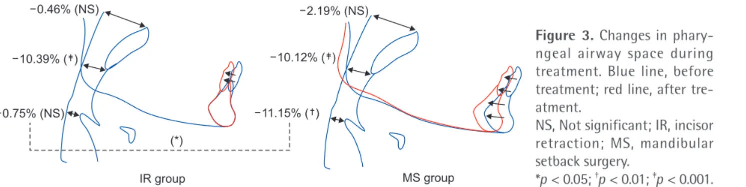
were observed in the SNB, B-VRP, and H-VRP. Additio-nally, no significant intergroup difference was observed in the E-IPW at T2. The hyoid bone moved more pos-teriorly in the MS group than in the IR group (p < 0.001), but no significant intergroup difference was observed in the vertical movement of the hyoid bone. No significant intergroup difference was observed in the superior pharyngeal airway (PNS-SPW) and the U-MPW after treatment. However, a greater decrease in the E-IPW was observed after surgery in the MS group than in the IR group (p < 0.05) (Figure 3). Moreover, no significant correlation was observed between changes in the PAS and the ske letal and dental cephalometric variables in either group (data not shown).
DISCUSSION
In this study, superior impaction of the posterior ma-xilla (about 3.3 mm) was not considered a criterion for exclusion because previous reports have shown that superior repositioning of the posterior nasal spine (about 3.5–4.5 mm) does not have an effect on the dimensional change of the PAS.5,17
The E-IPW in the MS group was about 1.24 mm larger than that in the IR group before treatment. Some studies have reported that the skeletal classification has no effect on the PAS.18,19 However, other studies have
revealed that patients with skeletal Class III malocclusion have a larger PAS than do patients with Class I or Class II malocclusion.20-22 In this study, neither the
PNS-SPW, U-MPW, nor E-IPW showed significant differences between the two groups at T2 because the PAS in the MS group decreased because of mandibular setback surgery.
In the IR group, the U-MPW decreased by 10.39% during treatment (Figure 3). In this study, unlike in a previous study,12 the posterior movement of the
mandibular incisor was measured at the estimated center of resis tance rather than at the anterior tip to ensure that the measurement reflected bodily retraction of the man dibular incisors.13,14 After treatment, the
movement of the mandibular incisors measured to
the mandibular plane showed that they had retracted by about 6.1o. Moreover, the posterior movement of
the center of resistance of the mandibular incisor was 6.64 ± 0.89 mm, indicating a 1:0.96 ratio of incisal edge-to-apex displacement. This ratio was considered acceptable to represent the bodily retraction of the mandibular incisor based on the findings of previous studies.23,24 Wang et al.12 reported that the U-MPW
decreased by 7.88% in patients with skeletal Class I malocclusion who had undergone four-bicuspid extraction, and showed a significant correlation between the retraction of the lower incisor and the airway behind the soft palate, uvula, and tongue. Because the maxillary incisor is located above the man dibular incisor, the bodily retraction of the maxillary incisor is not expected to affect the PAS change com pared with the bodily retraction of the mandibular incisor. Taken together, these results show that the PAS may decrease after mandibular incisor retraction, and it may occur because of a decrease in the intraoral volume and its compression by the posterior movement of the tongue and soft palate.
Many studies have described the decrease in the PAS after mandibular setback surgery, but the extent of decrease reported was slightly different in each study.3-6,15 Consistent with the results of previous studies,
the results of this study did not show any change in the PNS-SPW space in the MS group after surgery.6 However,
the U-MPW of those patients decreased by 10.12% and their E-IPW decreased by 11.15% after surgery (Figure 3).
After treatment, the E-IPW decreased in the MS group, but no significant intergroup difference in the E-IPW was observed at T2. Additionally, no signifi-cant intergroup difference was observed in the antero-posterior position of point B at T2. This result suggests a correlation between the anteroposterior position of the mandible body and the E-IPW.21,22 It is possible that
the U-MPW decreased in both groups because of the effect of the posterior displacement of the mandibular incisors on the tongue and soft palate, but the E-IPW decreased only in the MS group because of the posterior displacement of the mandibular body.
Figure 3. Changes in
phary-ngeal airway space during treatment. Blue line, before treatment; red line, after tre-atment.
NS, Not significant; IR, incisor retraction; MS, mandibular setback surgery. *p < 0.05; †p < 0.01; ‡p < 0.001. IR group MS group 0.46% (NS) 0.75% (NS) 10.39% ( ) (*) 11.15% ( ) 2.19% (NS) 10.12% ( )
Nevertheless, the extent of posterior movement of the incisors and/or the mandibular body does not have a direct correlation with the magnitude of decrease of the PAS. The U-MPW increased after treatment in 15.15% of the patients in the IR group (5 of 33) and 13.33% of the patients in the MS group (4 of 30). The E-IPW too increased in 23.33% of the patients in the MS group (7 of 30) after surgery. It is difficult to assume that, with increased posterior movement of the incisor and mandible, the PAS decreased correspondingly. This is because of the variations among individuals in the reaction of the tongue, pharyngeal airway, and adjacent muscles, as well as limitations in the precise evaluation of the PAS, as the transverse width of the pharyngeal airway could not be measured using lateral cephalometric radiograms.
In this study, the hyoid bone was observed to move more posteriorly, and inferiorly, in the MS group than in the IR group. However, no significant intergroup difference was observed in the extent of inferior move-ment of the hyoid bone. The impact of the posterior and inferior movement of the hyoid bone on the PAS remains controversial.25 Previous studies have reported
the posteroinferior movement of the hyoid bone im-mediately after mandibular setback surgery, and subsequent partial reversion to its original position.5,26-28
The position of the hyoid bone is determined by the balance of the muscles attached to the cranial base and mandibular symphysis,12 and the inferior movement of
the hyoid bone is an adaptation process to prevent the tongue from encroaching the PAS.3 Further long-term
evaluation of the stability of the hyoid bone is required. This study has several limitations, which should be taken into consideration when interpreting the data. The purpose of this study was to compare and evaluate the extent of PAS reduction by site when comparing the bodily retraction of the mandibular incisor to the posterior movement associated with the simul-taneous posterior displacement of the mandibular incisor and body through mandibular setback surgery. Unfortunately, the two groups had significantly different anteroposterior skeletal relationships of the jaws before treatment. This is because most cases requiring bodily retraction of the maxillary and mandibular incisors are primarily those of skeletal Class I or II malocclusion with bimaxillary protrusion, whereas mandibular setback surgery is mainly performed in cases of skeletal Class III malocclusion with mandibular prognathism. An additional consideration is that the lateral cephalogram, used for evaluation here, has the limitation of allowing only a two-dimensional evaluation of the PAS. Although many previous studies4,5 have used two-dimensional
lateral cephalograms to measure changes in tongue position, this study did not measure the change in
tongue position, size, or volume before and after treat-ment in each group. This is because the position of the tongue may be affected by subtle changes in head position, even though all radiograms were acquired in a reproducible manner with unstrained position of the head.29 In fact, systematic reports of individual variability
and reproducibility of the airway dimensions and tongue and hyoid position on lateral cephalometric radiograms at the same head position are scarce. However, initial tongue position and subsequent changes may predispose the effect of treatments on the upper airway. In the MS group, patients who underwent one-jaw and two-jaw surgeries were included. Although this study minimized the effect of maxillary surgery on the dimensional change of the PAS by limiting the anteroposterior move-ment of the maxilla, the MS group lacked homogeneity and this could constitute another limitation of this study. This study was designed retrospectively and evaluated only the PAS without subjective data such as patient questionnaires or objective data such as pul-monary function or polysomnography data. Further research is required to verify the conclusions drawn from this study. This should include a prospective design including the abovementioned subjective and objective data and should control for confounding factors or individual variations such as the reaction of the tongue, pharyngeal airway, and adjacent muscles, as well as the transverse width of the pharyngeal airway.
CONCLUSION
Within the limitations of this study, it still showed that the majority of patients studied experienced a decrease in the U-MPW because of the posterior displacement of the mandibular incisors and/or the mandibular body, thereby affecting the tongue and soft palate. The E-IPW decreased only in the MS group because of the posterior displacement of only the mandibular body. However, the amount of posterior movement of the incisors and/or mandibular body did not have a direct correlation with the amount of decrease of the PAS because the reactions of the tongue, pharyngeal airway, and adjacent muscles varied among subjects. Future prospective and well-controlled studies of individual variations are necessary to obtain more robust results.
REFERENCES
1. Hoekema A, Hovinga B, Stegenga B, De Bont LG. Craniofacial morphology and obstructive sleep apnoea: a cephalometric analysis. J Oral Rehabil 2003;30:690-6.
2. Alessandri-Bonetti G, Ippolito DR, Bartolucci ML, D'Antò V, Incerti-Parenti S. Cephalometric pre dictors
of treatment outcome with mandibular advan-cement devices in adult patients with obstructive sleep apnea: a systematic review. Korean J Orthod 2015;45:308-21.
3. Tselnik M, Pogrel MA. Assessment of the pharyngeal airway space after mandibular setback surgery. J Oral Maxillofac Surg 2000;58:282-5; discussion 285-7.
4. Kawakami M, Yamamoto K, Fujimoto M, Ohgi K, Inoue M, Kirita T. Changes in tongue and hyoid positions, and posterior airway space following ma-ndibular setback surgery. J Craniomaxillofac Surg 2005;33:107-10.
5. Hwang S, Chung CJ, Choi YJ, Huh JK, Kim KH. Changes of hyoid, tongue and pharyngeal airway after mandibular setback surgery by intraoral vertical ramus osteotomy. Angle Orthod 2010;80:302-8. 6. Gokce SM, Gorgulu S, Gokce HS, Bengi O,
Sabun-cuoglu F, Ozgen F, et al. Changes in posterior air way space, pulmonary function and sleep quality, follo-wing bimaxillary orthognathic surgery. Int J Oral Maxillofac Surg 2012;41:820-9.
7. Mattos CT, Vilani GN, Sant'Anna EF, Ruellas AC, Maia LC. Effects of orthognathic surgery on oropha-ryngeal airway: a meta-analysis. Int J Oral Maxillofac Surg 2011;40:1347-56.
8. Park HS, Yoon DY, Park CS, Jeoung SH. Treatment effects and anchorage potential of sliding mechanics with titanium screws compared with the Tweed-Me-rrifield technique. Am J Orthod Dentofacial Orthop 2008;133:593-600.
9. Liu YH, Ding WH, Liu J, Li Q. Comparison of the differences in cephalometric parameters after ac-tive orthodontic treatment applying mini-screw implants or transpalatal arches in adult patients with bialveolar dental protrusion. J Oral Rehabil 2009;36:687-95.
10. Germec-Cakan D, Taner T, Akan S. Uvulo-glossopha-ryngeal dimensions in non-extraction, extraction with minimum anchorage, and extraction with maximum anchorage. Eur J Orthod 2011;33:515-20. 11. Chen Y, Hong L, Wang CL, Zhang SJ, Cao C, Wei F, et al. Effect of large incisor retraction on upper airway morphology in adult bimaxillary protrusion patients. Angle Orthod 2012;82:964-70.
12. Wang Q, Jia P, Anderson NK, Wang L, Lin J. Chan-ges of pharyngeal airway size and hyoid bone posi-tion following orthodontic treatment of Class I bi-maxillary protrusion. Angle Orthod 2012;82:115-21. 13. Burstone CJ, Pryputniewicz RJ. Holographic deter-mination of centers of rotation produced by orthodontic forces. Am J Orthod 1980;77:396-409. 14. Koo YJ, Choi SH, Keum BT, Yu HS, Hwang CJ,
Me-lsen B, et al. Maxillomandibular arch width
diffe-rences at estimated centers of resistance: comparison between normal occlusion and skeletal Class III ma-locclusion. Korean J Orthod 2017;47:167-75. 15. Chen F, Terada K, Hua Y, Saito I. Effects of
bima-xillary surgery and mandibular setback surgery on pharyngeal airway measurements in patients with Class III skeletal deformities. Am J Orthod Den-tofacial Orthop 2007;131:372-7.
16. Degerliyurt K, Ueki K, Hashiba Y, Marukawa K, Na-kagawa K, Yamamoto E. A comparative CT evalu-ation of pharyngeal airway changes in Class III pa-ti ents receiving bimaxillary surgery or mandibular setback surgery. Oral Surg Oral Med Oral Pathol Oral Radiol Endod 2008;105:495-502.
17. Jakobsone G, Stenvik A, Espeland L. The effect of maxillary advancement and impaction on the upper airway after bimaxillary surgery to correct Class III malocclusion. Am J Orthod Dentofacial Orthop 2011;139(4 Suppl):e369-76.
18. Alves PV, Zhao L, O'Gara M, Patel PK, Bolognese AM. Three-dimensional cephalometric study of upper airway space in skeletal Class II and III healthy patients. J Craniofac Surg 2008;19:1497-507. 19. de Freitas MR, Alcazar NM, Janson G, de Freitas KM,
Henriques JF. Upper and lower pharyngeal airways in subjects with Class I and Class II malocclusions and different growth patterns. Am J Orthod Dentofacial Orthop 2006;130:742-5.
20. Martin O, Muelas L, Viñas MJ. Comparative study of nasopharyngeal soft-tissue characteristics in patients with Class III malocclusion. Am J Orthod Dentofacial Orthop 2011;139:242-51.
21. Hong JS, Oh KM, Kim BR, Kim YJ, Park YH. Three-dimensional analysis of pharyngeal airway volume in adults with anterior position of the mandible. Am J Orthod Dentofacial Orthop 2011;140:e161-9. 22. Zheng ZH, Yamaguchi T, Kurihara A, Li HF, Maki K.
Three-dimensional evaluation of upper airway in patients with different anteroposterior skeletal pat-terns. Orthod Craniofac Res 2014;17:38-48. 23. Yao CC, Lai EH, Chang JZ, Chen I, Chen YJ.
Com-parison of treatment outcomes between skeletal anchorage and extraoral anchorage in adults with maxillary dentoalveolar protrusion. Am J Orthod Dentofacial Orthop 2008;134:615-24.
24. Al-Sibaie S, Hajeer MY. Assessment of changes fo-llowing en-masse retraction with mini-implants anchorage compared to two-step retraction with conventional anchorage in patients with Class II division 1 malocclusion: a randomized controlled trial. Eur J Orthod 2014;36:275-83.
25. Hu Z, Yin X, Liao J, Zhou C, Yang Z, Zou S. The effect of teeth extraction for orthodontic treatment on the upper airway: a systematic review. Sleep
Breath 2015;19:441-51.
26. Gu G, Gu G, Nagata J, Suto M, Anraku Y, Nakamura K, et al. Hyoid position, pharyngeal airway and head posture in relation to relapse after the mandibular setback in skeletal Class III. Clin Orthod Res 2000; 3:67-77.
27. Güven O, Saraçoğlu U. Changes in pharyngeal air-way space and hyoid bone positions after body ostectomies and sagittal split ramus osteotomies. J Craniofac Surg 2005;16:23-30.
28. Athanasiou AE, Toutountzakis N, Mavreas D, Ritzau M, Wenzel A. Alterations of hyoid bone position and pharyngeal depth and their relationship after surgical correction of mandibular prognathism. Am J Orthod Dentofacial Orthop 1991;100:259-65. 29. Malkoc S, Usumez S, Nur M, Donaghy CE.
Repro-ducibility of airway dimensions and tongue and hyoid positions on lateral cephalograms. Am J Or-thod Dentofacial Orthop 2005;128:513-6.