Immediate effects of mandibular posterior
displacement on the pharyngeal airway space:
A preliminary study
Objective: This study aimed to evaluate the immediate effects of mandibular posterior displacement on the pharyngeal airway space (PAS) by using cephalometric evaluations and to investigate how the surrounding structures are schematically involved. Methods: In this retrospective study, 38 subjects with functional Class III malocclusion and two lateral cephalograms were selected.
The first lateral cephalogram was taken with the mandible in the habitual occlusal position, and the second in anterior edge-to-edge bite. Paired t-test was used to analyze changes in the PAS, hyoid bone, tongue, and soft palate, followed by mandibular posterior displacement. Pearson’s correlation analysis was used to determine the relationship between the amount of mandibular posterior displacement and other variables. Results: A statistically significant decrease was observed in the PAS following mandibular posterior displacement.
Along with mandibular posterior displacement, the tongue decreased in length (p < 0.001) and increased in height (p < 0.05), while the soft palate increased in length, decreased in thickness, and was posteriorly displaced (p < 0.001). The hyoid bone was also posteriorly displaced (p < 0.05). There was no correlation between the amount of mandibular posterior displacement and the measured variables. Conclusions: The PAS showed a statistically significant decrease following mandibular posterior displacement, which was a consequence of retraction of the surrounding structures. However, there were individual variances between the amount of mandibular posterior displacement and the measured variables.
[Korean J Orthod 2020;50(2):129-135]
Key words: Lateral cephalogram, Airway, Mandibular posterior displacement Yeonju Choi
Yong-Il Kim Seong-Sik Kim Soo-Byung Park Woo-Sung Son Sung-Hun Kim
Department of Orthodontics, Pusan National University Dental Hospital, Dental Research Institute, Yangsan, Korea
Received August 9, 2019; Revised December 13, 2019; Accepted December 23, 2019.
Corresponding author: Sung-Hun Kim.
Clinical Professor, Department of Orthodontics, Pusan National University Dental Hospital, 20, Geumo-ro, Mulgeum-eup, Yangsan 50612, Korea.
Tel +82-55-360-5153 e-mail [email protected]
How to cite this article: Choi Y, Kim YI, Kim SS, Park SB, Son WS, Kim SH. Immediate effects of mandibular posterior displacement on the pharyngeal airway space:
A preliminary study. Korean J Orthod 2020;50:129-135.
© 2020 The Korean Association of Orthodontists.
This is an Open Access article distributed under the terms of the Creative Commons Attribution Non-Commercial License (http://creativecommons.org/licenses/by-nc/4.0) which permits unrestricted non-commercial use, distribution, and reproduction in any medium, provided the original work is properly cited.
pISSN 2234-7518 • eISSN 2005-372X
https://doi.org/10.4041/kjod.2020.50.2.129
INTRODUCTION
The craniofacial structures are physiologically con- nected to each other, and their coordinated growth is essential for normal craniofacial development. Growth and function of the pharyngeal airway space (PAS) are especially intimately related to the normal development of the maxillofacial structures.
1Due to the importance of the PAS, various studies have assessed its growth and development.
2For example, narrowed dimensions of the PAS may cause breathing difficulties. Subsequent mouth breathing due to pharyngeal obstruction may also pres- ent symptoms like an “adenoid face” or a short, retract- ed mandible.
The pharynx is the main anatomical structure that constitutes the PAS and is an organ composed of mus- cles that originate from the adjacent skeletal structures.
Its size and shape are affected by the surrounding skel- etal structure.
3The nasopharynx and the oropharynx form a functional unit involved in breathing, swallow- ing, and pronunciation.
The dimensions of the PAS are affected by factors such as head posture, tongue position, age, body mass index (BMI),
4as well as by alterations in mandibular po- sition. When the mandibular position is altered, subse- quent changes occur in the positions of the hyoid bone, the tongue, and the soft palate, ultimately altering the dimensions of the PAS. For example, repositioning the mandible anteriorly or posteriorly in an orthognathic surgery,
5,6or using an intraoral device that advances the mandible forward,
7may alter the dimensions of the PAS.
Thus, we should consider the dimensional changes in the PAS when designing an orthodontic intraoral appli- ance.
Since multiple factors contribute to the dimensional changes in the PAS, it is difficult to properly predict the changes exclusively affected by mandibular displace- ment. Thus, in order to eliminate other undesired fac- tors affecting the PAS, the data must be taken at the same time and posture, only making modifications in the mandibular position. Previous studies,
5-7however, have used measurements taken from before and after orthodontic treatment, or while wearing a device, all of which presented limitations in maintaining consistent conditions with respect to growth, tongue position, and head posture. Thus, studies pertaining to changes in the PAS following mandibular posterior displacement that is unaffected by factors such as growth, tongue position, and head posture are unprecedented. The availability of two lateral cephalograms taken to diagnose functional Class III malocclusions may facilitate such studies.
The aim was to investigate the changes in the PAS following posterior displacement of the mandible by using cephalometric evaluations and to assess the inter-
relationships with the surrounding structures.
MATERIALS AND METHODS
Subjects
We selected 369 subjects among anterior crossbite pa- tients who visited the Department of Pediatric Dentistry and Orthodontics, Pusan National University Dental Hospital, between 2009 and 2018, and had taken two lateral cephalograms in altered mandibular positions for diagnostic purposes. Out of these patients, 38 (14 boys and 24 girls) patients satisfied the inclusion criteria (Table 1): aged 5 to 13 years, no orthodontic appliance, overbite less than 2 mm, nose breather, no obstructive sleep apnea syndrome (OSAS), BMI less than 30 kg/
m
2, no developmental syndromes, such as cleft lip and palate, anterior crossbite in the habitual occlusal posi- tion (HOP), the ability to guide the mandible to anterior edge-to-edge position, and no changes in cranio-cer- vical angle while taking two cephalograms. The sample size with a test power of 0.8 (α = 0.05) and a medium effect size of 0.5 proposed by Cohen
8included 34 sub- jects, whereas this study included 38 subjects to amply satisfy the sample size criteria. This study was approved by the Institutional Review Board of Pusan National University Dental Hospital (PNUDH-2019-008).
Data acquisition
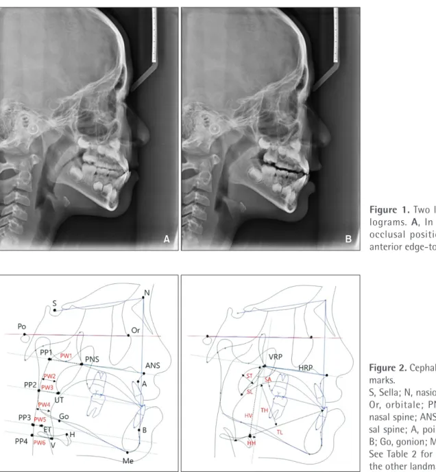
The subjects were cautioned to refrain from swallow- ing or changing their posture while the lateral cepha- lograms were taken. Two lateral cephalograms of each subject were taken consecutively while maintaining the same head posture and only making alterations in the mandibular position. The first cephalogram was taken with the mandible in the HOP and the second with the mandible in anterior edge-to-edge bite (Figure 1). The landmarks used were referenced on previous studies.
5-7Changes in the PAS and the surrounding structures fol-
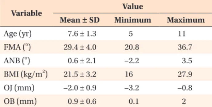
Table 1. Descriptive statistics
Variable Value
Mean ± SD Minimum Maximum
Age (yr) 7.6 ± 1.3 5 11
FMA (
o) 29.4 ± 4.0 20.8 36.7
ANB (
o) 0.6 ± 2.1 −2.2 3.5
BMI (kg/m
2) 21.5 ± 3.2 16 27.9
OJ (mm) −2.0 ± 0.9 −3.2 −0.8
OB (mm) 0.9 ± 0.6 0.1 2
SD, Standard deviation; FMA, Frankfort-mandibular plane
angle; ANB, A point-Nasion-B point angle; BMI, body mass
index; OJ, anterior overjet; OB, anterior overbite.
lowing mandibular posterior displacement were evalu- ated (Figure 2, Table 2). The dimensions in the PAS were quantified in six linear variables (PW1–6), the hyoid bone in two linear variables (HH and HV), the tongue in two linear variables (TL and TH), and the soft palate in two linear variables (ST and SL) and one angular vari- able (SA). Description of the measurements used in this study are depicited in Table 2.
Statistical analysis
All measurements were taken by one examiner. After 1 month, 15 randomly selected subjects underwent re- measurements in order to calculate the intra-examiner reliability using intraclass correlation coefficient (ICC) and Dahlberg’s method error formula. Shapiro-Wilk test
showed a normally distributed population. Therefore, the paired t-test was used in order to compare the ef- fects of mandibular posterior displacement in the PAS and the surrounding structures. Pearson’s correlation analysis was used to determine the relationship between the amount of mandibular posterior displacement and other measured variables.
RESULTS
Intra-examiner reliability was high. The average meth- od error was 0.24 mm and 0.34
oand the average ICC value was 0.892. The average mandibular posterior dis- placement at the incisal tip was 2.0 ± 0.9 mm. We ob- served a statistically significant decrease in all the vari-
A B
Figure 1. Two lateral cepha- lograms. A, In the habitual occlusal position. B, In the anterior edge-to-edge bite.
Figure 2. Cephalometric land- marks.
S, Sella; N, nasion; Po, porion;
Or, orbitale; PNS, posterior nasal spine; ANS, anterior na- sal spine; A, point A; B, point B; Go, gonion; Me, menton.
See Table 2 for definitions of
the other landmarks.
ables except PW1 (Table 3, Figure 3). In addition, male and female subjects showed no significant differences in airway changes. After mandibular posterior displace- ment, the tongue decreased in length and increased in height, and the soft palate increased in length, de- creased in thickness, and showed posterior displacement.
The hyoid bone also showed posterior displacement (Figure 4). No significant correlation was found among any of the analyzed variables following mandibular pos- terior displacement (Table 4).
DISCUSSION
The growth and development of the oral and maxil- lofacial system must be closely monitored before the growth rate reaches its peak in the adolescence period.
In particular, it is very important to recognize and iden- tify patients who have difficulty in nose-breathing in their early childhood. Orthodontists are responsible for preventing and eliminating any predisposing factors that may have adverse effects on growth direction and craniofacial patterns in the maxillofacial region. Since
the mandible and the pharynx are closely interrelated, prediction of PAS changes that might interfere with breathing is crucial while designing intraoral devices for orthodontic or orthopedic treatments.
Since the pharynx is known to undergo rapid growth until 13 years of age,
9it is necessary to take into ac- count developmental changes in order to accurately understand the dimensional changes in the PAS follow- ing mandibular displacement. Moreover, dimensional changes in the PAS may be biased by time discrepan- cies, resulting in discrepancies in head posture, tongue position, and so on. Therefore, it was difficult to clearly identify the changes exclusively pertaining to mandibu- lar posterior displacement. In this study, we evaluated the changes in the PAS following mandibular posterior displacement at the same point in time in the same pos- ture of the same patient, to accurately understand the correlations with the surrounding structures.
Statistical analysis showed reductions in the dimen- sional values in all regions of the PAS except the PW1 region since the mandible was posteriorly displaced.
As the tongue is posteriorly displaced, the contact area Table 2. Cephalometric landmarks and descriptions
Cephalometric landmark Description
H The most anterosuperior point of the hyoid bone
UT Tip of the uvula
ET Tip of the epiglottis
V Vallecula
PP1 The intersection point of the palatal plane at the posterior pharyngeal wall
PP2 The intersection point of the parallel plane drawn from UT to the posterior pharyngeal wall PP3 The intersection point of the parallel plane drawn from ET to the posterior pharyngeal wall PP4 The intersection point of the parallel plane drawn from V to the posterior pharyngeal wall PW1 Distance from PP1 to the posterior nasal spine (PNS)
PW2 Shortest distance from the soft palate to the posterior pharyngeal wall
PW3 Distance from PP2 to the UT
PW4 Shortest distance from the tongue to the posterior pharyngeal wall
PW5 Distance from PP3 to the ET
PW6 Distance from PP4 to V
Horizontal reference plane (HRP) The palatal plane
Vertical reference plane (VRP) Line through the PNS, perpendicular to the HRP
ST Soft palate thickness
SL Soft palate length
SA Inclination of the long axis of the soft palate to the palatal plane
TH Tongue height
TL Tongue length
HH Distance from H to VRP
HV Distance from H to HRP
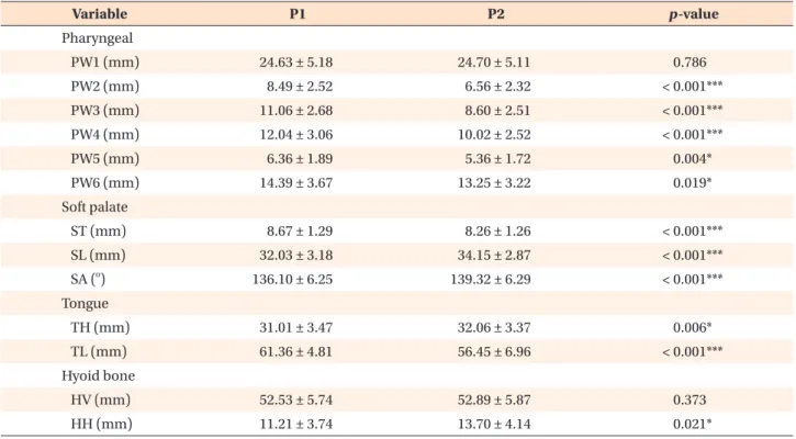
Table 3. Comparison of the changes in the pharyngeal, soft palate, tongue, and hyoid bone variables during mandibular posterior displacement
Variable P1 P2 p-value
Pharyngeal
PW1 (mm) 24.63 ± 5.18 24.70 ± 5.11 0.786
PW2 (mm) 8.49 ± 2.52 6.56 ± 2.32 < 0.001***
PW3 (mm) 11.06 ± 2.68 8.60 ± 2.51 < 0.001***
PW4 (mm) 12.04 ± 3.06 10.02 ± 2.52 < 0.001***
PW5 (mm) 6.36 ± 1.89 5.36 ± 1.72 0.004*
PW6 (mm) 14.39 ± 3.67 13.25 ± 3.22 0.019*
Soft palate
ST (mm) 8.67 ± 1.29 8.26 ± 1.26 < 0.001***
SL (mm) 32.03 ± 3.18 34.15 ± 2.87 < 0.001***
SA (
o) 136.10 ± 6.25 139.32 ± 6.29 < 0.001***
Tongue
TH (mm) 31.01 ± 3.47 32.06 ± 3.37 0.006*
TL (mm) 61.36 ± 4.81 56.45 ± 6.96 < 0.001***
Hyoid bone
HV (mm) 52.53 ± 5.74 52.89 ± 5.87 0.373
HH (mm) 11.21 ± 3.74 13.70 ± 4.14 0.021*
Values are presented as mean ± standard deviation.
P1, The mandible in the habitual occlusal position; P2, the mandible in the anterior edge-to-edge bite.
Paired t-test was used. *p < 0.05, ***p < 0.001.
See Table 2 for definitions of each landmark or mearurement.
Figure 3. Changes in the pharyngeal airway space after mandibular posterior displacement.
NS, Not significant.
*p < 0.05, ***p < 0.001.
+0.07 mm (NS)
-1.93 mm (***) -2.46 mm (***)
-2.02 mm (***) -1.00 mm (*)
-1.14 mm (*)
2.0 mm