Comparison of treatment effects between the
modified C-palatal plate and cervical pull headgear for total arch distalization in adults
Objective: The purpose of this study was to evaluate the dental and skeletal effects of the modified C-palatal plate (MCPP) for total arch distalization in adult patients with Class II malocclusion and compare the findings with those of cervical pull headgear. Methods: The study sample consisted of the lateral cephalograms of 44 adult patients with Class II Division 1 malocclusion, including 22 who received treatment with MCPP (age, 24.7 ± 7.7 years) and 22 who received treatment with cervical pull headgear (age, 23.0 ± 7.7 years).
Pre- (T1) and post-treatment (T2) cephalograms were analyzed for 24 linear and angular measurements. Multivariate analysis of variance was performed to evaluate the changes after treatment in each group and differences in treatment effects between the two groups. Results: The mean amount of distalization at the crown and root levels of the maxillary first molar and the amount of distal tipping was 4.2 mm, 3.5 mm, and 3.9
oin the MCPP group, and 2.3 mm, 0.6 mm, and 8.6
oin the headgear group, respectively. In addition, intrusion by 2.5 mm was observed in the MCPP group. In both groups, the distal movement of the upper lip and the increase in the nasolabial angle were statistically significant (p < 0.001). However, none of the skeletal and soft tissue variables exhibited significant differences between the two groups. Conclusions: The results of this study suggest that MCPP is an effective treatment modality for total arch distalization in adults.
[Korean J Orthod 2017;47(6):375-383]
Key words: Class II, Headgear, Orthodontic implant, Palatal plate Chong Ook Park
aNoor Laith Sa’aed
bMohamed Bayome
b,cJae Hyun Park
d,eYoon-Ah Kook
fYoung-Seok Park
gSeong Ho Han
ha
Private Practice; Department of Orthodontics, The Catholic University of Korea and Seoul National University, Seoul, Korea
b
Dental Department, Iraqi Armed Hospital, Ministry of Defense, Bagdad, Iraq
c
Department of Postgraduate Studies, the Universidad Autonóma del Paraguay, Asunción, Paraguay
d
Postgraduate Orthodontic Program, Arizona School of Dentistry & Oral Health, A.T. Still University, Mesa, AZ, USA
e
Graduate School of Dentistry, Kyung Hee University, Seoul, Korea
f
Department of Orthodontics, Seoul St.
Mary’s Hospital, College of Medicine, The Catholic University of Korea, Seoul, Korea
g
Department of Oral Anatomy, Dental Research Institute and School of Dentistry, Seoul National University, Seoul, Korea
h
Division of Orthodontics, Department of Dentistry, St. Vincent’s Hospital, College of Medicine, The Catholic University of Korea, Suwon, Korea
Received October 19, 2016; Revised June 6, 2017; Accepted June 21, 2017.
Corresponding author: Seong Ho Han.
Assistant Professor, Division of Orthodontics, Department of Dentistry, St. Vincent’s Hospital, College of Medicine, The Catholic University of Korea, 93 Jungbu-daero, Paldal-gu, Suwon 16247, Korea.
Tel +82-31-249-7670 e-mail [email protected]
*This study was partly supported by the funds of the Department of Dentistry and Graduate School of Clinical Dental Science, The Catholic University of Korea.
© 2017 The Korean Association of Orthodontists.
The authors report no commercial, proprietary, or financial interest in the products or companies described in this article.
This is an Open Access article distributed under the terms of the Creative Commons Attribution Non-Commercial License (http://creativecommons.org/licenses/by-nc/4.0) which permits unrestricted non-commercial use, distribution, and reproduction in any medium, provided the original work is properly cited.
pISSN 2234-7518 • eISSN 2005-372X
https://doi.org/10.4041/kjod.2017.47.6.375
INTRODUCTION
Distalization of the maxillary dentition has been recognized as an important treatment approach for the correction of Class II malocclusions and it has been traditionally performed using headgear. However, known disadvantages of headgear appliances include poor esthetics and dependence on patient compliance.
1,2In an attempt to overcome the limitations of headgear, several noncompliance devices such as distal jet and pendulum appliances were introduced.
3,4However, they often resulted in undesirable side effects such as extrusion and protrusion of the maxillary anterior teeth and extrusion, distal tipping, and distal rotation of the maxillary first molars.
5,6The advent of temporary anchorage devices (TADs) has allowed better control over unwanted reciprocal movement of the anchor units, although miniscrew insertion in the interradicular region is associated with serious limitations, including a short range of action, coupled with the risk of root injury. Miniplates have been advocated as an alternative to avoid contact with adjacent roots in the area of insertion; however, their placement and removal require more invasive surgical procedures.
Anatomically, the palatal area provides easy access, ample keratinized tissue, and adequate bone thickness and density for the placement of TADs.
7-10The modified C-palatal plate (MCPP) has been reported to result in successful distalization of the maxillary dentition without significant side effects in adolescents.
11,12In addition, it was recently reported that MCPPs and headgear resulted in similar sagittal skeletal and dental treatment effects in growing patients.
13Moreover, one study demonstrated that MCPPs can be used for maxillary distalization in adults
14; however, it was a single-arm study and did not compare the treatment effects of MCPP with those of other conventional devices. Such a comparison can provide a deeper insight into differences in treatment outcomes between appliances and allow for the determination of precise indications for each device on the basis of efficiency and side effects.
Even though the application of headgear in adults may not be very popular, they are widely used as appliances of choice for distalization of the maxillary dentition in patients with Class II malocclusion. Park et al.
15compared the effects of high-pull headgear with those of sliding mechanics aided by the concurrent use of miniscrews in adults. Other studies compared treatment outcomes between headgear and miniscrews or miniplates placed in maxillary posterior region in adults.
16-18In 2015, Chen et al.
19used headgear in adult patients with bimaxillary protrusion and compared the treatment effects with those of self-ligation brackets
coupled with miniscrew anchorage.
In the aforementioned studies, headgears were mainly used as anchorage devices for retraction of the anterior segment. However, no scientific investigations have compared the treatment effects of MCPP with those of headgear used as active distalization appliances in adult patients, where the efficacy of the appliances could be influenced by various factors such as skeletal maturity and eruption status of the maxillary molars.
Therefore, the purpose of the present study was to evaluate the dental and skeletal effects of MCPP for total arch distalization in adult patients with Class II malocclusion and compare the findings with those of cervical pull headgear.
MATERIALS AND METHODS
The study sample comprised the lateral cephalograms of 44 patients with Class II Division 1 malocclusion;
22 (6 men, 16 women; age, 24.7 ± 7.7 years) were treated with MCPP (Jeil Medical Co., Seoul, Korea) at the Department of Orthodontics, St. Mary’s Hospital, The Catholic University of Korea, while 22 were treated with cervical pull headgear (6 men, 16 women; age, 23.0 ± 7.7 years;) in a private clinic. The inclusion criteria were as follows: skeletal growth completion at the time of treatment initiation (cervical vertebral maturation stage V
20), a diagnosis of Class II Division 1 malocclusion, moderate maxillary arch crowding (Little’s Irregularity Index < 5 mm) with maxillary protrusion, mild mandibular arch crowding (Little’s Irregularity Index < 3 mm), no tooth extraction during treatment, maxillary molar distalization that had been exclusively accomplished by either MCPP or cervical pull headgear, absence of craniofacial syndromes, and availability of high-quality lateral cephalograms and treatment records.
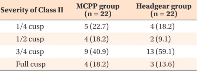
Table 1 shows the distribution of the severity of ma- locclusion with regard to the molar relationship in each group. MCPP was used to facilitate molar distalization
Table 1. Distribution of the severity of Class II malo- cclusion in adult patients treated with cervical pull head- gear or MCPP
Severity of Class II MCPP group
(n = 22) Headgear group (n = 22)
1/4 cusp 5 (22.7) 4 (18.2)
1/2 cusp 4 (18.2) 2 (9.1)
3/4 cusp 9 (40.9) 13 (59.1)
Full cusp 4 (18.2) 3 (13.6)
Values are presented as number (%).
MCPP, modified C-palatal plate.
p = 0.649; analyzed by chi-square test.
according to the method described in previous stu- dies.
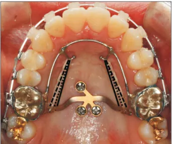
21,22The appliances were fitted on dental casts to adapt to the shape of the palatal surface, with their arms extending into the area between the first molars and second premolars. Adequate space was maintained between the arms and palatal slopes. The same operator (YAK) transferred MCPP from the cast to the oral cavity using a jig and fixed the appliance with three miniscrews measuring 8 mm in length and 2.0 mm in diameter (Jeil Medical Co.). Then, a palatal bar with two hooks extending along the gingival margins of the teeth was cemented on the right and left maxillary first molars.
Immediately after placement, distalization was initiated by engaging elastics or nickel-titanium closed coil springs between the MCPP arm notches and the hooks on the palatal bar, with an approximate force of 300 g per side. The outer bows of the cervical pull headgear were adjusted slightly upward so that they passed near to the center of resistance of the maxillary first molars.
Most MCPPs and headgear were delivered with fixed appliances during the same visit. The straight wire technique with 0.022-inch-slot brackets was used for both groups, with either In-Ovation C (Dentsply GAC
International, Islandia, NY, USA) or minitwin metal brackets (Ormco Co., Orange, CA, USA) depending on the patient preference. During the leveling phase, the size of the archwire was increased up to 0.018 × 0.025 or 0.019 × 0.025-inch stainless steel. Once the maxillary first molars were distalized to the intended overcorrected positions, MCPP and the cervical pull headgear were discontinued. Interarch elastics, including Class II elastics, were judiciously used, particularly during the later stages of treatment.
The study was approved by the institutional re- view board of the Catholic University of Korea (KC15- RISE0843). Informed consent was obtained according to the tenets of the Declaration of Helsinki. All late ral cephalograms, dental casts, facial and intraoral pho- tographs, and treatment charts were examined in detail.
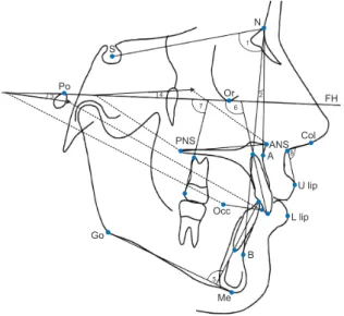
Cephalometric measurements
Lateral cephalograms obtained before the insertion of any appliance (T1) and after debonding (T2) were digitized using V-Ceph 5.5 software (Cybermed, Seoul, Korea). Two reference lines were constructed for hori- zontal and vertical measurements. The horizontal re- ference line was represented by the Frankfort horizontal
VRL
FH (HRL) Po
TVL
Pt Or
5 1
UL
LL Sn 2
3
4 6
7
8 9
10 11
12
Figure 1. Linear cephalometric variables used for analysis of the effects of cervical pull headgear or the modified C-palatal plate.
Po, Porion; Pt, pterygoid; Or, orbitale; Sn, subnasale; UL, upper lip; LL, lower lip; FH, Frankfort horizontal plane;
HRL, horizontal reference line; VRL, vertical reference line; TVL, true vertical line; 1, central incisor apex to HRL;
2, central incisor apex to VRL; 3, central incisor crown to HRL; 4, central incisor crown to VRL; 5, first molar apex to HRL; 6, first molar apex to VRL; 7, first molar crown to HRL; 8, first molar crown to VRL; 9, overjet; 10, overbite;
11, UL to TVL; 12, LL to TVL.
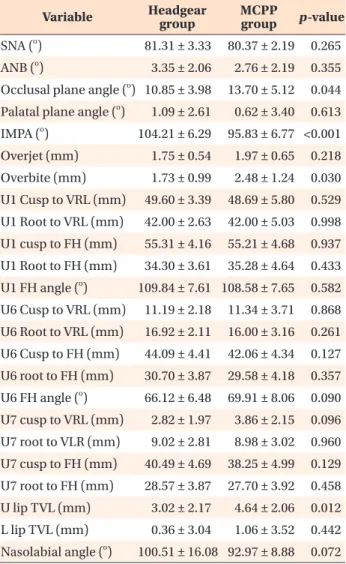
Figure 2. Angualr cephalometric variables used for analysis of the effects of cervical pull headgear or the modified C-palatal plate.
S, Sella; N, nasion; Po, porion; Or, orbitale; FH, Frankfort Horizontal plane; PNS, posterior nasal spine; ANS, anterior nasal spine; Col, columella; A, A point; U, upper; Occ, occlusal plane point; L, lower; Go, gonion; B, B point; Me, menton; 1, SNA; 2, ANB; 3, occlusal plane angle; 4, palatal plane angle; 5, incisor mandibular plane angle (IMPA); 6, central incisor inclination; 7, first molar angulation; 8, nasolabial angle.
Po Or
FH S
N
PNS
Go
Occ
Me B
L lip U lip
Col ANS A
1
3 4 2
5 7 6
8
(FH) plane, while the vertical reference line was defined to be perpendicular to the FH plane while passing through the pterygoid point. A total of 24 linear and angular measurements were recorded by one examiner, as shown in Figures 1 and 2. Differences between T1 and T2 findings were calculated (T2–T1) to assess the effects of treatment.
To identify systematic errors and compare measu- rement accuracy, 10 randomly selected cephalograms from each group were redigitized and measured at least twice on two separate occasions at an interval of 2 weeks by the same examiner. Intraexaminer reliability was evaluated using the intraclass correlation coefficient,
which was found to be > 0.90 for all variables.
Statistical analysis
All statistical analyses were performed using SPSS ver.
16.0 (SPSS Inc., Chicago, IL, USA). The Kolmogorov–
Smirnov method was used to confirm the normal dis- tribution of measurements. Paired t-tests were used to evaluate skeletal, dental, and soft tissue changes from T1 to T2 in each group. Multivariate analysis of variance (MANOVA) was performed to evaluate the changes after treatment in each group and compare the treatment effects between groups. A chi-square test showed no significant differences in the distribution
Table 2. Comparison of pretreatment cephalometric variables between the headgear and the modified C-pa- latal plate (MCPP) groups
Variable Headgear
group MCPP
group p-value SNA (
o) 81.73 ± 3.51 81.49 ± 2.38 0.785 ANB (
o) 3.49 ± 2.20 3.57 ± 1.97 0.901 Occlusal plane angle (
o) 7.95 ± 4.95 9.86 ± 4.73 0.193 Palatal plane angle (
o) 1.37 ± 2.74 0.44 ± 3.17 0.300 IMPA (
o) 95.86 ± 7.19 92.60 ± 7.16 0.135 Overjet (mm) 5.42 ± 2.15 4.67 ± 1.08 0.143 Overbite (mm) 3.23 ± 1.43 2.59 ± 1.33 0.129 U1 cusp to VRL (mm) 52.11 ± 4.09 51.87 ± 6.18 0.881 U1 root to VRL (mm) 42.44 ± 2.73 42.22 ± 4.66 0.845 U1 cusp to FH (mm) 54.40 ± 4.89 53.61 ± 4.45 0.578 U1 root to FH (mm) 33.80 ± 4.33 34.33 ± 4.69 0.693 U1 FH angle (
o) 115.13 ± 8.63 116.60 ± 9.23 0.585 U6 cusp to VRL (mm) 13.48 ± 2.06 15.56 ± 3.84 0.029 U6 root to VRL (mm) 17.53 ± 2.13 19.51 ± 3.75 0.036 U6 cusp to FH (mm) 43.73 ± 4.01 44.59 ± 4.08 0.480 U6 root to FH (mm) 29.76 ± 3.59 31.37 ± 4.00 0.163 U6 FH angle (
o) 74.15 ± 7.05 73.76 ± 6.46 0.849 U7 cusp to VRL (mm) 4.70 ± 2.35 6.30 ± 3.22 0.065 U7 root to VLR (mm) 11.28 ± 2.33 11.68 ± 3.48 0.650 U7 cusp to FH (mm) 39.76 ± 4.77 40.42 ± 4.00 0.614 U7 root to FH (mm) 27.75 ± 3.91 29.61 ± 4.11 0.128 U lip TVL (mm) 4.31 ± 2.15 5.78 ± 2.10 0.025 L lip TVL (mm) 0.46 ± 2.38 2.30 ± 3.68 0.055 Nasolabial angle (
o) 96.0 ± 15.37 87.90 ± 8.62 0.050 Values are presented as mean ± standard deviation.
Refer to the legends of Figures 1 and 2 for the definition of each measurement.
MANOVA: main effect (p = 0.017).
After Bonferroni correction; p < 0.002.
Table 3. Comparison of post-treatment cephalometric variables between the headgear and modified C-palatal plate (MCPP) groups
Variable Headgear
group MCPP
group p-value SNA (
o) 81.31 ± 3.33 80.37 ± 2.19 0.265 ANB (
o) 3.35 ± 2.06 2.76 ± 2.19 0.355 Occlusal plane angle (
o) 10.85 ± 3.98 13.70 ± 5.12 0.044 Palatal plane angle (
o) 1.09 ± 2.61 0.62 ± 3.40 0.613 IMPA (
o) 104.21 ± 6.29 95.83 ± 6.77 <0.001 Overjet (mm) 1.75 ± 0.54 1.97 ± 0.65 0.218 Overbite (mm) 1.73 ± 0.99 2.48 ± 1.24 0.030 U1 Cusp to VRL (mm) 49.60 ± 3.39 48.69 ± 5.80 0.529 U1 Root to VRL (mm) 42.00 ± 2.63 42.00 ± 5.03 0.998 U1 cusp to FH (mm) 55.31 ± 4.16 55.21 ± 4.68 0.937 U1 Root to FH (mm) 34.30 ± 3.61 35.28 ± 4.64 0.433 U1 FH angle (
o) 109.84 ± 7.61 108.58 ± 7.65 0.582 U6 Cusp to VRL (mm) 11.19 ± 2.18 11.34 ± 3.71 0.868 U6 Root to VRL (mm) 16.92 ± 2.11 16.00 ± 3.16 0.261 U6 Cusp to FH (mm) 44.09 ± 4.41 42.06 ± 4.34 0.127 U6 root to FH (mm) 30.70 ± 3.87 29.58 ± 4.18 0.357 U6 FH angle (
o) 66.12 ± 6.48 69.91 ± 8.06 0.090 U7 cusp to VRL (mm) 2.82 ± 1.97 3.86 ± 2.15 0.096 U7 root to VLR (mm) 9.02 ± 2.81 8.98 ± 3.02 0.960 U7 cusp to FH (mm) 40.49 ± 4.69 38.25 ± 4.99 0.129 U7 root to FH (mm) 28.57 ± 3.87 27.70 ± 3.92 0.458 U lip TVL (mm) 3.02 ± 2.17 4.64 ± 2.06 0.012 L lip TVL (mm) 0.36 ± 3.04 1.06 ± 3.52 0.442 Nasolabial angle (
o) 100.51 ± 16.08 92.97 ± 8.88 0.072 Values are presented as mean ± standard deviation.
Refer to the legends of Figures 1 and 2 for the definition of each measurement.
MANOVA: main effect (p = 0.010).
After Bonferroni correction: p < 0.002.
of malocclusion severity between the two groups (p = 0.649), while an independent samples t-test showed no significant difference in age between groups (p = 0.501).
The statistical significance was initially set at 0.05. After the application of Bonferroni correction, the significance level was 0.002.
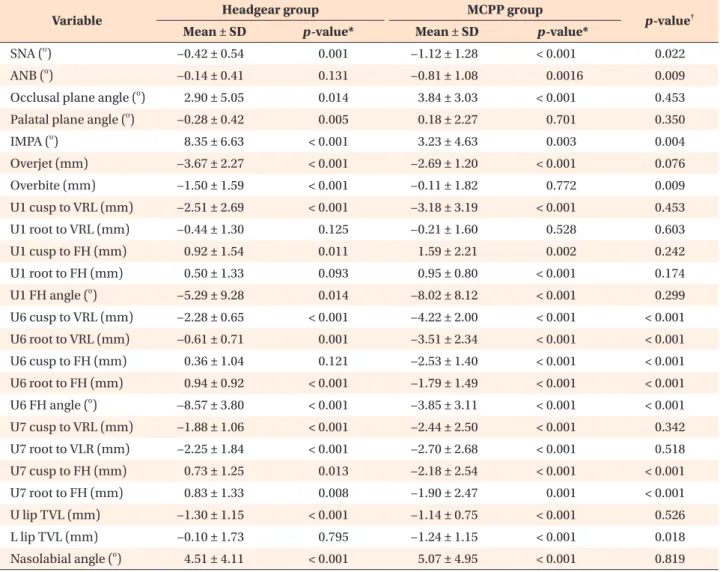
RESULTS
There were no significant differences in all pretreat- ment variables (Table 2) and all post-treatment variables except incisor mandibular plane angle (IMPA) (Table 3) between the two groups. In addition, there was no significant difference in the treatment duration between groups (MCPP, 29.9 ± 11.9 months; headgear, 24.1 ± 9.1 months; p = 0.099). Both groups demonstrated a
significant decrease in the overjet, whereas only the headgear group exhibited a decrease in the overbite, which showed no significant difference between the two groups (Table 4).
The MCPP group exhibited significant changes in the position of the maxillary first molar (p < 0.001).
The mean amount of distalization at the crown and root levels was 4.2 and 3.5 mm, respectively, with distal tipping of 3.9
oand intrusion by 2.5 mm. Meanwhile, in the headgear group, distalization was 2.3 mm at the crown level and 0.6 mm at the root level, with distal tipping of 8.6
oand extrusion by 0.4 mm. All variables were significantly different between the two groups ( p <
0.001, Table 4).
The maxillary second molar also showed significant distalization of 2.4 mm at the crown level and 2.7 mm
Table 4. Comparison of treatment effects between the headgear and modified C-palatal plate (MCPP) groups
Variable Headgear group MCPP group
p-value
†Mean ± SD p-value* Mean ± SD p-value*
SNA (
o) −0.42 ± 0.54 0.001 −1.12 ± 1.28 < 0.001 0.022
ANB (
o) −0.14 ± 0.41 0.131 −0.81 ± 1.08 0.0016 0.009
Occlusal plane angle (
o) 2.90 ± 5.05 0.014 3.84 ± 3.03 < 0.001 0.453
Palatal plane angle (
o) −0.28 ± 0.42 0.005 0.18 ± 2.27 0.701 0.350
IMPA (
o) 8.35 ± 6.63 < 0.001 3.23 ± 4.63 0.003 0.004
Overjet (mm) −3.67 ± 2.27 < 0.001 −2.69 ± 1.20 < 0.001 0.076
Overbite (mm) −1.50 ± 1.59 < 0.001 −0.11 ± 1.82 0.772 0.009
U1 cusp to VRL (mm) −2.51 ± 2.69 < 0.001 −3.18 ± 3.19 < 0.001 0.453
U1 root to VRL (mm) −0.44 ± 1.30 0.125 −0.21 ± 1.60 0.528 0.603
U1 cusp to FH (mm) 0.92 ± 1.54 0.011 1.59 ± 2.21 0.002 0.242
U1 root to FH (mm) 0.50 ± 1.33 0.093 0.95 ± 0.80 < 0.001 0.174
U1 FH angle (
o) −5.29 ± 9.28 0.014 −8.02 ± 8.12 < 0.001 0.299
U6 cusp to VRL (mm) −2.28 ± 0.65 < 0.001 −4.22 ± 2.00 < 0.001 < 0.001
U6 root to VRL (mm) −0.61 ± 0.71 0.001 −3.51 ± 2.34 < 0.001 < 0.001
U6 cusp to FH (mm) 0.36 ± 1.04 0.121 −2.53 ± 1.40 < 0.001 < 0.001
U6 root to FH (mm) 0.94 ± 0.92 < 0.001 −1.79 ± 1.49 < 0.001 < 0.001
U6 FH angle (
o) −8.57 ± 3.80 < 0.001 −3.85 ± 3.11 < 0.001 < 0.001
U7 cusp to VRL (mm) −1.88 ± 1.06 < 0.001 −2.44 ± 2.50 < 0.001 0.342
U7 root to VLR (mm) −2.25 ± 1.84 < 0.001 −2.70 ± 2.68 < 0.001 0.518
U7 cusp to FH (mm) 0.73 ± 1.25 0.013 −2.18 ± 2.54 < 0.001 < 0.001
U7 root to FH (mm) 0.83 ± 1.33 0.008 −1.90 ± 2.47 0.001 < 0.001
U lip TVL (mm) −1.30 ± 1.15 < 0.001 −1.14 ± 0.75 < 0.001 0.526
L lip TVL (mm) −0.10 ± 1.73 0.795 −1.24 ± 1.15 < 0.001 0.018
Nasolabial angle (
o) 4.51 ± 4.11 < 0.001 5.07 ± 4.95 < 0.001 0.819
SD, Standard deviation. Refer to the legends of Figures 1 and 2 for the definition of each measurement.
*Paired t-tests comparing pre- and post-treatment measurements within each group.
†