Cardiovascular disease (CVD)-related chronic diseases are the leading cause of death in societies with a large aged population such as Korea and Western
Contribution of Dietary Intakes of Antioxidants to Homocysteine-Induced Low Density Lipoprotein
(LDL) Oxidation in Atherosclerotic Patients
Hongseog Seo,1* Hyunhee Oh,2* Hyesoon Park,3Miyoung Park,4Yangsoo Jang,5and Myoungsook Lee4
1Cardiovascular Center, Department of Internal Medicine, Korea University College of Medicine, Seoul;
2Lee Gil Ya Cancer and Diabetes Institute, Gachon University of Medicine and Science, Incheon;
3Department of Family Medicine, University of Ulsan College of Medicine, Ulsan;
4Department of Food and Nutrition, Sungshin Women’s University, Seoul;
5Division of Cardiology, Cardiovascular Genome Center, Yonsei University College of Medicine, Seoul, Korea.
Purpose:Elevated circulating oxidized low density lipoprotein (Ox-LDL) levels are associated with increased risk of atherosclerosis, which may be due to high plasma homocysteine (Hcy) and low intakes of antioxidants. We investigated the contribution of dietary intakes of antioxidants to Hcy-induced LDL oxidation in atherosclerotic patients (AP) and controls. Materials and Methods:Male AP (n = 101) who were confirmed by coronary angiography and 91 controls were evaluated by blood biochemistry and dietary intakes. To determine whether homocysteine is an independent risk factor for atherosclerosis, subjects were divided into three groups; low- (≤ 6.9 uM/L), normal- (7 uM-12 uM/L) and high- (≥ 12.1 uM/L) Hcy.
Results: Plasm levels of homocysteine and LDL were higher, but plasma apo A-I in HDL and folate were lower in the AP group. The odds ratio (OR) for the risk of atherosclerosis was 3.002 [95% confidence interval (CI), 1.27-7.09] for patients in the highest tertile with homocysteine ≥ 12.1 uM/L. AP having high homocysteine levels had low intakes of vitamin A, β-carotene and vitamin C. By logistic regression analysis, age, body mass index (BMI), plasma LDL, plasma folate, and low intakes of vitamin A and β-carotene were found to be risk factors for atherosclerosis in patients with high-Hcy, but dietary B vitamins including folate were not. Conclusion: A high-Hcy level was a risk factor for atherosclerosis in patients with high Ox-LDL levels. High intakes of antioxidants appeared to be a protective factor for atherosclerosis, perhaps exerting a pro-oxidative effect on LDL when combined with high levels of Hcy and LDL. However, more evidence for the benefits of B vitamins as a homocysteine-lowering therapy is needed.
Key Words: Atherosclerosis, homocysteine, oxidized low density lipoprotein, folate, Vit A, β-carotene
Received: July 14, 2009 Revised: September 1, 2009 Accepted: September 2, 2009
Corresponding author: Dr. Myoungsook Lee, Department of Food and Nutrition, Sungshin Women's University, Dongseon-dong 3-ga, Seongbuk-gu, Seoul 136-742, Korea.
Tel: 82-2-920-7211, Fax: 82-2-926-3574 E-mail: [email protected] and Dr. Yangsoo Jang,
Division of Cardiology, Cardiovascular Genome Center, Yonsei University College of Medicine, 250 Seongsan-ro, Seodaemun-gu, Seoul 120-752, Korea.
Tel: 82-2-2228-8445, Fax: 82-2-393-2041 E-mail: [email protected]
*These authors contributed equally to this work.
∙The authors have no financial conflicts of interest.
© Copyright:
Yonsei University College of Medicine 2010 This is an Open Access article distributed under the terms of the Creative Commons Attribution Non- Commercial License (http://creativecommons.org/
licenses/by-nc/3.0) which permits unrestricted non- commercial use, distribution, and reproduction in any medium, provided the original work is properly cited.
INTRODUCTION
countries. Elevated levels of homocysteine (Hcy) above 12.1 µmol/L have been shown to double the risk of patho- physiological conditions such as atherosclerosis, myocar- dial infraction, cerebral or peripheral vascular diseases.1 Meta-analysis of 20 prospective studies indicated that the odds ratios (OR) for the risk of ischemic heart disease and stroke were 1.32 [95% confidence interval (CI), 1.19-1.45]
and 1.59 (95% CI, 1.29-1.96) for every 5 uM/L increase in Hcy.2 Elevated Hcy may contribute to progressive atheros- clerosis by several mechanisms, including arterial endo- thelial function impairment, oxidative stress induction, and the promotion of inflammation and thrombosis.1-4
A recent randomized clinical trial, however, did not support Hcy-lowering vitamin supplements to prevent heart diseases.5Since Hcy is a branch-point intermediate of cystathionine and methionine metabolism, folate, B6, and B12are essential to the conversion of Hcy. It has been shown that not only dietary B vitamin deficiency but also genetic disorders, certain drugs, and renal impairment are related to hyperhomocysteinemia.6,7 Folate and B12supplements decrease blood Hcy levels and carotid intima-media thick- nening, but they do not inhibit LDL oxidation, a marker of atherosclerosis.7,8Attempts to delineate causative factors of atherosclerosis have often investigated production of reac- tive oxygen species (ROS) and reduction of availabilities of endothelial nitric oxide (NO).9,10Hcy is known to induce oxidative stress and ROS or lipid peroxides (LPO) produc- tion, and decrease NO availability.11,12Hcy-induced ROS has been shown to be related to impaired mitochondrial NF-κB activation and NO bioavailability in endothelial cells, cardiac myocytes, fibroblasts and leukocytes, how- ever, they return to basal levels by antioxidants.9Therefore, Hcy affects the molecular structure or the activation of control agents which participate in the oxidative mecha- nism of atherosclerosis.
The aim of the current study was to investigate whether elevated Hcy levels are associated with oxidation of LDL, and further explore the contribution of dietary intakes of B vitamins or antioxidants to Hcy-induced LDL oxidation in atherosclerosis.
Subjects and samples
Healthy or atherosclerotic male subjects were recruited from among in- and out-patients of Korea University College of Medicine in Seoul, Korea, after approval from Biomedical Human Subjects Review Committee. After first screening (n = 340) with a health questionnaire (family history, exercise), subjects were further evaluated with a dietary survey and anthropometry [body mass index (BMI),
blood pressure (BP)]; biochemical tests were performed for 192 final subjects. The 192 subjects were divided into atherosclerosis patients (AP, n = 101) or control (n = 91) groups after AP were diagnosed by coronary angiography by a cardiologist. The coronary artery lesion was defined where stricture levels of Lt. main, Lt. anterior descending artery, left circumflex, or right coronary artery were over 50%. To determine whether Hcy is an independent risk factor for CVD, subjects were assigned to one of three Hcy levels, using the European Concerted Action Projects (ECAP) cut-off value.13Dietary surveys were performed using the CANpro 3.0 software (developed by Korean Nutrition Society). Survey results on dietary intakes and biochemistry were statistically analyzed. None of the parti- cipants were using vitamin and/or mineral supplements.
Biochemical measurements
Cholesterol and triglycerides (TG) were analyzed by enzy- matic Kits (Sigma Co, St Louis, MO, USA).14,15After HDL and LDL were separated by density gradient ultracentri- fugation, apo A-I in HDL and apo B-100 in LDL were quantified by Western blotting analysis (antibodies; Santa Cruz, CA, USA). In order to measure Ox-LDL, pure LDL was high-speed centrifugated using KBr, at a density of 1.019-1.063 g/mL. During centrifugation, 0.1% EDTA and 0.02% sodium azoid were added. Phosphate-buffered saline (PBS) was used for dialysis of centrifugated LDL.
SDS-polyacrylamine gel electrophoresis (PAGE) was used to verify LDL purification.16The purified LDL was dialyzed for a day, and then LPO [malondialdehyde (MDA)] pro- duction was analyzed. The thiobarbituric acid (TBA) method (modified Packer and Smith method) was used to obtain correct amount of lipid peroxide with the MDA produced. By using 1,1,3,3-tetraethoxypropane as the stan- dard reagent, comparisons were made between different treatments according to thiobarbituric acid reactive sub- stances (TBARS) values from different amounts of 1 mg protein. Butylhydroxytoluen (BHT), sulfuric acid, and Na2WO4were mixed with LDL, and the mixture was cen- trifugated. The liquid that rose was taken; NaOH and 1%
TBA were added and heated for 60 minutes at 100˚C. After centrifugation at 3,000 rpm for 10 minutes, absorbance was read.16-18After the protein content of LDL was mea- sured, units of LDL per mg protein were used to express the amount. Total plasma Hcy (sum of free-, protein- bound and mixed disulfide forms) in all samples was deter- mined by the use of “Abbott IMX system Homocysteine E/B3D390, 77-0650/R2” kit (ABBOTT Plasma Total Hcy Diagnostic kit; Abbott, Abbott Park, IL, USA) in one run.19 By processing Hcy molecules (protein, residual thiol, or sulfide combined) with DTT, a crystalline form of Hcy can be obtained. By applying an S-adenosyl-L-homocysteine
MATERIALS AND METHODS
(SAH) hydrolase to the crystalline with cholesterol, the amount of Hcy that combines with adenosine to form SAH was measured. For determination of vitamin B12and folate in serum and plasma, vitamin B12/folate kit (Diagnostic Products Corporation, Los Angeles, CA, USA) was used according to the method of Soild Phase No Boil Dual- couint.20: 200 µL of serum was mixed with sample buffer, and the mixture was left for 30 minutes at room temper- ature. The above samples were reacted with NaOH/KCN at 37˚C, and vitamin B12/folate binder was added into each tube. The supernatant after centrifugation at 2,000 g for 15 minutes was discarded. The radioactivity from 125I-folate and 57Co-vit B12 in the pellet was measured by Gamma counter Cobra II. For the amount of vitamin B12, pg/mL was multiplied by 0.7378 to convert to pmol/L. For folate, ng/mL was multiplied by 2.266 to convert to pmol/L. The clinical reference value for blood vitatimin B12is 200-950 pg/mL, and 3.0-17 ng/mL for blood folate.
Statistical analysis
Using SAS statistical software (Carey, NC, USA), ANOVA and independent t-test were performed. Statistical signi-
ficance was accepted at p-value < 0.05. Relationships among variables were assessed by correlation analysis. Univariate and multivariate logistic regression analyses were used to obtain the crude and adjusted OR (AOR) and 95% CI.
Demographics of subjects
Average age of the AP group was 56.2 years, while the average age of the control group was 42.4 years. The two groups exhibited no differences in blood pressure and BMI (Table 1). Over 60% of the AP subjects exhibited other co- morbidities also, whereas 73% of controls were disease- free. The incidences of hypertension and diabetes were 35% and 16%, respectively, in the AP (data not shown).
Smoking and drinking rates were similar in both groups, but the amount and length of the use were much higher in the AP; 47.7% of total AP smoked more than 1 pack of cigarettes/day (control: 32.2%) and 35.5% of the AP drank 2-4 times/week (control: 28.8%). The AP also had higher coffee consumption rate (5 cups/day) than the control (data
RESULTS
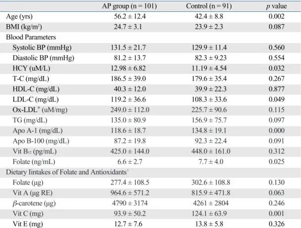
Table 1. Mean Values (± SD) of General Characteristics, Blood Parameters and Daily Nutrient Intakes of Folate and Antioxidants Related to Atherogenic Risk Factors in AP and Control Groups
AP group (n = 101) Control (n = 91) p value
Age (yrs) 56.2 ± 12.4 42.4 ± 8.8 0.002
BMI (kg/m2) 24.7 ± 3.1 23.9 ± 2.3 0.087
Blood Parameters
Systolic BP (mmHg) 131.5 ± 21.7 129.9 ± 11.4 0.560
Diastolic BP (mmHg) 81.2 ± 13.7 82.3 ± 9.23 0.554
HCY (uM/L) 12.98 ± 6.82 11.19 ± 4.54 0.032
T-C (mg/dL) 186.5 ± 39.0 179.6 ± 35.4 0.267
HDL-C (mg/dL) 40.3 ± 12.0 39.9 ± 22.3 0.877
LDL-C (mg/dL) 119.2 ± 36.6 108.3 ± 33.6 0.049
Ox-LDL*(uM/mg) 249.0 ± 112.0 225.7 ± 90.6 0.115
TG (mg/dL) 135.0 ± 80.9 156.9 ± 75.7 0.097
Apo A-1 (mg/dL) 118.6 ± 18.7 134.8 ± 19.1 0.000
Apo B-100 (mg/dL) 87.2 ± 19.8 92.3 ± 22.4 0.091
Vit B12(pg/mL) 425.0 ± 144.0 448.0 ± 161.0 0.312
Folate (ng/mL) 6.6 ± 2.7 7.7 ± 4.0 0.025
Dietary Iintakes of Folate and Antioxidants�
Folate (µg) 277.4 ± 108.5 302.6 ± 108.8 0.130
Vit A (µg RE) 964.6 ± 571.2 815.9 ± 471.8 0.063
β-carotene (µg) 4790 ± 3174 4261 ± 2804 0.246
Vit C (mg) 93.9 ± 50.2 124.1 ± 63.9 0.001
Vit E (mg) 12.7 ± 7.6 13.8 ± 5.8 0.326
BMI, body mass index; Hcy, homocysteine; T-C, total cholesterol; LDL, low density lipoprotein; HDL, high density lipoprotein; TG, triacylglycerol; Vit, vitamin; BP, blood pressure.
*Oxidized LDL; malondialdehyde (uM) concentration per mg LDL protein.
�Adjusted by the calorie intake.
not shown).
Blood lipid profiles, folate & B12in AP or hyperhomocysteinemia
In the AP, blood Hcy and LDL cholesterol levels were higher and apo A-I in HDL and folate were lower, exhibit- ing characteristic lipid spectrum of atherosclerosis (Table 1).
Plasma Hcy and LDL can be viewed as pro-arteriosclerosis factors, and HDL’s apo A-I as an anti-arteriosclerosis factor. However, no apparent differences in total choles- terol (T-C), HDL, TG, and apo B-100 were observed bet- ween AP and control. Blood Hcy and Ox-LDL were higher in AP, suggesting that Hcy plays a role in LDL oxidation.
Blood folate levels were lower in AP, whereas vitamin B12 levels were similar. In order to verify the changes in lipid profiles, resulting from the effects of vitamins and Ox- LDL on Hcy concentrations, subjects were divided into 3 groups depending on Hcy levels: low-Hcy (≤ 6.9 uM/L), normal (7 uM-12 uM/L) and high-Hcy (≥ 12.1 uM/L)
(Table 2). Ox-LDL levels were not different between AP and control, but signifi-cantly higher in the normal and high-Hcy groups than in the low-Hcy group (Table 2). The positive association of LDL with Ox-LDL was stronger in high-Hcy subjects than that in total subjects, normal or low Hcy subjects (data not shown). The increased Ox-LDL in the high-Hcy group can be explained as LDL oxidation caused by the increase in blood Hcy. Blood folate levels were significantly different (p < 0.05) among the 3 Hcy groups, and it was negatively correlated with Hcy (Fig. 1).
There was no correlation bet-ween vitamin B12 and Hcy even though blood folate and vitamin B12were positively correlated (data not shown). The blood lipid levels did not change according to Hcy levels and were not correlated with Hcy.
Dietary intakes in AP or hyperhomocysteinemia The relative percentages of carbohydrate, protein and fat intakes per total calories were similar in AP and controls, Table 2. Mean Values (± SD) of Blood Metabolic Variables and Daily Nutrient Intakes of Folate and Antioxi- dants According to the Classification of Plasma Hcy Levels
Low-Hcy Normal-Hcy High-Hcy
(≤ 6.9 µM/L) (7 - 12 µM/L) (≥ 12.1 µM/L) p value
(n = 9) (n = 112) (n = 53)
Age (yrs) 54.8 ± 17.1 47.9 ± 11.9 51.8 ± 13.3 0.077
BMI (kg/m2) 24.9 ± 3.7 24.4 ± 2.6 23.9 ± 2.8 0.430
Blood pressure and blood variables
Systolic (mmHg) 128.2 ± 28.3 132.4 ± 16.8 127.6 ± 16.3 0.229
Diastolic (mmHg) 82.1 ± 14.4 82.1 ± 10.9 80.8 ± 13.0 0.807
HCY (uM/L) 6.5 ± 0.3a) 9.5 ± 1.3b) 18.1 ± 7.2c) 0.000
T-C (mg/dL) 199.6 ± 29.9 183.1 ± 38.7 182.3 ± 36.3 0.566
HDL-C (mg/dL) 37.6 ± 20.7 38.5 ± 13.3 43.5 ± 22.4 0.251
LDL-C (mg/dL) 131.1 ± 28.4 144.8 ± 35.7 111.9 ± 36.4 0.461
Ox-LDL* (uM/mg) 161.6 ± 76.0a) 237.6 ± 108.2b) 255.3 ± 95.7b) 0.005
TG (mg/dL) 154.2 ± 66.7 148.8 ± 49.4 134.6 ± 79.9 0.577
Apo A-1 (mg/dL) 124.2 ± 24.7 128.1 ± 20.9 123.0 ± 18.7 0.280
Apo B-100 (mg/dL) 93.7 ± 17.2 90.9 ± 21.9 86.1 ± 19.8 0.318
Vit B12(pg/mL) 438.2 ± 139.6 452.4 ± 159.3 402.2 ± 136.5 0.121
Folate (ng/mL) 6.9 ± 3.7b) 7.6 ± 3.7c) 6.1 ± 2.4a) 0.023
Dietary intakes of folate and antioxidants�
Folate (µg) 351.0 ± 180.1 289.4 ± 107.6 282.6 ± 95.9 0.217
Vit A (µg RE) 1370 ± 1219b) 880 ± 484a) 820 ± 381a) 0.013
β-carotene (µg) 7166 ± 7436b) 4483 ± 2626a) 4137 ± 2267a) 0.018 Vit C (mg) 170.9 ± 66.4b) 105.0 ± 56.1a) 108.9 ± 60.6a) 0.006
Vit E (mg) 16.2 ± 8.3 13.4 ± 7.0 12.7 ± 5.9 0.329
BMI, body mass index; Hcy, homocysteine; T-C, total cholesterol; LDL, low density lipoprotein; HDL, high density lipoprotein; TG, triacylglycerol; Vit, vitamin.
a), b), c)
Values within the classification of plasma HCY levels with different superscripts are significant at p < 0.05.
*Oxidized LDL; malondialdehyde (uM) concentration per mg LDL protein.
�Adjusted by the calorie intake.
even though AP had lower energy intakes (data not shown), suggesting that AP might have attempted to lose weight, since they had slightly, but not significantly, higher BMIs.
Dietary intakes of other B vitamins were not different between the two groups, except vitamin B1and B2which were lower in AP. Vitamin B12intake could not be evaluated because of insufficient data. There were no differences in any dietary intakes except vitamin A, β-carotene and vitamin C among the three Hcy groups (Table 2). Hcy was not affected by dietary folate intakes in this study, and the pattern of correlation between dietary folate and Hcy or plasma folate were not obvious (Fig. 1). Therefore, low dietary folate in AP might have not induced hyperhomo- cysteinemia in this study. However, dietary intakes of vitamin A, and its precursor, and vitamin C were signifi- cantly lower with increasing Hcy. The negative correla- tions between dietary vitamin A (r2= - 0.226) or β-carotene (r2= - 0.233) antioxidants and Ox-LDL were also signi- ficant in AP (p < 0.001) compared to no correlations in controls (data not shown).
Risk factors for atherosclerosis according to Hcy levels Using the ECAP cut-off value (≥ 12.1 uM/L), 26.6% of the total Korean male subjects had hyperhomocysteinemia, and 69.3% and 4.2% had normal and low-Hcy levels, respectively. The frequency of atherosclerosis was sub- stantially increased if the blood Hcy was higher than ≥ 12.1 uM/L (OR, 2.887; 95% CI, 1.51-5.53) and this status was stronger after adjusting for age and BMI (AOR, 3.002;
95% CI, 1.27-7.09)(data not shown). To identify links bet- ween risk factors for atherosclerosis and high blood Hcy levels, all anthropometric and biochemistry variables and daily nutrient intakes were subjected to logistic regression analysis (Table 3). The AOR for atherosclerosis was 4.420 (95% CI, 1.542-12.663) with high blood Hcy and LDL levels compared to when LDL was not included as a factor
Table 3. Logistic Regression Analysis of Independent Risk Factors for Korean Male Atherosclerosis with High Hcy Levels
Variables Adjusted OR (95% CI)�
I II III IV
Blood HCY§ 3.002 (1.269 - 7.088)� 3.037 (1.113 - 8.292)* 4.420 (1.542 - 12.663)� 4.206 (1.457 - 12.142)�
Dietary Folate 1.001 (0.997 - 1.005)
Plasma Folate 0.797 (0.670 - 0.949)�
LDL 1.014 (1.000 - 1.028) * 1.013 (1.000 - 1.028)*
Dietary β-carotene
1.000 (1.000 - 1.000) (or vit A)‖
Hcy, homocysteine; LDL, lowdency lipoprotein; BMI, body mass index.
*p < 0.05.
�p < 0.01.
�Risk factors for atherosclerosis were analyzed by logistic regression analysis. All variables of odds ratios for atherosclerosis were adjusted by age and BMI.
§High blood Hcy (serum HCY levels ≥ 12.1 uM/dL).
‖Same when vit A variable was included.
Fig. 1. Correlation between plasma folate or dietary folate and Hcy concentration in adult Korean men. Plasma folate levels were significantly and negatively correlated with Hcy concentration, but not with dietary folate.
(Model I & II). When dietary folate and plasma were added, the AOR of high-Hcy for atherosclerosis was not changed. After dietary vitamin A and β-carotene were included as antioxidants, the AOR of high-Hcy for ather- osclerosis was reduced to 4.206 (95% CI, 1.457-12.142).
We demonstrated that the most important risk factor for atherosclerosis was LDL and the least important risk factor was plasma folate in hyperhomocysteinemia. When par- ticipants were divided into tertiles of mean Hcy and LDL concentrations, 20.7% of subjects in the 3rd tertile of Ox- LDL (276.13-745 uM/mg LDL) came under two combined categories with the highest Hcy (11.89-50 uM/L) and LDL levels (124.81-244 mg/dL) (Fig. 2). The probability of sub- jects having the highest Ox-LDL while also having low Hcy and low LDL levels was only 3.4%. We found that the greatest progression of atherosclerosis was observed with the highest Ox-LDL simultaneously with both the highest Hcy and LDL concentrations. This means that a high cir- culating Hcy level was a risk factor for atherosclerosis only when the subject had also elevated LDL levels
In this study, AP exhibited multiple complications causing co-morbidities of metabolic syndrome such as hyper- tension and NIDDM, which were similar to the distri- bution of complications seen in Korean hyperlipopro- teinemia.21The ECAP study found that elevated Hcy was
an independent risk factor for Hcy and CVD, causing multiple effects on the risks of smoking, hypertension, and hypercholesterolemia.13It has been reported that 85.2% of Korean AP smoke 20 cigarettes/day or more, and 85.4% of AP smoked more than one pack of cigarettes/day for the last month.1Since elevated Hcy strongly correlates with smoking and low blood folate levels were found in the PRIME case-control study, Hcy levels might be a secon- dary risk factor to low blood folate and smoking.22Elevated Hcy may also be involved in hypertension because it is associated with impaired endothelial function due to the generation of oxidative stress, VCAM-1 and reduction of NO bioavailability.23,24High dietary intake of sodium is also a cause of hypertension. Since Koreans customarily consume large amounts of traditional salty fermented foods such as kimchi or soybean paste, sodium intake is almost 3 times higher than the DRI established by WHO (2 g/day). In American adults, blood Hcy levels are 5-15 µmol/L, and Hcy concentration of the control (healthy) group is around 7-12 µM/L.25In Jacques’ study,26male coronary artery disease patients (age, 43-57 years) had Hcy concentrations of 13.66 uM/L while the control had 10.93 uM/L concentration. If the total blood Hcy is low, the death rate of CVD is low. Every 5 µM/L increase in the Hcy con- centration increases the risk of CVD by 50%, and TC levels by 20 mg/dL.1,2,25,26While doubts were raised concerning the cause-effect relationship between the Hcy and CVD, we found that Hcy is definitely higher in AP than in controls.
The mechanisms possibly responsible for causing endothelial dysfunction include changes in LDL, and Ox- LDL, since higher LDL, which is associated with oxida- tion of LDL was observed in the AP. We also found that elevated Hcy makes the correlation between LDL and Ox- LDL much stronger. In Mansoor’s research, Hcy levels and Ox-LDL were higher in subjects with CAD than in control. The oxidation of LDL is increased by the com- bination of thiolactone and apo B’s free lysyl epsilon amino residue.10,27,28When LDL is reacted with Hcy- thiolactone in methionine, which is an explicit initiator of arteriosclerosis, LDL-binding thiol is increased by 250 nM per mg of LDL protein.29The free amino- or thiol-adducted LDL causes aggregation, and increases LDL uptake in macrophages and atheroma production by lipids.30In Marek’s research, LDL oxidation in the aortic cell walls was 40% higher in hyper-homocysteinemia (≥ 14.0 µM/L) compared to control.31Another mechanism by which Hcy may cause LDL oxidation is a possible deformation of LDL through Hcy autoxidation, which causes the oxida- tion of side chains of LDL such as fatty acids or apo B- 100.7Both Hcy and Ox-LDL could participate in throm- bosis by increasing VCAM-1 and ICAM-1, caused by endothelial cell activation due to fibrinogen-platelet GPIIb-
DISCUSSION
Fig. 2. The participants (n = 192) were divided into tertiles of mean Hcy and LDL levels. The greatest progression of coronary atherosclerosis was observed in subjects with Ox-LDL 3rd tertile corresponding to highest Hcy and LDL levels.
Left, The frequencies (%) in subjects having highest Ox-LDL according to tertiles of Hcy and LDL concentrations. Hcy (uM/L): small tertile, < 9.3; medium tertile, 9.31 to 11.89; large tertile, > 11.90. LDL levels (mg/dL): low tertile, < 96.9; medium tertile, 97 to 124.8; high tertile, > 124.81. Ox-LDL (uM/mg): low tertile, < 183.58;
medium tertile, 183.59 to 276.12; high tertile, > 276.13. LDL, low density lipoprotein;
Ox-LDL, oxidized LDL; Hcy, homocysteine.
lIIa formation. Ox-LDL affects both initial and progressive stages of arteriosclerosis.11,12,23,32On the other hand, circulat- ing Hcy reduced NO-induced detoxification, vasodilation, and endothelial function.10NO participates in a metabolic pathway (S-nitroso-HCY) that is able to protect against Hcy-induced endothelial oxidative damage.
Since Hcy is a branch-point intermediate of cystathionine and methionine metabolism, the deficiency of B vitamins (folate, vitamin B6and B12) might cause hyperhomocystei- nemia.4,33In animal and clinical tests, vitamin B12and folate deficiency is related to increased blood Hcy levels, but the correlation between Hcy and B vitamins is not clear.34,35In the Hcy- Lowering Trial’s Collaboration Research, an additional 7% supplement of vitamin B12 reduced fasting Hcy levels and decreased hypercysteinemia by 25%, however, vitamin B6had no effect on Hcy levels.5,25In Ver- meulen’s research, where AP was given 5 mg of folate and 250 mg of vitamin B6, the Hcy level decreased from 14.7 µM/L to 7.4 µM/L, showing a higher reduction rate than in the placebo group (12.0 µM/L).36Supplemention of folate (2.5 mg), vitamin B6(25 mg), and B12(500 µg) for one year reduced Hcy from 10.5 to 6.56 µM/L and carotid intima-media thickness by 5.3%.8,37However, Young and Woodside reported that supplementary folate and vitamin B12lower blood Hcy levels, but they cannot stop LDL oxidation.7Vitamins B12or B6and the C677T NTHFR poly- morphism have not been associated with carotid intima- media thickness.8Food-based feeding trials have shown a reduction in blood Hcy in subjects who consume fortified cereals or whole grains in combination with fruits, vege- tables and low fat dairy products.38,39However, more evi- dence is needed for the association of vitamin B12and Hcy levels with the pro-oxidative mechanism of LDL and the dietary benefits of Hcy-lowering therapy for atheros- clerosis. The effects of antioxidants on decreasing the oxidation of LDL may be an important observation when blood LDL and Hcy levels are increased. Because low intakes of vitamin A, β-carotene and vitamin C are asso- ciated with Hcy levels, even though the biochemical data did not support that the Hcy-induced oxidative damage is protected by antioxidants. Both hydro- and lipid-soluble antioxidants have been shown to be depleted in oxidative endothelial damage.10,40 Our findings confirmed a role of antioxidant compounds, particularly β-carotene, against the oxidative damage promoted by Hcy and elevated LDL levels. Nevertheless, it needs to be further evaluated in conjunction with markers of oxidative damage.
The study was supported by a grant (10526) from the
Korean Seoul City Research & Business Development (R
& BD) Program and the Ministry of Health and Welfare, Republic of Korea (A000385).
1. Castro R, Rivera I, Blom HJ, Jakobs C, Tavares de Almeida I.
Homocysteine metabolism, hyperhomocystenemia and vascular disease: an overview. J Inherit Metab Dis 2006;29:3-20.
2. Wald DS, Law M, Morris JK. Homocysteine and cardiovascular disease: evidence on causality from a meta-analysis. BMJ 2002;
325:1202.
3. Jakubowski H. Pathophysiological consequences of homocysteine excess. J Nutr 2006;136:1741S-9.
4. Wald DS, Law M, Morris JK. The dose-response relation between serum homocysteine and cardiovascular disease: implications for treatment and screening. Eur J Cardiovasc Prev Rehabil 2004;11:
250-3.
5. Lonn E, Yusuf S, Arnold MJ, Sheridan P, Pogue J, Micks M, et al. Homocysteine lowering with folic acid and B vitamins in vascular disease. N Engl J Med 2006;354:1567-77.
6. Boushey CJ, Beresford SA, Omenn GS, Motulsky AG. A quan- titative assessment of plasma homocysteine as a risk factor for vascular disease. Probable benefits of increasing folic acid intakes.
JAMA 1995;274:1049-57.
7. Young IS, Woodside JV. Folate and homocysteine. Curr Opin Clin Nutr Metab Care 2000;3:427-32.
8. Durga J, Verhoef P, Bots ML, Schouten E. Homocysteine and carotid intima-media thickness: a critical appraisal of the evidence.
Atherosclerosis 2004;176:1-19.
9. Perez-de-Arce K, Foncea R, Leighton F. Reactive oxygen species mediates homocysteine-induced mitochondrial biogenesis in human endothelial cells: modulation by antioxidants. Biochem Biophys Res Commun 2005;338:1103-9.
10. Rocchi E, Bursi F, Ventura P, Ronzoni A, Gozzi C, Casalgrandi G, et al. Anti- and pro-oxidant factors and endothelial dysfunction in chronic cigarette smokers with coronary heart disease. Eur J Intern Med 2007;18:314-20.
11. Jakubowski H. Homocysteine thiolactone: metabolic origin and protein homocysteinylation in humans. J Nutr 2000;130:377S-81.
12. Dardik R, Varon D, Tamarin I, Zivelin A, Salomon O, Shenkman B, et al. Homocysteine and oxidized low density lipoprotein enhanced platelet adhesion to endothelial cells under flow con- ditions: distinct mechanisms of thrombogenic modulation. Thromb Haemost 2000;83:338-44.
13. Graham IM, Daly LE, Refsum HM, Robinson K, Brattström LE, Ueland PM, et al. Plasma homocysteine as a risk factor for vascular disease. The European Concerted Action Project. JAMA 1997;
277:1775-81.
14. Allain CC, Poon LS, Chan CS, Richmond W, Fu PC. Enzymatic determination of total serum cholesterol. Clin Chem 1974;20:
470-5.
15. Foster LB, Dunn RT. Stable reagents for determination of serum triglycerides by a colorimetric Hantzsch condensation method.
Clin Chem 1973;19:338-40.
16. Nagyová A, Raslová K, Ginter E. Oxidation resistance of LDL in hypertriglyceridaemic patients treated with ciprofibrate. Physiol Res 1998;47:185-90.
REFERENCES
ACKNOWLEDGEMENTS
17. Pritchard KA Jr, Groszek L, Smalley DM, Sessa WC, Wu M, Villalon P, et al. Native low-density lipoprotein increases endo- thelial cell nitric oxide synthase generation of superoxide anion.
Circ Res 1995;77:510-8.
18. Edelstein C, Pfaffinger D, Scanu AM. Advantages and limita- tions of density gradient ultracentrifugation in the fractionation of human serum lipoproteins: role of salts and sucrose. J Lipid Res 1984;25:630-7.
19. Brunelli T, Pepe G, Marcucci R, Giusti B, Prisco D, Abbate R, et al. Comparison of three methods for total homocysteine plasma determination. Clin Lab 2001;47:393-7.
20. Woo KS, Chook P, Lolin YI, Sanderson JE, Metreweli C, Celer- majer DS. Folic acid improves arterial endothelial function in adults with hyperhomocystinemia. J Am Coll Cardiol 1999;34:
2002-6.
21. Lee M, Kim JQ, Kim J, Oh H, Park M. Studies on the plasma lipid profiles, and LCAT and CETP activities according to hyper- lipoproteinemia phenotypes (HLP). Atherosclerosis 2001;159:
381-9.
22. Troughton JA, Woodside JV, Young IS, Arveiler D, Amouyel P, Ferrières J, et al. Homocysteine and coronary heart disease risk in the PRIME study. Atherosclerosis 2007;191:90-7.
23. Vadachkoria S, Sanchez SE, Qiu C, Muy-Rivera M, Malinow MR, Williams MA. Hyperhomocyst(e)inemia and elevated soluble vascular cell adhesion molecule-1 concentrations are associated with an increased risk of preeclampsia. Gynecol Obstet Invest 2004;58:133-9.
24. Zhang X, Li H, Jin H, Ebin Z, Brodsky S, Goligorsky MS.
Effects of homocysteine on endothelial nitric oxide production.
Am J Physiol Renal Physiol 2000;279:F671-8.
25. Nygård O, Refsum H, Ueland PM, Vollset SE. Major lifestyle determinants of plasma total homocysteine distribution: the Hor- daland Homocysteine Study. Am J Clin Nutr 1998;67:263-70.
26. Genest JJ Jr, McNamara JR, Salem DN, Wilson PW, Schaefer EJ, Malinow MR. Plasma homocyst(e)ine levels in men with pre- mature coronary artery disease. J Am Coll Cardiol 1990;16:1114-9.
27. Mansoor MA, Ueland PM, Aarsland A, Svardal AM. Redox status and protein binding of plasma homocysteine and other aminothiols in patients with homocystinuria. Metabolism 1993;
42:1481-5.
28. Mansoor MA, Bergmark C, Haswell SJ, Savage IF, Evans PH, Berge RK, et al. Correlation between plasma total homocysteine and copper in patients with peripheral vascular disease. Clin Chem 2000;46:385-91.
29. Ferguson E, Hogg N, Antholine WE, Joseph J, Singh RJ, Partha- sarathy S, et al. Characterization of the adduct formed from the reaction between homocysteine thiolactone and low-density lipo- protein: antioxidant implications. Free Radic Biol Med 1999;26:
968-77.
30. Perna AF, Ingrosso D, Lombardi C, Acanfora F, Satta E, Cesare CM, et al. Possible mechanisms of homocysteine toxicity. Kidney Int Suppl 2003;(84):S137-40.
31. Naruszewicz M. Elevated homocysteine increases the ability of human aortic cells to oxidize LDL, Atherosclerosis XI Sym- posium. Paris, France; 1997.p.493-6.
32. Erl W, Weber PC, Weber C. Monocytic cell adhesion to endothe- lial cells stimulated by oxidized low density lipoprotein is mediated by distinct endothelial ligands. Atherosclerosis 1998;136:297-303.
33. Lewis CA, Pancharuniti N, Sauberlich HE. Plasma folate ade- quacy as determined by homocysteine level. Ann N Y Acad Sci 1992;669:360-2.
34. Ubbink JB. Vitamin nutrition status and homocysteine: an athero- genic risk factor. Nutr Rev 1994;52:383-7.
35. Boushey CJ, Beresford SA, Omenn GS, Motulsky AG. A quanti- tative assessment of plasma homocysteine as a risk factor for vascular disease. Probable benefits of increasing folic acid intakes.
JAMA 1995;274:1049-57.
36. Vermeulen EG, Stehouwer CD, Twisk JW, van den Berg M, de Jong SC, Mackaay AJ, et al. Effect of homocysteine-lowering treatment with folic acid plus vitamin B6 on progression of sub- clinical atherosclerosis: a randomised, placebo-controlled trial.
Lancet 2000;355:517-22.
37. Till U, Röhl P, Jentsch A, Till H, Müller A, Bellstedt K, et al.
Decrease of carotid intima-media thickness in patients at risk to cerebral ischemia after supplementation with folic acid, Vitamins B6 and B12. Atherosclerosis 2005;181:131-5.
38. Lutsey PL, Steffen LM, Feldman HA, Hoelscher DH, Webber LS, Luepker RV, et al. Serum homocysteine is related to food intake in adolescents: the Child and Adolescent Trial for Cardio- vascular Health. Am J Clin Nutr 2006;83:1380-6.
39. Appel LJ, Miller ER 3rd, Jee SH, Stolzenberg-Solomon R, Lin PH, Erlinger T, et al. Effect of dietary patterns on serum homocy- steine: results of a randomized, controlled feeding study. Circula- tion 2000;102:852-7.
40. Hirano K, Ogihara T, Miki M, Yasuda H, Tamai H, Kawamura N, et al. Homocysteine induces iron-catalyzed lipid peroxidation of low-density lipoprotein that is prevented by alpha-tocopherol.
Free Radic Res 1994;21:267-76.