• 검색 결과가 없습니다.

Acute ST Elevated Myocardial Injury due to Coronary Thrombosis during Thoracic Endovascular Aortic Repair in Patient with Protein S Deficiency

N/A
N/A
Protected

Academic year: 2022

Share "Acute ST Elevated Myocardial Injury due to Coronary Thrombosis during Thoracic Endovascular Aortic Repair in Patient with Protein S Deficiency"

Copied!
5
0
0

로드 중.... (전체 텍스트 보기)

전체 글

(1)

Korean Circulation Journal

Introduction

With a lower complication rate and satisfactory results, thoracic endovascular therapy has become a suitable alternative treatment for thoracic aneurysm.

1)

However, complications of the procedure including stroke, spinal cord ischemia or myocardial infarction are still in existence, leading problematic sequelae.

2)

A protein S defi- ciency is a well known genetic tendency to the venous thrombo- embolism.

3)

It can also be the risk factors for arterial occlusive dis- eases including coronary artery occlusion.

4)5)

However, it is not ge- nerally concerned as a procedural risk factor for endovascular treat- ment, thus special preparation for this thrombophilic disorder is not usually achieved during the peri-procedural period. In this report, we describe a rare case of peri-procedural thrombotic occlusion of

Print ISSN 1738-5520 • On-line ISSN 1738-5555

Acute ST Elevated Myocardial Injury due to Coronary Thrombosis during Thoracic Endovascular Aortic Repair in Patient with Protein S Deficiency

Tae-Hoon Kim, MD, Young-Soo Oh, MD, Moon-Yong Eom, MD, Young-Lee Jung, MD, Hyun-A Cho, MD, Woong Choi, MD, and Won-Heum Shim, MD

Division of Cardiology, Department of Internal Medicine, Sejong General Hospital, Bucheon, Korea

A 71-year-old woman who had suffered from pulmonary thromboembolism with deep vein thrombosis for 12 years presented the hospital with a huge thoracic aortic aneurysm. During thoracic endovascular therapy, she had a sudden coronary artery occlusion without having organized stenosis or plaque rupture even under the dual antiplatelet treatment and heparinization. She turned out to be having a protein S deficiency. A procedure related thrombotic adverse event in patient with protein S deficiency is very rare, so we report a case with litera- ture review. (Korean Circ J 2014;44(6):429-433)

KEY WORDS: Protein S deficiency; Endovascular procedures; Coronary thrombosis; Aortic aneurysm, thoracic.

Received: November 11, 2013 Revision Received: December 9, 2013 Accepted: December 18, 2013

Correspondence: Won-Heum Shim, MD, Division of Cardiology, Sejong General Hospital, 28 Hohyeon-ro 489beon-gil, Sosa-gu, Bucheon 422-711, Korea

Tel: 82-32-340-1101, Fax: 82-32-349-3005 E-mail: [email protected]

• The authors have no financial conflicts of interest.

This is an Open Access article distributed under the terms of the Creative Commons Attribution Non-Commercial License (http://creativecommons.

org/licenses/by-nc/3.0) which permits unrestricted non-commercial use, distribution, and reproduction in any medium, provided the original work is properly cited.

coronary artery during thoracic endovascular aortic repair (TEVAR) in protein S deficiency and a literature review.

Case

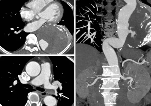
A 71 year-old woman who did not have a history of smoking was admitted to our hospital for treatment of the thoracic aortic aneu- rysm. She underwent a radical hysterectomy with bilateral salpingo oophorectomy in 1996 due to stage Ib of cervical cancer. She has also had a medical history of anticoagulation due to the pulmonary thromboembolism with deep vein thrombosis in both legs since 2001. However, she had quit her medication for 1 month without doctor’s permission prior to this admission. There was a 11 cm di- ameter of thoracic aortic aneurysm containing mural thrombus with contrast leakage at the descending aorta on pre-procedural CT (Fig.

1A and B). A chronic eccentric embolism on the left pulmonary ar-

tery was also found in the CT angiography (Fig. 1C). An echocardiog-

raphy indicated normal systolic function of the left ventricle without

any intra-cardiac thrombi. On laboratory study, her anti-phospholip-

id IgG antibody and the tumor markers of CEA and CA19-9 were

within the normal value. A protein C antigen level of the patient was

also normal, but the protein S antigen was lower at 44.2% (normal

range; 60–150%). Since the size of the patient’s aneurysm was very

large despite the absence of any symptoms, there might have been

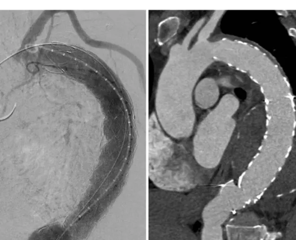
a risk of aortic rupture. Therefore, we planned TEVAR for her aneu-

rysm.

(2)

A 300 mg of aspirin and the 600 mg of clopidogrel were admin- istrated orally on the day of the procedure. A Rt. common femoral artery (CFA) was prepared for the 22 Fr large sheath insertion by preclose technique,

6)

and the 7 Fr sheath was inserted after punc- ture via Lt. CFA. Then, 5000 IU (100 IU/kg) of heparin was adminis- trated intravenously for endovascular treatment. After the marker pig tail catheter was placed at the aortic arch via Lt. CFA, a 5 Fr mul- tipurpose catheter with 0.035 coating guide wire (J tip, Terumo,

Tokyo, Japan) was located at the ascending artery via Rt. CFA. Then, the lunderquist extra-stiff guide wire (Cook, Inc., Bloomington, IL, USA) was inserted into the multipurpose catheter after coating guide wire was removed from it. However, during the wire exchang- ing procedure, the patient complained about a severe chest pain.

The ECG of the monitor showed ST elevation of avF limb leads, and the bradycardia with hypotension was documented (Fig. 2B). A prompt coronary angiography was performed and it revealed a large

A

B C

Fig. 1. Pre-procedural aorta CT of presented patient. A: axial CT image shows the huge aneurysm (11 cm of diameter) containing the thrombus. B: recon- structed CT image shows the huge aneurysm containing the mural thrombus and the contrast leakage is seen inside the aneurysm as well. C: axial CT im- age shows the organized thrombi at the left main pulmonary artery.

A B

Fig. 2. Baseline and the event polygraphic records show the 3 limb leads ECG and the pulse pressure at the right common femoral artery sheath. A: base-

line ECG shows 70 beats per minute of heart rate without significant ST height difference and blood pressure is approximately 110/60 mm Hg. B: an ECG at

the time of chest pain indicates newly developed ST elevation at avF limb lead compared to the baseline ECG with 45 beats per minute of heart rate and

the blood pressure is approximately 80/50 mm Hg. ECG: electrocardiography.

(3)

A B

Fig. 3. Right coronary artery angiography before and after the thrombus aspiration. A: right coronary angiography shows the mobile multiple thrombi in- side of the vessels and the TIMI grade 1 of blood flow. B: right coronary angiography after the thrombus aspiration shows no organic stenosis or remnant thrombi inside the vessel and the TIMI grade 4 of blood flow is restored. TIMI: Thrombolysis in Myocardial Infarction.

A B

Fig. 4. A pig-tail aortogram and CT angiography after stent-graft insertion. A: a aortogram shows the good position of stent-graft without any kind of

leakage. B: a CT angiography taken before discharge shows complete coverage of aneurysm.

(4)

amount of mobile thrombi with Thrombolysis in Myocardial Infarc- tion 1 coronary blood flow in right coronary artery (Fig. 3A). How- ever, neither coronary artery stenosis nor the plaque rupture was found in the coronary artery (Fig. 3B) after the thrombus aspiration (Eliminate aspiration catheter, Terumo, Tokyo, Japan). After achieving the patient’s stabilization, 30 mm by 150 mm of thoracic stent graft was first deployed at the descending aorta. Then, a 34 mm by 200 mm of the stent graft was deployed at the aortic arch just distally to the left subclavian artery (Seal thoracic stent graft, S&G Inc, Seongnam, Korea) (Fig. 4A). A creatine kinase-MB was increased 1.9 to 5.4 ng/mL 6 hours after the procedure without increase of tropo- nin-T. Post-procedural CT angiography showed a complete cover- age of aneurysm without any leakage (Fig. 4B).

Discussion

Thoracic endovascular aortic repair for descending thoracic aortic aneurysm is becoming the standard alternative therapy because of good applicability of endogarfting. As it is remarkably less morbid than surgical treatment, it leads to more expanded inclusion for the patient who has many medical conditions.

2)

Yet, still the peri-proce- dural complications after TEVAR “including” myocardial infarction, major stroke and the spinal cord infarction “are” the considerable demerits making the patient permanent sequalea.

Having an aortic aneurysm can be a risk factor for the peri-opera- tive adverse outcome in itself because most aortic aneurysms are resulted from atherosclerotic degeneration. Therefore, it seems to be firmly associated with other types of atherosclerosis which are manifested by the coronary or peripheral arterial disease.

7)

For this reason, the risk of peri-procedural or peri-operative myocardial in- farction rate was reported as high as 6% during aneurysm repair.

8)

It is not surprising that patients with more comorbidities had a signifi- cantly higher rate of major adverse event than those who did not have the risk factors.

9)

However, protein S deficiency was not generally considered as a risk factor for intervention. Inherited thrombophilia including anti- coagulant protein deficiency is known as a genetic tendency to ve- nous thromboembolism. An anticoagulant protein S deficiency were reported in 7.3% of the patients who had deep vein thrombosis,

3)

and 74% and 38% of the patients with protein S deficiency had sustained deep venous thrombosis or pulmonary embolism, respec- tively, which is similarly to the patients in the current report.

10)

Un- like venous thrombosis, there is a lack of evidence for proving a risk factor for the development of arterial thrombosis, only a few stud- ies of protein S deficiency demonstrated a high prevalence of arte- rial occlusive disease, ischemic stroke or cerebral ischemia.

4)5)11)

It is interesting that while several reports indicated coronary thrombo-

sis in patients with protein S deficiency, their coronary arteries did not have organic stenosis or evidence of plaque rupture.

12-14)

Al- though the patient in current report was medicated antiplatelet agents and administered heparin before the procedure, the current phenomenon is likely to arise from embolic event if considering the pathophysiology of protein S deficiency. The source of throm- bus might be from the coated wire surface and then could be dis- lodged while catheters exchange procedure. Thus, checking the ac- tivated coagulation time is mandatory before initiation of procedure and more cautious peri-procedural device handling under heparinized fluid is required for high risk patients having throm- bophilia.

Another important consideration for the anticoagulant protein deficiency is the timing of testing in the laboratory evaluation since erroneous diagnoses can be made by the clinical situations as acute thrombosis, serious illness or anticoagulant therapy.

15)

However, the patient in the current report had good physical activity without severe illness at the time of admission and had discontinued warfa- rin arbitrarily for 1 month prior to the admission. The sample was taken at the time of the admission when the coumadization was not ini- tiated.

We experienced a rare thrombotic coronary occlusion during the TEVAR and overcame the situation with immediate response, which yielded/produced favorable results preventing myocardial infarction.

We prescribed warfarin and dual antiplatelet including the aspirin and clopidogrel, then continued the regimen for 6 months without any clinical events. Current case report contributes to a better un- derstanding of the thrombotic complications during endovascular procedure and also thrombophilic diseases including protein S de- ficiency to the arterial occlusive adverse events. Hence, extra cau- tion is required when performing endovascular treatment in pa- tients with a thrombophlic disease.

References

1. Walsh SR, Tang TY, Sadat U, et al. Endovascular stenting versus open surgery for thoracic aortic disease: systematic review and meta-anal- ysis of perioperative results. J Vasc Surg 2008;47:1094-8.

2. Desai ND, Burtch K, Moser W, et al. Long-term comparison of thoracic endovascular aortic repair (TEVAR) to open surgery for the treatment of thoracic aortic aneurysms. J Thorac Cardiovasc Surg 2012;144:

604-9; discussion 609-11.

3. Mateo J, Oliver A, Borrell M, Sala N, Fontcuberta J. Laboratory evalua- tion and clinical characteristics of 2,132 consecutive unselected pa- tients with venous thromboembolism--results of the Spanish Multi- centric Study on Thrombophilia (EMET-Study). Thromb Haemost 1997;

77:444-51.

4. Allaart CF, Aronson DC, Ruys T, et al. Hereditary protein S deficiency in

young adults with arterial occlusive disease. Thromb Haemost 1990;64:

(5)

206-10.

5. Douay X, Lucas C, Caron C, Goudemand J, Leys D. Antithrombin, pro- tein C and protein S levels in 127 consecutive young adults with isch- emic stroke. Acta Neurol Scand 1998;98:124-7.

6. Lee WA, Brown MP, Nelson PR, Huber TS. Total percutaneous access for endovascular aortic aneurysm repair (“Preclose” technique). J Vasc Surg 2007;45:1095-101.

7. Sukhija R, Aronow WS, Yalamanchili K, Sinha N, Babu S. Prevalence of coronary artery disease, lower extremity peripheral arterial disease, and cerebrovascular disease in 110 men with an abdominal aortic aneu- rysm. Am J Cardiol 2004;94:1358-9.

8. Bub GL, Greenberg RK, Mastracci TM, et al. Perioperative cardiac events in endovascular repair of complex aortic aneurysms and association with preoperative studies. J Vasc Surg 2011;53:21-7.e1-2.

9. Wang GJ, Fairman RM, Jackson BM, Szeto WY, Pochettino A, Woo EY.

The outcome of thoracic endovascular aortic repair (TEVAR) in patients with renal insufficiency. J Vasc Surg 2009;49:42-6.

10. Engesser L, Broekmans AW, Briët E, Brommer EJ, Bertina RM. Heredi-

tary protein S deficiency: clinical manifestations. Ann Intern Med 1987;106:677-82.

11. Munts AG, van Genderen PJ, Dippel DW, van Kooten F, Koudstaal PJ.

Coagulation disorders in young adults with acute cerebral ischaemia.

J Neurol 1998;245:21-5.

12. Beattie S, Norton M, Doll D. Coronary thrombosis associated with in- herited protein S deficiency: a case report. Heart Lung 1997;26:76-9.

13. Carrié D, Béard T, Sié P, Boudjemaa B, Delay M, Bernadet P. [Simulta- neous thrombosis of the left anterior interventricular and right coro- nary arteries in a 27 year-old patient with protein S deficiency]. Arch Mal Coeur Vaiss 1993;86:921-4.

14. Manzar KJ, Padder FA, Conrad AR, Freeman I, Jonas EA. Acute myocar- dial infarction with normal coronary artery: a case report and review of literature. Am J Med Sci 1997;314:342-5.

15. Schwarz HP, Fischer M, Hopmeier P, Batard MA, Griffin JH. Plasma pro-

tein S deficiency in familial thrombotic disease. Blood 1984;64:1297-

300.

수치

Fig. 1. Pre-procedural aorta CT of presented patient. A: axial CT image shows the huge aneurysm (11 cm of diameter) containing the thrombus
Fig. 4. A pig-tail aortogram and CT angiography after stent-graft insertion. A: a aortogram shows the good position of stent-graft without any kind of  leakage

참조

관련 문서

Thoracic aortogram following thoracic endovascular aortic repair shows a delayed filling of the aneurysmal sac with blood flow from the left seventh intercostal artery, suggesting

Endovascular aneurysm repair versus open repair in patients with abdominal aortic aneurysm (EVAR trial 1): randomised controlled trial. Type III and type IV

The indications for endovascular repair were abdominal aortic aneurysm in 5 patients, descending thoracic aortic aneurysm in 6 patients and acute type B aortic dissection in

Aortic Valve Replacement Using Balloon Catheter for Thoracic Endovascular Aortic Repair to Patient with Calcified Aorta.. In Sook Kim,

Because the patient strongly refused surgery, we performed thoracic endovascular aortic repair (TEVAR) with left subclavian artery (LSCA) to left common carotid

Background: Endovascular aneurysm repair (EVAR) has dramatically changed the management of abdominal aortic aneurysms (AAAs) as the number of open aneurysm repairs

After computed tomography (CT) for an episode of hemoptysis, the patient underwent elective thoracic endovascular aortic repair (TEVAR) because of a degenerative

Objective: This study aimed to assess the surgical morbidity and mortality of thoracic endovascular repair (TEVAR) as compared with open surgical repair (OSR) for isolated