• 검색 결과가 없습니다.

The Risk Factors and Outcomes of Acute Kidney Injury after Thoracic Endovascular Aortic Repair

N/A
N/A
Protected

Academic year: 2021

Share "The Risk Factors and Outcomes of Acute Kidney Injury after Thoracic Endovascular Aortic Repair"

Copied!
7
0
0

로드 중.... (전체 텍스트 보기)

전체 글

(1)

ISSN: 2233-601X (Print) ISSN: 2093-6516 (Online)

Department of Thoracic and Cardiovascular Surgery, Catholic University of Daegu School of Medicine

Received: July 6, 2015, Revised: September 2, 2015, Accepted: September 14, 2015, Published online: February 5, 2016

Corresponding author: Chi-Hoon Bae, Department of Thoracic and Cardiovascular Surgery, Catholic University of Daegu School of Medicine, 33 Duryugongwon-ro 17-gil, Nam-gu, Daegu 42472, Korea

(Tel) 82-53-650-3420 (Fax) 82-53-629-6963 (E-mail) [email protected]

C

The Korean Society for Thoracic and Cardiovascular Surgery. 2016. All right reserved.

CC

This is an open access article distributed under the terms of the Creative Commons Attribution Non-Commercial License (http://creative- commons.org/licenses/by-nc/4.0) which permits unrestricted non-commercial use, distribution, and reproduction in any medium, provided the original work is properly cited.

The Risk Factors and Outcomes of Acute Kidney Injury after Thoracic Endovascular Aortic Repair

Yun-Ho Jeon, M.D., Chi-Hoon Bae, M.D.

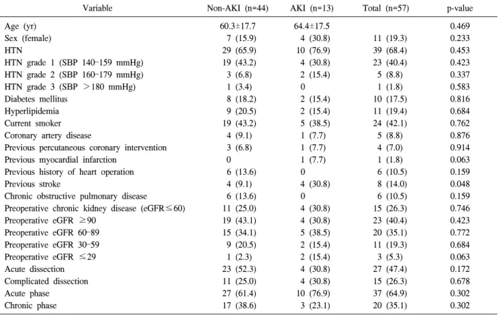
Background: We aimed to evaluate the incidence, predictive factors, and impact of acute kidney injury (AKI) after thoracic endovascular aortic repair (TEVAR). Methods: A total of 53 patients who underwent 57 TEVAR operations between 2008 and 2015 were reviewed for the incidence of AKI as defined by the RIFLE (risk, injury, failure, loss, and end-stage kidney disease risk) consensus criteria. The estimated glomerular filtration rate was determined in the perioperative period. Comorbidities and postoperative outcomes were retrospectively reviewed. Results: Under- lying aortic pathologies included 21 degenerative aortic aneurysms, 20 blunt traumatic aortic injuries, six type B aortic dissections, five type B intramural hematomas, three endoleaks and two miscellaneous diseases. The mean age of the patients was 61.2±17.5 years (range, 15 to 85 years). AKI was identified in 13 (22.8%) of 57 patients.

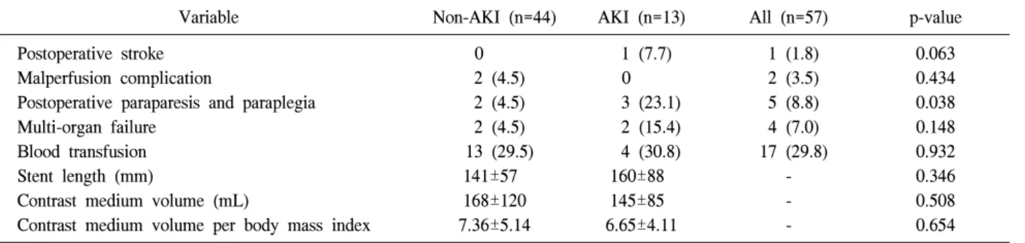
There was an association of preoperative stroke and postoperative paraparesis and paraplegia with AKI. The aver- age intensive care unit (ICU) stay in patients with AKI was significantly longer than in patients without AKI (5.3 vs.

12.7 days, p=0.017). The 30-day mortality rate in patients with AKI was significantly higher than patients without AKI (23.1% vs. 4.5%, p=0.038); however, AKI did not impact long-term survival. Conclusion: Preoperative stroke and postoperative paraparesis and paraplegia were identified as predictors for AKI. Patients with AKI experienced longer average ICU stays and greater 30-day mortality than those without AKI. Perioperative identification of high- risk patients, as well as nephroprotective strategies to reduce the incidence of AKI, should be considered as im- portant aspects of a successful TEVAR procedure.

Key words: 1. Thoracic endovascular aortic repair 2. Acute kidney injury

3. RIFLE

INTRODUCTION

Thoracic endovascular aortic repair (TEVAR) has emerged as an alternative to conventional open surgery for treatment of various thoracic aortic diseases such as degenerative aortic aneurysm, type B aortic dissection, and type B intramural hematoma. The suggested advantages of TEVAR include shorter operative time, reduced duration of general anesthesia, shorter

hospital stay, less blood loss, and avoidance of cardiopulmo-

nary bypass, aortic cross clamping, invasive thoracotomy, tho-

racoabdominal incision, and hypothermic arrest [1-3]. The in-

cidence of acute kidney injury (AKI) after TEVAR has been

reported to be 1% to 34% [4]. This relatively common com-

plication of TEVAR is associated with prolonged hospital

stays and increased risk of mortality [5-7]. The goal of this

clinical research was to investigate the risk factors and impact

http://dx.doi.org/10.5090/kjtcs.2016.49.1.15

(2)

Table 1. Underlying pathology in patients undergoing thoracic en- dovascular aortic repair (N=57)

Aortic pathology Value

Aortic aneurysm 21 (36.8)

Traumatic aortic injury 20 (35.1) Type B aortic dissection 6 (10.5)

Intramural hematoma 5 (8.8)

Endoleak 3 (5.3)

Miscellaneous 2 (3.5)

Values are presented as number (%).

Table 2. Aortic aneurysm classification according to location (N=21)

Aneurysm location Value

Aortic arch 8 (38.1)

Proximal descending aorta 2 (9.5) Descending thoracic aorta 11 (52.4)

Thoracoabdominal aorta 0

Values are presented as number (%).

Table 3. RIFLE

a)

criteria for the classification of acute kidney injury

Class GFR criteria

Risk Plasma creatinine increase 1.5×from baseline or GFR decline >25%

Injury Plasma creatinine increase 2×from baseline or GFR decline >50%

Failure Plasma creatinine increase 3×from baseline or GFR decline >75% or acute plasma creatinine >4 mg/dL Loss Persistent acute renal failure=complete loss of kidney function requiring dialysis for >4 weeks but <3 months End-stage End-stage kidney disease requiring dialysis for >3 months

GFR, glomerular filtration rate.

a)

Risk, injury, failure, loss, and end-stage kidney disease risk.

of AKI after TEVAR.

METHODS

1) Patient population and procedural details

Between April 2008 and February 2015, 53 patients under- went 57 TEVAR operations at our institution. Two of these 53 patients suffered from post-TEVAR endoleak and two oth- er patients were operated on for progressive new-onset pene- trating aortic ulceration. The 57 TEVAR cases consisted of 21 aortic aneurysms, 20 traumatic aortic injuries, six type B aortic dissections, five Type B intramural hematomas, three endoleaks and two miscellaneous diseases (Behcet’s aortitis and anastomotic pseudoaneurysm); these details and the loca- tion of the TEVAR are described in Tables 1 and 2.

To reduce postoperative AKI, patients were given 60 mL/

hr hydration fluid from the day before surgery, and 600 mg N-acetylcysteine was intravenously injected 12 hours before and just prior to surgery. Iodixanol (Visipaque; GE Health- care, Milwaukee, WI, USA), an iso-osmolar-contrast medium with reduced nephrotoxicity, was injected during angiogram

by contrast media injector (Mark V Provis; Medrad Inc., Indi- anola, PA, USA).

All patients underwent TEVAR operations using a portable

C-arm fluoroscopic device (OEC 9900 Elite, GE Healthcare)

under general anesthesia. To reduce postoperative paraparesis

or paraplegia, lumbar cerebrospinal fluid (CSF) drainage was

performed in all except three patients: in one case, the patient’s

bleeding tendency was very high and the other two had cer-

vical and lumbar spine fractures. During the operation, CSF

pressure was sustained at 12 cm H

2

O and CSF drainage was

maintained postoperatively for three days. The femoral artery

was cannulated with a percutaneous sheath through a small

longitudinal incision. A sheath was introduced percutaneously

on the contralateral side. To accurately identify the site of de-

ployment, a pigtail catheter was introduced into a percuta-

neous sheath and a soft guidewire was introduced into the

cannulated sheath. Using a Bern catheter, the soft guide wire

was then exchanged for a stiff one; subsequently, an endovas-

cular stent graft (S & G Biotech, Seoul, Korea) was in-

troduced to the target lesion. To prevent the stent graft from

being pushed back, systolic blood pressure (BP) was main-

tained below 100 mmHg during the deployment. The prox-

imal landing zone was secured at least 2 cm proximal to the

site of the intimal tear. After stent graft deployment, an an-

(3)

Table 4. Demographics and comorbidities

Variable Non-AKI (n=44) AKI (n=13) Total (n=57) p-value

Age (yr) 60.3±17.7 64.4±17.5 0.469

Sex (female) 7 (15.9) 4 (30.8) 11 (19.3) 0.233

HTN 29 (65.9) 10 (76.9) 39 (68.4) 0.453

HTN grade 1 (SBP 140 –159 mmHg) 19 (43.2) 4 (30.8) 23 (40.4) 0.423

HTN grade 2 (SBP 160–179 mmHg) 3 (6.8) 2 (15.4) 5 (8.8) 0.337

HTN grade 3 (SBP >180 mmHg) 1 (3.4) 0 1 (1.8) 0.583

Diabetes mellitus 8 (18.2) 2 (15.4) 10 (17.5) 0.816

Hyperlipidemia 9 (20.5) 2 (15.4) 11 (19.4) 0.684

Current smoker 19 (43.2) 5 (38.5) 24 (42.1) 0.762

Coronary artery disease 4 (9.1) 1 (7.7) 5 (8.8) 0.876

Previous percutaneous coronary intervention 3 (6.8) 1 (7.7) 4 (7.0) 0.914

Previous myocardial infarction 0 1 (7.7) 1 (1.8) 0.063

Previous history of heart operation 6 (13.6) 0 6 (10.5) 0.159

Previous stroke 4 (9.1) 4 (30.8) 8 (14.0) 0.048

Chronic obstructive pulmonary disease 6 (13.6) 0 6 (10.5) 0.159

Preoperative chronic kidney disease (eGFR ≤60) 11 (25.0) 4 (30.8) 15 (26.3) 0.746

Preoperative eGFR ≥90 19 (43.1) 4 (30.8) 23 (40.4) 0.423

Preoperative eGFR 60–89 15 (34.1) 5 (38.5) 20 (35.1) 0.772

Preoperative eGFR 30–59 9 (20.5) 2 (15.4) 11 (19.3) 0.684

Preoperative eGFR ≤29 1 (2.3) 2 (15.4) 3 (5.3) 0.063

Acute dissection 23 (52.3) 4 (30.8) 27 (47.4) 0.172

Complicated dissection 11 (25.0) 4 (30.8) 15 (26.3) 0.678

Acute phase 27 (61.4) 10 (76.9) 37 (64.9) 0.302

Chronic phase 17 (38.6) 3 (23.1) 20 (35.1) 0.302

Values are presented as mean±standard deviation or number (%).

AKI, acute kidney injury; HTN, hypertension; SBP, systolic blood pressure; eGFR, estimated glomerular filtration rate.

giogram was performed to assess the outcome and mean BP was maintained above 90 mmHg to ensure cerebrospinal perfusion. After the procedure, patients were transferred to the intensive care unit (ICU).

2) Definitions

The baseline estimated glomerular filtration rate (eGFR) was calculated by the Cockcroft-Gault equation (eGFR=

sex×[140-age]/serum creatinine×weight/72, where male sex=1 and female=0.85). Patients with AKI were defined by a 25%

decrease in eGFR or a ≥1.5-fold increase in serum crea- tinine, compared with baseline, up to 48 hours following the procedure (risk, injury, failure, loss, and end-stage kidney dis- ease [RIFLE] classification) (Table 3), or the need for con- tinuous renal replacement therapy during hospitalization.

Chronic renal failure was defined as a baseline eGFR ≤60 mL/min/1.73 m

2

. Symptoms were classified as acute phase if

they occurred within the 14 days prior to TEVAR and as chronic phase if TEVAR was conducted 14 days after symptoms first occurred. A complicated dissection was defined as a case in which the patient had continuous or repeated back pain de- spite maximal antihypertensive therapy, uncontrolled hyper- tension, malperfusion syndromes, or (imminent) rupture [8].

3) Statistical analysis

IBM SPSS Statistics software ver. 21.0 (IBM Co., Armonk, NY, USA) was used for statistical analysis of the data. Cate- gorical variables were analyzed using a chi-square test and continuous variables were analyzed using a two-sample t-test.

Variables with a p-value of less than 0.1 in univariate analy-

sis were included in a multivariate analysis. Survival analysis

was performed using the Kaplan-Meier log-rank method.

(4)

Table 5. Intraoperative and postoperative risk factors

Variable Non-AKI (n=44) AKI (n=13) All (n=57) p-value

Postoperative stroke 0 1 (7.7) 1 (1.8) 0.063

Malperfusion complication 2 (4.5) 0 2 (3.5) 0.434

Postoperative paraparesis and paraplegia 2 (4.5) 3 (23.1) 5 (8.8) 0.038

Multi-organ failure 2 (4.5) 2 (15.4) 4 (7.0) 0.148

Blood transfusion 13 (29.5) 4 (30.8) 17 (29.8) 0.932

Stent length (mm) 141±57 160±88 - 0.346

Contrast medium volume (mL) 168±120 145±85 - 0.508

Contrast medium volume per body mass index 7.36±5.14 6.65±4.11 - 0.654

Values are presented as number (%) or mean±standard deviation.

AKI, acute kidney injury.

Table 6. Impact of postoperative AKI

Variable Non-AKI (n=44) AKI (n=13) All (total=57) p-value

Intensive care unit stay (day) 5.3±7.9 12.7±20.5 7.0±12.2 0.017

Hospital stay (day) 22.0±23.1 45.8±93.6 27.4±48.9 0.607

30-Day mortality 2 (4.5) 3 (23.1) 5 (8.8) 0.038

Values are presented as mean±standard deviation or number (%).

AKI, acute kidney injury.

RESULTS 1) Patient characteristics

The mean age of the patients was 61.2±17.5 years (range, 15 to 85 years) and 46 of them were male. The mean pre- operative eGFR was 79.4±32.1 mL/min/1.73 m

2

. The under- lying disease(s) and baseline characteristics of patients under- going TEVAR are shown in Table 4. Common morbidities were hypertension (68.4%), current smoking (42.1%), hyper- lipidemia (19.3%), diabetes (17.5%), previous stroke (14.0%), and coronary artery disease (8.8%).

Postoperative paraparesis and paraplegia developed in five patients (8.8%; three developed paraparesis and two para- plegia). Among these patients, two (40%) were in the non- AKI group. Four of them recovered completely but one pa- tient with paraplegia only recovered partially, with paraparesis remaining.

2) Acute kidney injury predictors and incidence AKI was identified in 13 of 57 cases (overall incidence, 22.8%; risk, 11; injury, 2; failure/loss/end-stage, 0), with 4 pa- tients (7%) requiring temporary renal replacement therapy.

Univariate analysis identified previous stroke (p=0.048) and postoperative paraparesis and paraplegia (p=0.038) as pre- dictors of postoperative AKI. Hypertension grade, preopera- tive chronic kidney disease (CKD), complicated dissection, and acute and chronic phase were not significantly different between AKI and non-AKI groups (Table 4).

Surprisingly, no significant difference in the volume of contrast medium used was observed between the two groups (Table 5). Multivariate analysis did not identify any statisti- cally significant risk factors for the development of AKI.

3) Impact of postoperative acute kidney injury The average ICU stay in patients without AKI was 5.3 days, which was significantly lower than the average of 12.7 days for those with AKI (p=0.017) (Table 6). The average hospital stay in patients without AKI was 22 days, compared with 45.8 days in those with AKI.

The overall 30-day mortality was 8.8% (5 of 57 cases).

The 30-day mortality occurred in two of the 44 cases without AKI (4.5%) and three of the 13 cases with AKI (23.1%); this difference was statistically significant (p=0.038) (Table 6).

However, Kaplan-Meier analysis showed survival of 91.9%,

(5)

Fig. 1. The impact of postoperative AKI on late mortality. AKI, acute kidney injury.

82.0%, and 63.3% at 1, 3, and 5 years, respectively, for patients with AKI and 87.5%, 75.0%, and 75.0% at 1, 3, and 5 years for patients without AKI; these data were not significantly dif- ferent between the two groups (log-rank=0.922) (Fig. 1).

DISCUSSION

Thoracic endovascular aortic repair has emerged as a less invasive alternative to open aortic surgery for the treatment of thoracic aortic disease [3,4,9-11]. The occurrence of AKI af- ter TEVAR markedly increases hospital stay and is independ- ently associated with the risk of mortality [4]. However, data on the risk factors and impact of AKI after TEVAR have not yet been published by The Korean Society for Thoracic and Cardiovascular Surgery.

We used the RIFLE classification, a sensitive index of re- nal function used to predict long-term survival that was pro- posed by the Acute Dialysis Quality Initiative [5,12] that is the preferred diagnostic tool for monitoring the progression and the severity of AKI after cardiothoracic surgery [13-16].

In previous studies, the incidence of AKI varies from 14%

to 30.8%. In our study, AKI incidence was 22.8% and the perioperative risk factors for AKI after TEVAR were preope- rative stroke and postoperative paraparesis and paraplegia.

AKI developed in half of the patients with preoperative stroke. The pathophysiology of AKI in these patients with preoperative stroke is unclear; however, stroke and renal dys- function share vascular risk factors such as hypertension, dia-

betes, dyslipidemia, aging, and obesity [17,18]. Moreover, the kidney and brain are vulnerable to arteriosclerotic injury with similar microvascular functional and anatomical aspects [17,19].

In our study, three of five patients with postoperative para- paresis and paraplegia developed AKI. Postoperative para- paresis or paraplegia after TEVAR is a serious complication caused by spinal cord ischemia. The rate of paraplegia ranges from 0% to 13.3% among reported case series [20,21]. Probable mechanisms of ischemic damage to the spinal cord include perioperative hypotension and embolization during device in- troduction and deployment, and long stent graft coverage blocking critical extrinsic vertebral, intercostal, and lumbar supply to the anterior spinal artery [22]. The reasons for the frequent occurrence of AKI in patients with postoperative paraplegia are unclear; however, insufficient renal blood flow is a likely element of the pathophysiology of AKI.

Significant risk factors for AKI after TEVAR reported in other studies include preoperative CKD, thoracoabdominal ex- tent, postoperative transfusion [12], intraoperative hypotension, stroke, sepsis, lengthy procedures and number of stents [1], acute dissection, complicated dissection, and malperfusion complications.

It is unsurprising that preoperative CKD is a significant risk factor in previous studies; however, in our study, pre- operative eGFR ≤29 was not associated with significant risk for AKI. This may be because interventions to prevent AKI such as minimizing contrast use, stopping nephrotoxic agents, hydration, and permissive hypertension were more common in patients with low eGFR.

Postoperative transfusion has been previously associated with AKI. During transfusion of red blood cells, cellular and molecular components of allogeneic blood induce and intensify inflammatory responses, including the release of pro-inflam- matory cytokines, which may cause kidney damage [12,23].

Contrast-induced nephropathy is a well-known iatrogenic cause of renal dysfunction. Contrast media induces vasocon- striction by inhibiting nitric oxide-mediated vasodilation and sustains vasoconstriction by altering intracellular calcium and adenosine concentrations in kidney smooth muscle cells [24].

In our study, we used the minimum amount of contrast me-

dium possible, and our results indicate that the volume of

contrast medium used was not a significant risk factor for

(6)

AKI.

Limitations of this study include its retrospective design and the relatively small number of patients. Our results are in concordance with those of other studies, and further research will investigate more extensively the impact of factors such as preoperative eGFR of ≤29, previous MI, and postopera- tive stroke, which were not statistically significantly asso- ciated with risk for AKI in the present study.

In conclusion, preoperative stroke and postoperative para- paresis and paraplegia are significant risk factors for AKI af- ter TEVAR. Optimizing perioperative blood pressure, blood glucose, and blood lipid levels, avoiding hypotension to max- imize organ perfusion, and careful manipulation of endovas- cular devices to reduce embolization of atherosclerotic debris to various organs including the kidney represent important protective strategies to reduce the incidence of AKI. Because AKI increases the duration of hospital stays as well as mor- bidity and mortality, an awareness of the various risk factors for AKI and aggressive efforts to minimize the incidence, se- verity, and duration of AKI in patients with these risk factors represent the cornerstone of successful TEVAR intervention.

CONFLICT OF INTEREST

No potential conflict of interest relevant to this article was reported.

ACKNOWLEDGMENTS

This study was supported by a Grant of the Samsung Vein Clinic Network (Daejeon, Anyang, Cheongju, Cheonan; Fund No. KTCS04-040).

REFERENCES

1. Pisimisis GT, Khoynezhad A, Bashir K, Kruse MJ, Donayre CE, White RA. Incidence and risk factors of renal dysfunc- tion after thoracic endovascular aortic repair. J Thorac Cardiovasc Surg 2010;140(6 Suppl):S161-7.

2. Song TK, Donayre CE, Walot I, et al. Endograft exclusion of acute and chronic descending thoracic aortic dissections.

J Vasc Surg 2006;43:247-58.

3. Greenberg RK, O’Neill S, Walker E, et al. Endovascular re- pair of thoracic aortic lesions with the Zenith TX1 and TX2

thoracic grafts: intermediate-term results. J Vasc Surg 2005;41:589-96.

4. Drews JD, Patel HJ, Williams DM, Dasika NL, Deeb GM.

The impact of acute renal failure on early and late outcomes after thoracic aortic endovascular repair. Ann Thorac Surg 2014;97:2027-33.

5. Zhu JC, Chen SL, Jin GZ, et al. Acute renal injury after thoracic endovascular aortic repair of Stanford type B aortic dissection: incidence, risk factors, and prognosis. J Formos Med Assoc 2014;113:612-9.

6. Martin MC, Giles KA, Pomposelli FB, Hamdan AD, Wyers MC, Schermerhorn ML. National outcomes after open repair of abdominal aortic aneurysms with visceral or renal bypass.

Ann Vasc Surg 2010;24:106-12.

7. Ellenberger C, Schweizer A, Diaper J, et al. Incidence, risk factors and prognosis of changes in serum creatinine early after aortic abdominal surgery. Intensive Care Med 2006;32:

1808-16.

8. Khoynezhad A, Donayre CE, Smith J, Kopchok GE, Walot I, White RA. Risk factors for early and late mortality after thoracic endovascular aortic repair. J Thorac Cardiovasc Surg 2008;135:1103-9.

9. Bavaria JE, Appoo JJ, Makaroun MS, et al. Endovascular stent grafting versus open surgical repair of descending thoracic aortic aneurysms in low-risk patients: a multicenter comparative trial. J Thorac Cardiovasc Surg 2007;133:369-77.

10. Patel HJ, Williams DM, Upchurch GR Jr, et al. Long-term results from a 12-year experience with endovascular therapy for thoracic aortic disease. Ann Thorac Surg 2006;82:2147-53.

11. Patel HJ, Sood V, Williams DM, Dasika NL, Diener AC, Deeb GM. Late outcomes with repair of penetrating thoracic aortic ulcers: the merits of an endovascular approach. Ann Thorac Surg 2012;94:516-22.

12. Barbash IM, Ben-Dor I, Dvir D, et al. Incidence and pre- dictors of acute kidney injury after transcatheter aortic valve replacement. Am Heart J 2012;163:1031-6.

13. Piffaretti G, Mariscalco G, Bonardelli S, et al. Predictors and outcomes of acute kidney injury after thoracic aortic en- dograft repair. J Vasc Surg 2012;56:1527-34.

14. Bellomo R, Kellum JA, Ronco C. Defining and classifying acute renal failure: from advocacy to consensus and vali- dation of the RIFLE criteria. Intensive Care Med 2007;33:

409-13.

15. Levey AS, Stevens LA, Schmid CH, et al. A new equation to estimate glomerular filtration rate. Ann Intern Med 2009;

150:604-12.

16. Mariscalco G, Lorusso R, Dominici C, Renzulli A, Sala A.

Acute kidney injury: a relevant complication after cardiac surgery. Ann Thorac Surg 2011;92:1539-47.

17. Ma S, Zhao H, Ji X, Luo Y. Peripheral to central: organ interactions in stroke pathophysiology. Exp Neurol 2015;272:

41-9.

(7)

18. Ninomiya T. Risk of stroke in kidney disease. Contrib Nephrol 2013;179:58-66.

19. Mogi M, Horiuchi M. Clinical Interaction between Brain and Kidney in Small Vessel Disease. Cardiol Res Pract 2011;

2011:306189.

20. Bobadilla JL, Wynn M, Tefera G, Acher CW. Low incidence of paraplegia after thoracic endovascular aneurysm repair with proactive spinal cord protective protocols. J Vasc Surg 2013;57:1537-42.

21. Rizvi AZ, Sullivan TM. Incidence, prevention, and manage- ment in spinal cord protection during TEVAR. J Vasc Surg 2010;52(4 Suppl):86S-90S.

22. Drinkwater SL, Goebells A, Haydar A, et al. The incidence of spinal cord ischaemia following thoracic and thoracoa- bdominal aortic endovascular intervention. Eur J Vasc Endovasc Surg 2010;40:729-35.

23. Nuis RJ, Rodes-Cabau J, Sinning JM, et al. Blood trans- fusion and the risk of acute kidney injury after transcatheter aortic valve implantation. Circ Cardiovasc Interv 2012;5:680-8.

24. Azzalini L, Spagnoli V, Ly HQ. Contrast-induced nephrop- athy: from pathophysiology to preventive strategies. Can J Cardiol 2015 May 23 [Epub]. http://dx.doi.org/10.1016/j.cjca.

2015.05.013.

수치

Table 3. RIFLE a)  criteria for the classification of acute kidney injury
Table 4. Demographics and comorbidities
Table 5. Intraoperative and postoperative risk factors
Fig. 1. The impact of postoperative AKI on late mortality. AKI,  acute kidney injury.

참조

관련 문서

This study aimed to evaluate the site and extent of injury, injury mechanism, player position, and the reinjury incidence in the hamstring by using magnetic

(Background) Gallbladder wall thickening(GWT) and gallbladder contraction are often observed in patients with acute hepatitis.. The incidence of acute hepatitis A

The risk of complicated acute appendicitis should be evaluated in terms of medical factors, including psychi- atric disease entity, psychotropic prescription, and

Age at diagnosis after 30 years, baseline serum creatinine levels, and cyst infection are the independent risk factors for kidney failure, as found in this

Further research on other intraoperative risk factors, such as flap thickness and length of incision, in addition to the incision type, may help in improving the postoperative

ENDOSTROKE, Endovascular Stroke Treatment; BAO, basilar artery occlusion; EVT, endovascular treatment; ASITN/SIR, The American Society of Interventional

The ‘Investigation of outcomes after GOLimumab Discon- tinuation,’ or GOLD, was an observational study involving 10 centers nationwide that analyzed clinical data

Prevalence and Factors of Osteoporosis and High Risk of Osteoporotic Fracture in Patients with Ankylosing Spondylitis: A Multicenter Comparative Study of Bone Mineral Density