ABSTRACT
Purpose: Reflux esophagitis is a disease caused by the reflux of stomach contents and
stomach acid etc. into the esophagus due to defect in the lower esophageal sphincter and is currently increasing worldwide. This study was conducted to evaluate the effect of a mixture of Citrus Reticulata and Scutellariae Radix (CS) extract on acute reflux esophagitis in rats.
Methods: Rats were divided into five groups for examination: normal group (Normal, n = 8),
water-treated acute reflux esophagitis rats (Control, n = 8), tocopherol 30 mg/kg body weight-treated acute reflux esophagitis rats (Toco, n = 8), CS 100 mg/kg body weight-treated acute reflux esophagitis rats (CS100, n = 8), CS 200 mg/kg body weight-treated acute reflux esophagitis rats (CS200, n = 8). The experimental groups were administrated of each treatment compounds and after 90 min, acute reflux esophagitis was induced through surgery. Rats were sacrificed 5 h after surgery. We measured the level of reactive oxygen species (ROS) in serum and analyzed the expression of nicotinamide adenine dinucleotide phosphate, inflammatory, and tight junction-related proteins by western blot in the esophageal tissues.
Results:
CS administration significantly protected the esophageal mucosal damage due to reflux esophagitis, and the level of ROS in the serum was significantly reduced with CS administration as compared to Control. In addition, CS administration significantly suppressed mitogen-activated protein kinase (MAPK or MAP kinase) and nuclear factor- kappa B (NF-κB) pathways and increased protein expressions of tight junction protein.
Conclusion: These results suggest that the CS not only regulates the expression of
inflammatory proteins by inhibiting oxidative stress, but also reduces damage to the esophageal mucosa by inhibiting the expression of tight junction proteins.
Keywords: esophagitis, Citrus Reticulata, Scutellariae Radix, oxidative stress, inflammation
Research Article
Received: Feb 19, 2021 Revised: Apr 13, 2021 Accepted: Apr 30, 2021 Correspondence to Hae-Jin Park
DHU Bio Convergence Testing Center, 1 Hanuidae-ro, Gyeongsan 38610, Korea.
Tel: +82-53-819-7870 E-mail: [email protected]
© 2021 The Korean Nutrition Society This is an Open Access article distributed under the terms of the Creative Commons Attribution Non-Commercial License (http://
creativecommons.org/licenses/by-nc/3.0/) which permits unrestricted non-commercial use, distribution, and reproduction in any medium, provided the original work is properly cited.
ORCID iDs Jin A Lee
https://orcid.org/0000-0002-5615-4557 Mi-Rae Shin
https://orcid.org/0000-0002-4365-6988 Seong-Soo Roh
https://orcid.org/0000-0002-4162-6849 Hae-Jin Park
https://orcid.org/0000-0002-4283-0809 Funding
This research was supported by the National Research Foundation of Korea (NRF) grant funded by the Korea Government (MSIP) (No. 2019R1I1A1A01064068 and No.
2017R1A2B2006858).
Conflict of Interest
There are no financial or other issues that might lead to conflict of interest.
Effects of a mixture of Citri
Pericarpium and Scutellariae Radix on acute reflux esophagitis in rats
Jin A Lee
1, Mi-Rae Shin
1, Seong-Soo Roh
1, and Hae-Jin Park
21Department of Herbology, College of Korean Medicine, Daegu Haany University, Daegu 42158, Korea
2DHU Bio Convergence Testing Center, Gyeongsan 38610, Korea
진피 - 황금 혼합물이 급성 역류성 식도염
흰쥐에 미치는 효과
이진아
1, 신미래
1, 노성수
1, 박해진
21대구한의대학교 한의과대학 본초약리학교실
2대구한의대학교 DHU 바이오융복합시험센터
서론
위식도 역류질환
(gastroesophageal reflux disease)은 식도 내의 위산이 역류해 형태학적 변화
및 임상증상을 나타내는 경우를 말하며
,위식도 역류질환 중 하나인 역류성 식도염
(refluxesophagitis)
은 위
-식도 괄약근의 불안정으로 위 속 내용물이 역류하여 염증 및 궤양 등의 병변
이 유발되는 질환으로 가슴쓰림
,만성 기침
,목 이물감
,천식
,흉통 등의 증상을 동반한다
[1,2].고열량식과 고지방식
,음주
,흡연 등의 원인으로 서양의 경우
5%이상에서 발병하는 흔한 질 병이며
,현재 우리나라를 비롯한 아시아 지역에서도 매년 발병률이 증가하는 추세이다
[3].역류성 식도염 치료를 위하여 흔히 사용되고 있는 약물로는
proton pump inhibitor (PPI)와
histamine type 2 receptor antagonist
제제가 있으며
,이러한 약물들은 산 분비를 억제하는 약 물로 산 분비량을 줄여 위산 역류 시 발생하는 증상을 완화시켜 손상된 식도 점막을 치유하
여 합병증을 막아주는 역할을 한다
[4].하지만 이러한 약물들은 일반적인 치료에 반응하지
않는 경우가 나타나며
,초기에 치유되었더라도 약 복용을 중단할 경우
1년 이내 재발률이
50–80%
에 이르기 때문에 장기간 치료가 필요하다
[5].이에 부작용이 적으면서 역류성 식도
염을 효과적으로 치료할 수 있는 새로운 기전의 치료제 개발이 필요한 실정이다
.진피
(陳皮, Citrus Reticulata)는 운형과에 속하는 상록 소교목인 감귤나무의 성숙한 감귤 과
실의 껍질을 말린 것이다
.귤은 구연산
,비타민
C,비타민
P,헤스페리딘 등의 성분을 함유한 다고 알려져 있는데 이러한 성분들은 귤 껍질에 풍부하게 함유되어 있으며
,특히 비타민
C의 함량은 과육보다 귤 껍질에서
4배 이상 높게 나타난다고 알려져 있다
[6].또한
,진피의 지표 성분 중 하나인 나린진은 플라보노이드계 성분으로써
,플라보노이드계 성분은 많은 연구를
통해 항염증
,항궤양
,항산화 활성을 가진다고 알려져 있다
[7].진피의 약리 효능으로는 항산
화
[8],항균
[9],항염증
[10],항비만
[11]효과 등이 알려져 있으며
,한방에서 진피는 습담
(濕痰)
으로 인한 병증에 사용되는 약재로써 역류성 식도염의 원인인 담음
(痰飮)및 기체
(氣滯)증상에 사용될 수 있다고 하였다
[12].또한
,진피에는 유기산
,유리당
,비타민 등
60여종의 영
양성분을 함유하고 있어 잼
,젤리 등으로 개발되어 소비량이 증가하고 있는 추세이며
,최근 에는 다이어트식품으로써의 중요성뿐만 아니라 고혈압
,순환계 질환 예방 및 개선을 위한 건 강기능식품으로 개발되어 관심을 받고 있다
[13].황금
(黃芩, Scutellariae Radix)은 꿀풀과에 속하는 다년생 초본인 황금의 주피를 벗긴 뿌리이
다
.황금은 식품으로 분류된 한약재로써 바이칼레인
,바이칼린
,우고닌 및 플라보노이드 등
30
여종의 생리활성 성분이 분리되어 있으며
[14],항균
[15],멜라닌 생성 저해 효과
[16],암세
포 증식 억제 효과
[17],항염증
[18]효과 등이 알려져 있다
.또한
,황금은 천연 항균제로 알려
져 있어 식품의 갈변을 저해할 뿐 아니라 저장 기간에도 긍정적인 영향을 주며
,오랜 기간 저
장이 필요한 약주
,막걸리 및 전통주 개발에도 사용되고 있다
[19-22].본 연구에서는 급성 역류성 식도염 동물 모델에서 항균 및 항염증 효과가 있다고 알려진 진피
와 황금을 혼합한 혼합물의 식도 점막 보호 효과를 확인하고자 하였으며
,식도 점막 손상 여
부
,염증 및 식도 기능 관련 단백질의 발현에서 유의한 결과를 얻었기에 이를 보고하는 바이다
.연구방법
시료추출
본 실험에서 사용한 진피와 황금은 옹기한약국
(Daegu, Korea)에서 구입한 것을 생약규격집 에 맞추어 관능검사 후 약전규격에 적합한 것만을 정산하여 사용하였다
.황금과 진피를 각각
200 g분쇄하여 물을
2,000 mL첨가한 후 열탕추출기
(DW-790, Daewoongbio, Seoul, Korea)에 서
2시간 추출하였으며
,얻어진 추출물은 회전 감압 추출장치
(N-1100, Eyela, Tokyo, Japan)를 이용하여 농축 후
,동결 건조기
(FD5508, IlShin, Seoul, Korea)를 이용해 완전 건조시켜 파우더
(진피
, 35%;황금
, 29%)를 얻었다
.추출 파우더는 실험 전까지
−80°C에서 보관하였고
,실험 당
일 진피와 황금을
1:1비율로 혼합
(CS)하여 사용하였다
.시약
본 실험에 사용된
potassium phosphate monobasic, potassium phosphate dibasic, 2′, 7′dichlo- rofluorescein diacetate (DCFH-DA)는
Sigma-Aldrich Co. (St. Louis, MO, USA)에서 구입하여 사 용하였다
. Nitrocellulose membrane은
Amersham GE Healthcare (Little Chalfont, UK)에서 구 입하였고
, NADPH oxidase 4 (NOX4), p47phox, p22phox, phospho-p38 MAPK (p-p38), phospho-ex- tracellular signal-regulated kinase (p-ERK), phospho-c-Jun N-terminal kinase (p-JNK), phos- phorylation of nuclear factor-kappa B p65 (NF-κBp65), inhibitor of nuclear factor kappa B alpha (IκBα), phosphorylation inhibitor of nuclear factor kappa B alpha (p-IκBα), cyclooxygenase-2 (COX-2), tumor necrosis factor-alpha (TNF-α), interleukin-6 (IL-6), claudin-1, claudin-4, histone, β-actin은
Santa Cruz Biotechnology (Dallas, CA, USA)로부터 구입하였다
. c-Fos와
c-Jun은
Cell Signaling Technology, Inc. (Beverly, MA, USA)에서 구입하여 사용하였으며
, 2차 항체는
Ge- neTex, Inc. (Irvine, LA, USA)에서 구입하여 사용하였다
. Protease inhibitor mixture, ethylene- diaminetetraacetic acid (EDTA)는
Wako Pure Chemical Industries, Ltd. (Osaka, Japan)에서 구입 하였다
. ECL western blotting detection reagents는
GE Healthcare로부터 구입하여 사용하였 으며
,단백질 정량을 위한
BCA protein assay kit는
Thermo Scientific (Rockford, IL, USA)에서 구입하였다
.실험동물
Sprague-Dawley (SD) rat
수컷
(180–200 g)을 대한바이오링크
(Eumseong, Korea)에서 구입하 여
1주일 동안 실험실 환경에 적응시킨 후 실험에 사용하였다
.동물 사육실 조건은 온도
(23 ± 2°C),습도
(50 ± 10%)및 명암주기
(12/12 hr)로 조절하였으며
,사료
(NIH-41, Zeigler Bros, Inc., USA)와 물은 자유식으로 급여하였다
(Table 1).본 실험은 동물실험의 윤리적
,과학적 타당성 검토 및 효율적인 관리를 위하여 대구한의대학교 동물실험윤리위원회
(Institution Animal Care and Use Committee; IACUC)의 승인
(DHU2020-067)을 받아 진행하였다
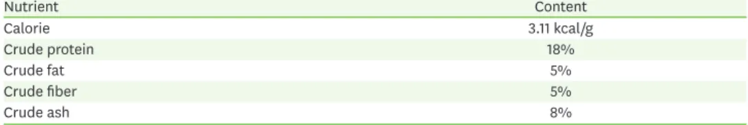
.Table 1. Nutritional component of NIH-41 diet
Nutrient Content
Calorie 3.11 kcal/g
Crude protein 18%
Crude fat 5%
Crude fiber 5%
Crude ash 8%
급성역류성식도염유발및동물처치
실험동물은 아무런 처치를 하지 않은 정상군
(Normal),증류수 경구투여 후 급성 역류성 식
도염을 유발한 대조군
(Control), tocopherol 30 mg/kg경구투여 후 급성 역류성 식도염을 유
발한 양성대조군
(Toco),진피
-황금 혼합물
100 mg/kg경구투여 후 급성 역류성 식도염을 유
발한 군
(CS100),진피
-황금 혼합물
200 mg/kg경구투여 후 급성 역류성 식도염을 유발한 군
(CS200)
총
5군으로 그룹 당
8마리씩 무작위 분류하였다
.실험
18시간 전 절식하였으며
,수술 당일 각 시료를
DW에 녹여 단회 경구투여하고
1시간
30분 후 이소플루레인
(isoflurane; Troikaa Pharmaceuticals Ltd., Gujarat, India)으로 흡입마취 하여 수술을 진행하였다
.본 실험에서 사용한 수술 방법은 급성 역류성 식도염 유발을 위해
Omura
등
[23]의 수술법을 변형한 방법으로써
,선행연구에서 다음과 같은 수술을 통해 역류
성 식도염을 유발하였으며
,역류성 식도염이 유발될 경우 위산의 역류로 인하여 식도 전체에 서 식도 점막이 손상되는 것을 확인하였다
[12,24].역류성 식도염 유발을 위하여 복부를 약
2 cm개복하여 위 조직의 위저부 및 날분무를 블랙실
크
(2-0)실로 묶은 후 복막과 피부를 봉합하였으며
, 5시간 후 희생하여 복대정맥에서 혈액을
채취하고 식도 조직을 적출하였다
[23].채취한 혈액은 원심분리
(4,000 rpm, 4°C, 10 min)하 여 혈청을 분리하여 사용하였으며
,분리한 혈청과 식도 조직은
−80°C에서 보관하였다
. 식도점막손상수술용 가위를 이용하여 적출한 식도를 세로로 절단하였다
.절단된 식도 내부를 생리식염수
으로 세척한 후 고정하여 광학 디지털 카메라
(DSCHX50V, Sony, Tokyo, Japan)를 이용하여 촬영
하였다
.손상된 식도 점막 측정은
I-Solution lite (Innerview Co., Seongnam, Korea)프로그램을 이용하여 실제 손상 부위의 면적을 측정한 후
,아래 식을 이용하여 손상 면적을 나타내었다
.식도 손상 비율 = 식도 손상 면적 식도 전체 면적 × 100
조직학적분석
식도 점막의 손상을 평가하기 위하여 현미경으로 조직학적 검사를 수행하였다
.식도 조직
을
24시간 동안
10%중성 완충 포르말린을 이용하여 고정하고
5 μm섹션으로 절단 후
hema-toxylin & eosin (H&E)
조직 염색 방법으로 염색하였으며
,염색된 슬라이드를 광학현미경으
로 관찰하였다
.혈청내활성산소종
(reactive oxygen species, ROS)
측정ROS
값은
Ali등
[25]방법에 의해 측정되었다
. 5 μL의 혈청에
50 mM sodium phosphate buffer (pH 7.4)와
0.125 mM DCFH-DA를 첨가하여 혼합한 후
,형광 광도계를 이용해
0분부터 매
5분 씩
emission파장
530 nm와
excitation파장
485 nm를 이용하여
30분간 누적 값의 변화를 측정 하였으며
,아래 식을 이용하여 값을 산출하였다
.ROS (fluorescence/mL) = Absorbance30min− Absorbance0min
30 × 200
식도조직
western blotting
식도 조직의 세포질을 얻기 위하여
100 mM Tris-HCl (pH 7.4), 5 mM Tris-HCl (pH 7.5), 15 mM CaCl2, 2 mM MgCl2, 1.5 M sucrose, 0.1 M DTT, protease inhibitor cocktail을 첨가한
buffer A를 넣고 조직분쇄기
(Biospec Product, Bartlesville, OK, USA)로 분쇄한 후
10% NP-40용액을 첨
가하여 아이스 위에서
30 min정치시킨 후 원심분리
(12,000 rpm, 4°C, 2 min)하여 세포질을
포함하고 있는 상층액을 분리하였다
.핵을 얻기 위하여
10% NP-40용액이 더해진
buffer A로
2회 세척하고
100 μL의
buffer C (50 mM HEPES, 50 mM KCl, 0.1 mM EDTA, 0.3 mM NaCl, 10% glycerol, 1 mM DTT, 0.1 mM PMSF)를 첨가하여 재부유시킨 다음
10분마다
3회
vortex하
였으며
,원심분리
(12,000 rpm, 4°C, 10 min)하여 핵을 포함한 상층액을 분리하였다
.얻어진
세포질과 핵은
−80°C에서 냉동 보관하였다
.식도 조직 세포질의
NOX4, p47phox, p22phox, p-p38, p-ERK, p-JNK, IκBα, p-IκBα, COX-2, TNF-α, IL-6, claudin-1, claudin-4, ꞵ-actin단백질과 핵에서 의
c-Fos, c-Jun, NF-κBp65, histone단백질 발현을 측정하기 위하여
12 μg의 단백질을
10-12%SDS polyacrylamide gel
을 이용하여 전기연동 후
, acrylamide gel을
nitrocellulose membrane으 로 이동시켰다
.준비된
membrane에 각각의
1차
antibody를 처리하여
4°C에서
overnight시 킨 다음
PBS-T로
6분마다
5회 세척하고
,각각 처리된
1차 항체에 사용되는
2차 항체
(PBS-T로
1:3,000
로 희석해서 사용
)를 사용하여 상온에서
2시간 반응시킨 후
, PBS-T로
6분마다
5회 세
척하였다
.그 후
membrane을
enhanced chemiluminescence (ECL)용액에 노출시킨 후
, Sen- si-Q2000 Chemidoc (Lugen Sci Co., Ltd., Seoul, Korea)에 감광시켜 단백질 발현을 확인한 후
,해당
band를
ATTO Densitograph Software (ATTO Corporation, Tokyo, Japan)프로그램을 사용 하여 정량하였다
.통계분석
In vivo
의 수치는 평균과 표준편차로 나타냈으며
, SPSS (Version 25.0, IBM, Armonk, NY, USA)를 사용하여
one-way analysis of variance (ANOVA) test를 실시한 후
least significant difference(LSD) test
로 사후검증을 실시하였으며
,통계적 유의성을
p-value < 0.05유의수준에서 검증
하였다
.결과
식도점막손상도측정
식도 점막의 손상을 육안으로 확인한 결과
,아무런 처치를 하지 않은
Normal군에서는 식도
점막의 손상이 발견되지 않았으나
,증류수 경구투여 후 급성 역류성 식도염을 유발한
Con-trol
군에서는 전반적인 식도의 손상 및 병변을 확인하였다
.반면에
Control군에 비하여
Toco군에서는 병변이
35% (p < 0.01)유의하게 감소하였다
.또한
, CS100군에서는
30% (p < 0.01), CS200군에서는
45% (p < 0.001)유의하게 감소하였으며
, CS200군에서는 양성대조군인
Toco군보다 더욱 뛰어난 식도 점막 보호 효과를 나타냈다
(Fig. 1).조직학적분석
H&E
염색을 통해 조직학적 변화를 관찰한 결과
,아무런 처치를 하지 않은
Normal군에서는
점막 상피층의 손상이 확인되지 않았으나
,급성 역류성 식도염을 유발한 후 증류수를 처리
한
Control군에서는 두꺼워진 조직층 및 식도 상피의 탈락
,염증세포의 침윤을 확인하였다
.CS100
군에서는
Control군에서 두껍게 나타났던 조직층이 완화된 것을 확인하였으며
, CS200군에서는 식도 상피의 탈락 및 염증세포의 침윤이 현저하게 감소한 것을 확인하였다
(Fig. 2).혈청내
ROS
측정혈청 내 산화적 스트레스 바이오마커인
ROS수치를 확인한 결과
, Normal군에 비하여
Control군에서
96% (p < 0.001)유의하게 증가하여
2배가량 높은 수치를 나타냈으며
, Control군에 비 하여
Toco군
35% (p < 0.001), CS100군에서
31% (p < 0.01), CS200군에서
37% (p < 0.001)유의 하게 감소하였다
(Fig. 3).식도조직내
NADPH
단백질발현량분석식도 조직 내에서
NADPH단백질인
NOX4, p47phox및
p22phox의 발현량을 확인하였다
. NOX4확인 결과
, Normal군에 비하여
Control군에서
38% (p < 0.001)유의하게 증가한 반면
Control군에 비하
###
** ***
60
40
20 50
30
10 0
B A
Gross mucosal damage ratio (%)
Normal Control Toco CS100 CS200
Normal Control Toco CS100 CS200
**
Fig. 1. Surgical induction of acute reflux esophagitis. A representative gross image; (A), esophageal ulcer ratio; (B). Esophageal ulcer ratio = damaged area of esophagus/total area of esophagus × 100. All data are expressed means ± SD (n = 8).
Normal, normal rats; Control, acute reflux esophagitis rats; Toco, tocopherol 30 mg/kg body weight/day-treated acute reflux esophagitis rats; CS100, mixture of Citrus Reticulata and Scutellariae Radix 100 mg/kg body weight/day-treated acute reflux esophagitis rats; CS200, mixture of Citrus Reticulata and Scutellariae Radix 200 mg/kg body weight/day-treated acute reflux esophagitis rats.
Significance: ###p < 0.001 vs. Normal rats and **p < 0.01, ***p < 0.001 vs. Control rats.
Normal Control Toco
CS100 CS200
Fig. 2. Esophagus histological examination through hematoxylin and eosin staining (magnification ×200).
Normal, normal rats; Control, acute reflux esophagitis rats; Toco, tocopherol 30 mg/kg body weight/day-treated acute reflux esophagitis rats; CS100, mixture of Citrus Reticulata and Scutellariae Radix 100 mg/kg body weight/
day-treated acute reflux esophagitis rats; CS200, mixture of Citrus Reticulata and Scutellariae Radix 200 mg/kg body weight/day-treated acute reflux esophagitis rats.
여
CS100군에서
20% (p < 0.001), CS200군에서
25% (p < 0.001)유의하게 감소하였다
. p47phox확 인 결과
, Normal군에 비하여
Control군에서
43% (p < 0.001)유의하게 증가한 반면
Control군에 비하여
CS200군에서
31% (p < 0.001)유의하게 감소하여
Normal군과 비슷한 수치를 나타냈다
.또한
p22phox확인 결과
, Normal군에 비하여
Control군에서
30% (p < 0.001)유의하게 증가하였으 며
, Control군에 비하여
CS200군에서
16% (p < 0.01)유의하게 감소하였다
(Fig. 4).###
*** ***
25,000
15,000 20,000
10,000 5,000 ROS (fluorescence/mL) 0
Normal Control Toco CS100 CS200
**
Fig. 3. ROS level in serum. ROS level = Absorbance30min − Absorbance0min/30 × 200. All data are expressed means
± SD (n = 8).
ROS, reactive oxygen species; Normal, normal rats; Control, acute reflux esophagitis rats; Toco, tocopherol 30 mg/kg body weight/day-treated acute reflux esophagitis rats; CS100, mixture of Citrus Reticulata and Scutellariae Radix 100 mg/kg body weight/day-treated acute reflux esophagitis rats; CS200, mixture of Citrus Reticulata and Scutellariae Radix 200 mg/kg body weight/day-treated acute reflux esophagitis rats.
Significance: ###p < 0.001 vs. Normal rats and **p < 0.01, ***p < 0.001 vs. Control rats.
NOX4 p47phox p22phox β-actin
70 kDa 47 kDa 22 kDa 43 kDa
###
***
2.0
1.0 1.5
0.5
0
D
p22phox (fold of normal)
Normal Control Toco CS100 CS200
###
2.0
1.0 1.5
0.5
0
C
p47phox (fold of normal)
Normal Control Toco CS100 CS200
###
** ** *** * **
***
2.0
1.0 1.5
0.5
0
B A
NOX4 (fold of normal)
Normal Control Toco CS100 CS200
***
Fig. 4. Expression of NADPH oxidase proteins in esophagus. Representative images of NOX4, p47phox, p22phox, and β-actin proteins (A). The expression level of NOX4 (B), p47phox (C), p22phox (D) were quantified. All data are expressed mean ± SD (n = 8).
Normal, normal rats; Control, acute reflux esophagitis rats; Toco, tocopherol 30 mg/kg body weight/day-treated acute reflux esophagitis rats; CS100, mixture of Citrus Reticulata and Scutellariae Radix 100 mg/kg body weight/
day-treated acute reflux esophagitis rats; CS200, mixture of Citrus Reticulata and Scutellariae Radix 200 mg/kg body weight/day-treated acute reflux esophagitis rats.
Significance: ###p < 0.001 vs. Normal rats and *p < 0.05, **p < 0.01, ***p < 0.001 vs. Control rats.
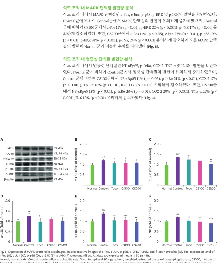
식도조직내
MAPK
단백질발현량분석식도 조직 내에서
MAPK단백질인
c-Fos, c-Jun, p-p38, p-ERK및
p-JNK의 발현을 확인하였다
.Normal
군에 비하여
Control군에서
MAPK단백질의 발현이 유의하게 증가하였으며
, Control군에 비하여
CS100군에서
c-Fos 11% (p < 0.05), p-ERK 22% (p < 0.001), p-JNK 17% (p < 0.01)유 의하게 감소하였다
.또한
, CS200군에서
c-Fos 11% (p < 0.05), c-Jun 23% (p < 0.01), p-p38 19%(p < 0.01), p-ERK 31% (p < 0.001), p-JNK 24% (p < 0.001)
유의하게 감소하여 모든
MAPK단백
질의 발현이
Normal군과 비슷한 수치를 나타냈다
(Fig. 5).식도조직내염증성단백질발현량분석
식도 조직 내에서 염증성 단백질인
NF-κBp65, p-IκBα, COX-2, TNF-α및
IL-6의 발현을 확인하
였다
. Normal군에 비하여
Control군에서 염증성 단백질의 발현이 유의하게 증가하였으며
,Control
군에 비하여
CS100군에서
NF-κBp65 11% (p < 0.05), p-IκBα 21% (p < 0.01), COX-2 17%(p < 0.001), TNF-α 16% (p < 0.01), IL-6 13% (p < 0.05)
유의하게 감소하였다
.또한
, CS200군 에서
NF-κBp65 13% (p < 0.01), p-IκBα 23% (p < 0.01), COX-2 20% (p < 0.001), TNF-α 22% (p <0.001), IL-6 18% (p < 0.01)
유의하게 감소하였다
(Fig. 6).## *
2.0
1.0 1.5
0.5
0
D
p-p38 (fold of normal)
Normal Control Toco CS100 CS200
2.0
1.0 1.5
0.5
0
E
p-ERK (fold of normal)
Normal Control Toco CS100 CS200
2.0
1.0 1.5
0.5
0
F
p-JNK (fold of normal)
Normal Control Toco CS100 CS200
## ###
** ** *** *** *** ## ** ** ***
* **
2.0
1.0 1.5
0.5
0
B A
c-Fos (fold of normal)
Normal Control Toco CS100 CS200
###
2.0
1.0 1.5
0.5
0
C
c-Jun (fold of normal)
Normal Control Toco CS100 CS200
* c-Fos
c-Jun Histone p-p38
β-actin p-JNK p-ERK
62 kDa 43, 48 kDa 32–33 kDa 38 kDa
46, 54 kDa 43 kDa 42, 44 kDa
Fig. 5. Expression of MAPK proteins in esophagus. Representative images of c-Fos, c-Jun, p-p38, p-ERK, P-JNK, and β-actin proteins (A). The expression level of c-Fos (B), c-Jun (C), p-p38 (D), p-ERK (E), p-JNK (F) were quantified. All data are expressed means ± SD (n = 8).
Normal, normal rats; Control, acute reflux esophagitis rats; Toco, tocopherol 30 mg/kg body weight/day-treated acute reflux esophagitis rats; CS100, mixture of Citrus Reticulata and Scutellariae Radix 100 mg/kg body weight/day-treated acute reflux esophagitis rats; CS200, mixture of Citrus Reticulata and Scutellariae Radix 200 mg/kg body weight/day-treated acute reflux esophagitis rats.
Significance: ##p < 0.01, ###p < 0.001 vs. Normal rats and *p < 0.05, **p < 0.01, ***p < 0.001 vs. Control rats.
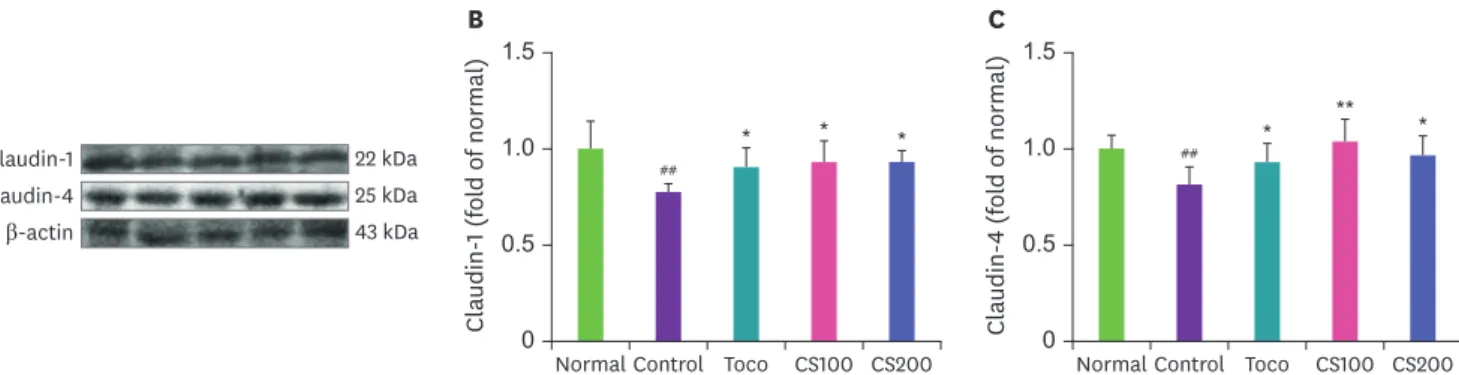
식도조직내
tight junction
단백질발현량분석식도 조직 내에서
tight junction단백질인
claudin-1및
claudin-4의 발현을 확인하였다
. Clau- din-1의 발현을 확인한 결과
, Normal군에 비하여
Control군에서
22% (p < 0.01)유의하게 감 소한 반면
CS100군
20% (p < 0.05), CS200군
20% (p < 0.05)유의하게 증가하였다
.또한
, clau- din-4의 발현은
Normal군에 비하여
Control군에서
19% (p < 0.01)유의하게 감소하였으며
, CS100군
28% (p < 0.01), CS200군
18% (p < 0.05)유의하게 증가하였다
(Fig. 7).###
**
2.0
1.0 1.5
0.5
0
D
COX-2 (fold of normal)
Normal Control Toco CS100 CS200
2.0
1.0 1.5
0.5
0
E
TNF-α (fold of normal)
Normal Control Toco CS100 CS200
2.0
1.0 1.5
0.5
0
F
IL-6 (fold of normal)
Normal Control Toco CS100 CS200
###
###
*** *** *** *** ** *** #
** * **
** **
** **
2.0
1.0 1.5
0.5
0
B A
NF-κBp65 (fold of normal)
Normal Control Toco CS100 CS200
##
2.0
1.0 1.5
0.5
0
C
p-IκBα (fold of normal)
Normal Control Toco CS100 CS200
* NF-κBp65
Histone p-IκBα IκBα
β-actin TNF-α IL-6 COX-2
65 kDa 32–33 kDa 41 kDa 45 kDa
43 kDa 26 kDa 21 kDa 70–72 kDa
Fig. 6. Expression of inflammation related proteins in esophagus. Representative images of NF-κBp65, p-IκBα, COX-2, TNF-α, IL-6, and β-actin proteins (A). The expression level of NF-κBp65 (B), p-IκBα (C), COX-2 (D), TNF-α (E), IL-6 (F) were quantified.
Normal, normal rats; Control, acute reflux esophagitis rats; Toco, tocopherol 30 mg/kg body weight/day-treated acute reflux esophagitis rats; CS100, mixture of Citrus Reticulata and Scutellariae Radix 100 mg/kg body weight/day-treated acute reflux esophagitis rats; CS200, mixture of Citrus Reticulata and Scutellariae Radix 200 mg/kg body weight/day-treated acute reflux esophagitis rats.
Significance: #p < 0.05, ##p < 0.01, ###p < 0.001 vs. Normal rats and *p < 0.05, **p < 0.01, ***p < 0.001 vs. Control rats.
##
* * * ** *
1.5
1.0
0.5
0
B A
Claudin-1 (fold of normal)
Normal Control Toco CS100 CS200
##
1.5
1.0
0.5
0
C
Claudin-4 (fold of normal)
Normal Control Toco CS100 CS200 Claudin-1 *
Claudin-4 β-actin
22 kDa 25 kDa 43 kDa
Fig. 7. Expression of tight junction proteins in esophagus. Representative images of Claudin-1, Claudin-4, and β-actin proteins (A). The expression level of Claudin-1 (B) and Claudin-4 (C) were quantified. All data are expressed means ± SD (n = 8).
Normal, normal rats; Control, acute reflux esophagitis rats; Toco, tocopherol 30 mg/kg body weight/day-treated acute reflux esophagitis rats; CS100, mixture of Citrus Reticulata and Scutellariae Radix 100 mg/kg body weight/day-treated acute reflux esophagitis rats; CS200, mixture of Citrus Reticulata and Scutellariae Radix 200 mg/kg body weight/day-treated acute reflux esophagitis rats.
Significance: ##p < 0.01 vs. Normal rats and *p < 0.05, **p < 0.01 vs. Control rats.
고찰
역류성 식도염은 산과 펩신 같은 위 내용물이 식도로 역류하여 상복부 통증 및 속쓰림 등 다
양한 증상을 유발하는 질환이다
[26].서양의 경우
5%이상의 유병률을 보이는 매우 흔한 질
환으로 알려져 있으며
,우리나라를 비롯한 아시아 지역에서도 고지방과 고열량식
,흡연
,음
주 등으로 발병률이 증가하고 있는 추세이다
[3,26].현재 역류성 식도염 치료를 위해서 사
용되는 치료제로는 역류성 식도염의 주요 원인으로 알려진 위산의 분비를 억제하는
proton pump inhibitor와
histamine type 2 receptor antagonist제제가 있으며
,이러한 치료제는 재발
률이 높은 등 여러 부작용이 나타나고 있다
[27,28].이에 현재 진행되고 있는 연구에서는 부
작용이 적으면서 역류성 식도염을 효과적으로 치료할 수 있는 치료제 개발이 다양한 방면으 로 이루어지고 있다
.본 실험에서 사용한 진피와 황금은 플라보노이드계 성분을 다량 함유 하고 있으며
,플라보노이드계 성분은 노화
,염증 및 암의 원인이 되는
ROS를 제거하여 산화 적 스트레스를 완화한다고 알려져 있다
[29].또한
,선행연구 결과 급성 역류성 식도염에서
대황
-황금 혼합물이 산화적 스트레스 조절
,염증 및 항염증 단백질과
tight junction단백질의
발현을 조절하였으며
[24],떫은감
-진피 혼합물 또한 산화적 스트레스 조절
,항산화 및 염증 관련 단백질의 발현을 조절하여 식도 점막을 보호하였다
[12].이에 본 연구에서는 선행연구 결과 역류성 식도염에서 식도 점막 보호 효과를 나타냈던 진피와 황금을 혼합하여 진피
-황 금 혼합물이 식도 점막에 미치는 효능을 평가하였으며
,천연 항산화제로 알려진
tocopherol을 양성대조군으로 사용하였다
.진피와 황금을
1:1로 혼합하여
100 mg/kg와
200 mg/kg농도 로 경구투여 후 수술을 통해 급성 역류성 식도염을 유발하였으며
,본 실험에서
100 mg/kg와
200 mg/kg농도는 일반 성인 기준
1 g/60 kg/day와
2 g/60 kg/day농도이다
.급성 역류성 식도
염을 유발한 동물의 식도 조직을 적출하여 육안으로 관찰한 결과
, Normal군에서는 식도 점
막의 손상이 발견되지 않았으나
Control군에서는 전반적인 식도의 손상 및 병변을 확인하였
으며
,진피
-황금 혼합물 투여군에서는 식도 점막의 병변이 현저하게 감소한 것을 확인하였 다
(Fig. 1).또한
, H&E염색을 통해 조직학적 변화를 관찰한 결과
,진피
-황금 혼합물 투여가
Control
군에서 두껍게 나타났던 조직층과 염증세포의 침윤을 현저하게 감소시킨 것을 확인
하였다
(Fig. 2).최근 역류성 식도염의 원인으로 산의 역류뿐만 아니라 산화적 스트레스
(oxidative stress)또
한 유효한 것으로 알려졌다
. NADPH oxidase (NOX)효소는 외부 스트레스 요인에 의해 반응
하여 빠르게 활성화 되며
, ROS를 발생시킨다
. NOX효소에 의해 생성된
ROS는 산화적 스트레
스를 유도할 뿐 아니라 단백질
,지질 및
DNA등과 같은 세포의 구성성분에 비가역적인 손상
을 야기하며
,과도하게 생성된
ROS는 염증을 유발할 수 있어 염증 경로를 조절하기 위해서는
ROS조절이 필수적이라 할 수 있다
[30-32].역류성 식도염 동물 모델에서 진피
-황금 혼합물 투여는 산화적 스트레스 관련 인자의 발현을 유의적으로 감소시켰으며
,특히
NOX4와
p47phox의 발현을 정상군과 비슷한 수준까지 조절하였다
(Figs. 3 and 4).염증반응은 많은 염증 매개인자의 유도 및 여러 사이토카인의 상호작용에 의해 야기되는 복
잡한 과정이다
.염증 경로 중 하나인
MAPK경로는 세포증식
,분화
,염증반응 및 세포 사멸 등
의 생리학적 반응을 유도하며
, MAPK의 활성화는 염증 전사 인자인
NF-κB의 활성을 유도한
다
[33,34]. NF-κB는 억제제인
IκB와 결합하여 비활성 상태로 존재하다가 활성화되면
IκB가
인산화되면서
NF-κB는 핵 속으로 이동하여 타켓 유전자의 전사를 야기하며
,다양한 사이토
카인의 발현을 유도하여 급성 및 만성 염증을 매개하는 것으로 알려져 있다
[35,36].본 실험
에서 진피
-황금 혼합물 투여는
MARK및
NF-κB경로를 억제하였으며
,특히 진피
-황금 혼합물
200 mg/kg
투여군에서
MARK및
NF-κB경로를 유의적으로 억제하여 식도 점막의 손상을 감
소시켰다
(Figs. 5 and 6).이러한 결과는 진피
-황금 혼합물이 산화적 스트레스를 억제시킴으
로써 식도 점막의 염증을 감소시키는 것으로 판단된다
.세포
-세포 사이의 연결 요소 중 하나인
tight junction은 상피세포의 극성 및 세포막의 선택적
투과에 있어서 매우 중요한 역할을 가지며
[37], tight junction의 대표적인 단백질인
claudin은
transmembrane domain을 포함하고 있다
[38]. Claudin은
actin cytoskeleton과 결합하여 수 분의 이동 조절 및 세포 내 신호 전달을 조절하는 다양한 생물학적 기능을 가지며
,역류성 식
도염에서 식도 점막이 손상되면
claudin을 포함한
tight junction단백질의 발현이 감소한다
고 알려져 있다
[38,39].본 실험에서는 진피
-황금 혼합물 투여가 급성 역류성 식도염을 유발
한 동물에서
tight junction단백질인
claudin-1및
claudin-4의 발현이 유의하게 증가시키는 것 을 확인하였으며
(Fig. 7),이는 진피
-황금 혼합물 투여가 상피세포의 결합조직을 보호하였음 을 나타낸다
.이상의 결과를 종합해보면 진피
-황금 추출물은 산화적 스트레스를 억제함으로써 염증성 단
백질의 발현을 조절할 뿐 아니라
tight junction단백질의 발현을 조절하여 식도 점막을 보호
하는 것으로 판단되며
,이러한 결과는 향후 역류성 식도염을 앓고 있는 인구를 위한 기능성 식품 개발에 있어서 중요한 근거가 될 것이라 사료된다
.요약
본 연구에서는 진피
-황금 혼합물
(CS)이 급성 역류성 식도염에 미치는 식도 점막 보호 효과를 평가하기 위하여
CS를 경구투여한 후 수술을 통해 역류성 식도염을 유발하였으며
,실험 종료 후 혈액 채취 및 식도 조직을 적출하였다
.동물에게서 적출한 식도 점막의 손상 정도를 육안 으로 확인한 결과
CS투여군에서 식도 점막의 손상이 유의하게 감소하였으며
, H&E staining을 통해 관찰한 결과 마찬가지로
CS투여군에서 식도 상피의 탈락 및 염증세포의 침윤이 현저 하게 감소한 것을 확인하였다
.혈액을 이용하여 역류성 식도염의 원인으로 유효하다고 알려 진
ROS의 수치를 확인한 결과
, CS투여군에서
ROS수치가 유의적으로 감소하였으며
, western blotting을 통해
NADPH oxidase인
NOX4, p47phox, p22phox의 발현을 확인한 결과
,마찬가지로
CS투여군에서 유의하게 감소하였고
,특히
CS200투여군에서
Normal군과 비슷한 수치를 나타
냈다
.또한
, CS투여는 염증성 단백질인
MAPK와
NF-κB경로를 유의적으로 억제하였을 뿐 아
니라
tight junction단백질인
claudin-1과
claudin-4의 발현을 유의하게 조절한 것을 확인하였 다
.이상의 결과를 종합해보면 진피
-황금 추출물은 산화적 스트레스를 억제함으로써 염증성
단백질의 발현을 조절할 뿐 아니라
tight junction단백질의 발현을 조절하여 식도 점막을 보
호하는 것으로 판단되나 역류성 식도염은 음식물의 섭취와 밀접한 관련이 있는 만큼 추후 동
물의 식이 섭취량을 조사하는 등 세부적인 추가 연구가 필요할 것으로 보인다
.REFERENCES
1. Lee JA, Shin MR, Lee JH, Roh SS. Effect of Coptidis Rhizoma and Evodiae Fructus mixture on esophageal mucosa in chronic reflux esophagitis. Korean J Pharmacogn 2020; 51(4): 349-359.
2. Song CH, Baek TH. A comparative study of Seplae Os, Arcae Concha, Ostreae Concha and esomeprazole in a mouse model of reflux esophagitis. J Korean Med 2018; 39(2): 92-105.
CROSSREF
3. Lee JA, Park HJ, Kim SH, Kim MJ, Kim KJ, Shin MR, et al. Evaluation of Evodiae Fructus extract on the chronic acid reflux esophagitis in rats. Korean J Herbol 2019; 34(2): 15-23.
4. Hwang GY, Kim DJ, Byun JS. Effects of Yijin-tang-gamibang extracts on reflux esophagitis. J Physiol Pathol Korean Med 2009; 23(5): 1073-1079.
5. Lee SH, Baik TH. Comparative study on the effects of Pinellia ternata, Zingiber officinale and Sobanhatang on reflux esophagitis. J Korean Med 2019; 40(2): 17-34.
CROSSREF
6. Park CH, Jung HK, Jeong YS, Hong JH, Lee GD, Park CD. Effects of citrus peel ethanol extract on the serum lipid and body fat of high-fat-diet-fed rats. Korean J Food Preserv 2011; 18(4): 567-574.
CROSSREF
7. Yoo YH, Song BJ, Back HM, Lee JY, Jeong SM, Hwang SY, et al. Quantitative content analysis of analytical marker in citrus unshiu peel by LC-MS/MS. J Pharm Sci 2016; 31: 76-80.
8. Lee SG, Oh SC, Jang JS. Antioxidant activities of Citrus unshiu extracts obtained from different solvents.
Korean J Food Nutr 2015; 28(3): 458-464.
CROSSREF
9. Chun JM, Bae JH. Preparation of fermented citrus peels extracts for their antimicrobial activity against campylobacter jejuni. J Korean Soc Food Cult 2015; 30(4): 475-480.
CROSSREF
10. Park SY, Han EJ, Kim MJ, Ahn CB, Jung KS, Ahn GN. Evaluation of anti-inflammatory activity of water extract from sterilized Citrus unshiu macrow peels in 12-0-tetradecanoylphorbol acetate (TPA)-induced mouse ear edema model. J Chitin Chitosan 2019; 24(3): 186-191.
CROSSREF
11. Jo HK, Han MH, Hong SH, Choi YH, Park C. Ethanol extracts of citrus peel inhibits adipogenesis through AMPK signaling pathway in 3T3-L1 preadipocytes. J Life Sci 2015; 25(3): 285-292.
CROSSREF
12. Kwon OJ, Lee AR, Roh SS. Improving effects on rats with reflux esophagitis treated with combined extract of young persimmon fruit and citrus peel. Kor J Herbol 2016; 31(1): 25-31.
CROSSREF
13. Hyeon JS, Kang SM, Senevirathne M, Koh WJ, Yang TS, Oh MC, et al. Antioxidative activities of extracts from dried Citrus sunki and C. unshiu peels. Korean Soc Food Sci Nutr 2010; 39(1): 1-7.
CROSSREF
14. Kim JM, Lee CW, Ahn YT, Lee H, Kim C, Kim HW, et al. Antimicrobial effect of scutellariae radix and its thermal stability. J Physiol Pathol Korean Med 2012; 26(3): 325-329.
15. Cho SH, Kim YR. Antimicrobial effects of scutellariae radix extract against listeria monocytogenes. J Korean Soc Food Sci Nutr 2001; 30(5): 959-963.
16. Kim NY. Effect of antioxidation and inhibition of melanogenesis from Scutellaria baicalensis extract.
Korean J Aesthet Cosmetol 2014; 12(1): 41-47.
17. Lim MJ, Gu YR, Hong JH. Extraction solvent-dependent antioxidant activities and cancer cell growth inhibitory effects of Scutellaria baicalensis extracts. Korean J Food Preserv 2019; 26(5): 566-575.
CROSSREF
18. Choi WS, Kwon HS, No RH, Choi GP, Lee HY. Enhancement of anti-inflammatory activities of fermented Scutellaria baicalensis extracts using Lactobacillus rhamnosus. J Soc Cosmet Sci Korea 2013; 39(4): 303-311.
CROSSREF
19. Park MK, Jung KS, In MJ. Effects of Scutellaria baicalensis and Phellodendron amurense extracts on growth of lactic acid bacteria and Kimchi fermentation. J Korean Soc Food Sci Nutr 2004; 33(2): 420-426.
CROSSREF
20. Park MJ, Chang MS, Jeong MC, Kim GH. Scutellaria baicalensis extracts as natural inhibitors of food browning. J Korean Soc Food Sci Nutr 2013; 42(5): 792-799.
CROSSREF
21. Park SH, Lee S, Jin HS. Antimicrobal activity of Sutellaria baicalensis·Coptidis rhizoma extract on the preservation of Makgeolli. Korean J Food Nutr 2012; 25(4): 974-979.
CROSSREF
22. Lee SJ, Kim EH, Lee HG. Development of rice wines using cornus officinalis and Scutellaria baicalensis by antioxidant activity tests. Korean J Food Sci Technol 2008; 40(1): 21-30.
CROSSREF
23. Omura N, Kashiwagi H, Chen G, Suzuki Y, Yano F, Aoki T. Establishment of surgically induced chronic acid reflux esophagitis in rats. Scand J Gastroenterol 1999; 34(10): 948-953.
PUBMED | CROSSREF
24. Lee JA, Shin MR, Lee SN, Park SA, Park HJ. Effect of a mixture of rhei rhizoma and scutellariae radix extract on acute reflux esophagitis rats. Korean J Herbol 2020; 35(6): 43-53.
25. Ali SF, LeBel CP, Bondy SC. Reactive oxygen species formation as a biomarker of methylmercury and trimethyltin neurotoxicity. Neurotoxicology 1992; 13(3): 637-648.
PUBMED
26. Lee YJ, Park JH, Roh SS. Effects on rats with reflux esophagitis treated with lonicerae flos extract. J Physiol Pathol Korean Med 2010; 24(6): 970-975.
27. Kim JW, Kim HS, Lee DK, Suk KT, Kim JM, Baik SK, et al. Therapeutic effect of low-dose omeprazole vs.
standard-dose ranitidine in mild to moderate reflux esophagitis. Korean J Gastroenterol 2004; 43(3): 153-159.
PUBMED
28. Lee ST, Kwak MH. Effects of individual herbal components of Yijintang-gamibang in the rat reflux esophagitis. Korean J Orient Int Med 2013; 34(2): 165-177.
29. Lee KS, Park MH, Cheon ME, Hong JW, Cho SI. Review of pharmacological effects of Scutellaria baicalensis and its bioactive compounds. Korean J Orient Prev Med Soc 2011; 15(2): 69-99.
30. Oh TY, Lee JS, Ahn BO, Cho H, Kim WB, Kim YB, et al. Oxidative damages are critical in pathogenesis of reflux esophagitis: implication of antioxidants in its treatment. Free Radic Biol Med 2001; 30(8): 905-915.
PUBMED | CROSSREF
31. Jeong HJ, Kim ST, Park JJ, Kim KH, Kim KM, Jun WJ. Antioxidant activities and protective effects of hot water extract from Curcuma longa L. on oxidative stress-induced C2C12 myoblasts. J Korean Soc Food Sci Nutr 2017; 46(11): 1408-1413.
32. Zeng MY, Miralda I, Armstrong CL, Uriarte SM, Bagaitkar J. The roles of NADPH oxidase in modulating neutrophil effector responses. Mol Oral Microbiol 2019; 34(2): 27-38.
PUBMED | CROSSREF
33. Zhang W, Liu HT. MAPK signal pathways in the regulation of cell proliferation in mammalian cells. Cell Res 2002; 12(1): 9-18.
PUBMED | CROSSREF
34. Lee JY, Park SS. The effect of allergic inflammation by sophora flavescens aiton extract ion through inhibition of the NFκB, JNK and p38 pathway. J Sasang Const Med 2009; 21(1): 139-149.
35. Han CW, Kim YC, Lee JH, Woo HJ. The effect of Chungganhaeju-tang(Qingganjiejiu-tang) on NFkB activation and apoptosis of kupffer cells. Korean J Orient Int Med 2004; 25(1): 59-70.
36. Kim MJ, Bae NY, Kim KBWR, Park SH, Jang MR, Ahn DH. Anti-inflammatory activity of ethanol extract of Sargassum miyabei Yendo via Inhibition of NF-κB and MAPK Activation. Microbiol Biotechnol Lett 2016;
44(4): 442-451.
37. Shin DY, Choi YH. Glutamine deprivation inhibits invasion of human prostate carcinoma LnCap cells through inactivation of matrix metalloproteinases and modulation of tight junctions. J Korean Soc Food Sci Nutr 2013; 42(8): 1167-1174.
CROSSREF
38. Cheon SH, Choi SK, Cho NJ, Kim KK, Lee WH, Hwang HS, et al. Investigation of the effect of Sappan Lignum and Brazilin on expression of tight junction related-genes in human keratinocyte. J Physiol Pathol Korean Med 2018; 32(2): 106-112.
CROSSREF
39. Lee JA, Shin MR, Lee JH, Roh SS. Effect of Coptidis Rhizoma and Evodiae Fructus mixture on esophageal mucosa in chronic reflux esophagitis. Korean J Pharmacogn 2020; 51(4): 349-359.