• 검색 결과가 없습니다.

Protective Effects of <i>Humulus japonicus</i> Extracts against Gastroesophageal Reflux Esophagitis in Rats

N/A
N/A
Protected

Academic year: 2021

Share "Protective Effects of <i>Humulus japonicus</i> Extracts against Gastroesophageal Reflux Esophagitis in Rats"

Copied!
8
0
0

로드 중.... (전체 텍스트 보기)

전체 글

(1)

Yakhak Hoeji Vol. 64, No. 1 DOI 10.17480/psk.2020.64.1.87

Protective Effects of Humulus japonicus Extracts against Gastroesophageal Reflux Esophagitis in Rats

Gil Hyung Kim*, Young Sil Min**, and Uy Dong Sohn*

,#

*Department of Pharmacology, College of Pharmacy, Chung-Ang University

**Department of Pharmaceutical Science, College of Science and Engineering, Jung Won University

(Received December 19, 2019; Revised January 30, 2020; Accepted February 17, 2020)

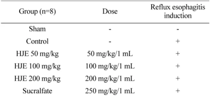
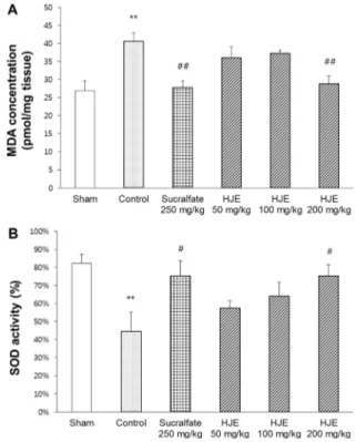
Abstract The incidence of gastroesophageal reflux disease (GERD) is increasing in Asia. Considering that GERD is a highly recurrent and chronic disease, there is an increasing demand for new therapeutic options. Humulus japonicus has antioxidant and anti-inflammatory properties. In this study, the protective effects of H. japonicus extracts (HJE) were evaluated in a surgically-induced reflux esophagitis rat model. Firstly, the esophageal mucosa was damaged in the control group and lesions were developed. Administration of HJE markedly reduced the lesions in the esophageal mucosa macroscopically. The higher acidity and volume of gastric contents in the control group was decreased by the administration of HJE. Malondialdehyde level, associated with lipid peroxidation due to oxidative stress, was reduced in the HJE-treated group. The level of superoxide dismutase, an antioxidant enzyme, was increased in the HJE-treated group. The levels of the inflammatory cytokines (tumor necrosis factor- α and interleukin-1β) were increased in the control group; however, HJE administration significantly lowered these levels. This implied that reflux esophagitis is caused by the combined action of oxidative stress and inflammatory response. HJE has shown protective effects against reflux esophagitis by its antioxidant, anti-inflammatory, and anti-secretory activities. These results suggested that HJE could have therapeutic effects in reflux esophagitis.

Keywords Anti-inflammation, antioxidation, gastroesophageal reflux disease, Humulus japonicus

Introduction

The prevalence of gastroesophageal reflux disease (GERD) is high in the countries of the western hemisphere. But it has been shown that its prevalence is increasing in Asia.

1)

GERD is a recurrent and chronic disease, which is accompanied by the symptoms of heartburn, chest tightness, dysphagia, laryngitis, and cough. Among these, heartburn and chest tightness are the most painful symptoms.

2)

Reflux esophagitis occurs when the gastric contents are chronically refluxed into the esophagus. Identifying the cause of reflux esophagitis is complex, but typical causes are the weakness of the low esophageal sphincter (LES) and the esophageal mucosa.

3)

LES is one of the most important barrier to acid reflux into the esophagus.

4)

The LES tension plays a key role in maintaining the LES function. As various factors weaken the tone

and tension of the LES, gastric acid gets refluxed into the esophagus. If the LES pressure is kept low, severe complications, such as chronic esophageal inflammation, aspiration pneumonia, or Barrett’s esophagus can occur.

5)

The pressure of the LES alone, however, does not accurately denote the stage of reflux esophagitis, which suggests that there are several other risk factors involved, such as weakening esophageal mucosa.

6)

The underlying mechanism of reflux esophagitis has not been elucidated.

7)

There are two aspects in the mechanism of reflux esophagitis. Traditionally, oxidative stress is known to be an important factor in GERD.

8)

In this aspect, many studies have shown that esophageal lesions are associated with reactive oxygen species (ROS) leading to lipid peroxidation.

9)

Recent studies have also shown that inflammatory cytokines, such as tumor necrosis factor- α (TNF-α), interleukin-1β (IL-1β) and interleukin-6, are associated with reflux esophagitis.

10)

These findings suggest that there are multiple factors which play important roles in the development of reflux esophagitis.

Excessive production of ROS, one of the pathophysiological causes of reflux esophagitis, happens due to a redox imbalance.

The redox imbalance worsens as ROS production increases. The excessive production of ROS causes oxidative stress in the

#

Corresponding author

Uy Dong Sohn, PhD, Department of Pharmacology, College of Phar- macy, Chung-Ang University, Seoul 06974, Republic of Korea Tel: +82-2-820-5614

Fax: +82-2-826-8752

E-mail: udsohn@cau.ac.kr

(2)

esophageal epithelium resulting in its damage.

11)

Substances with antioxidant activities can effectively eliminate excess ROS and thereby prevent damage to the esophageal tissue.

Studies have shown that gastric acid causes inflammation.

thereby increasing the expression of TNF-α and IL-1β.

12)

In particular, these inflammatory mediators have been reported to affect the progression of mucosal inflammation.

13)

Therefore, the protective effects of a drug against reflux esophagitis can be evaluated from the levels of these inflammatory mediators.

At the present, various treatments and surgery are available in the area of reflux esophagitis. Medications that act as antioxidants and anti-inflammatories are frequently used as pharmacological therapy in reflux esophagitis. For example, sucralfate, which inhibits pro-inflammatory cytokines and enhances the release of cytoprotectants such as mucus. Also, sucralfate has antioxidative effect and it was selected as the positive control in this study.

14,15)

Humulus japonicus, also known as Japanese hop, is a medicinal herb belonging to the Cannabaceae family. The plant is widely distributed throughout the temperate regions. H. japonicus has been used for the treatment of hypertension, pulmonary tuberculosis, and tuberculous cervical lymphadenitis. The plant is also reported to have antioxidant, anti-inflammatory, anti-aging, anti-mutagenic effects, and anti-bacterial properties.

16)

Because of the antioxidant and anti-inflammatory properties, the protective effects of H.

japonicus extracts (HJE) was investigated in a reflux esophagitis model in experimental rats. The aim of this study was to evaluate the protective effects of HJE on surgically-induced reflux esophagitis.

Experimental Methods

Animals

Six-weeks-old male Sprague-Dawley rats weighing 200220 g were provided by Raon Bio (Yongin, Gyeonggi, Korea). The animals were acclimatized for a day and were provided with food and water ad libitum. The animals were provided with water 24 h before the initiation of the experiment. The animal experiments were performed after receiving approval from the Institutional Animal Care and Use Committee (IACUC 2018-00030), Chung- Ang University.

Drugs

The leaves of H. japonicus were provided by Seorim Bio (Chuncheon, Gangwon, Korea). The leaves were chopped into multiple pieces and extracted with distilled water (×10). Sucralfate and sucrose octasulfate-aluminum complex were provided by Sigma (St. Louis, MO, USA). HJE was dissolved in distilled

water at different concentrations, and sucralfate was dissolved in saline. The total volume of the drug and vehicle administered was 1 mL.

Preparation of the H. japonicas leaf extracts

H. japonicus leaves were extracted with 99% water (10x) at room temperature for 8 h. The extracted stock solution was precipitated at room temperature for 8 h. The extracted stock solution was passed through a 25 μm diameter filter paper. The filtered extract was concentrated to 26 Brix using a reduced pressure concentrator. The condensed HJE was obtained using a spray dryer with a final yield of 17%. The HJE had a medium viscosity and was water soluble. It was stored at 4

o

C. All these processes were executed at Samjin Ginseng Natural Food Company (Hongchun, Gangwon, Korea).

Establishment of reflux esophagitis model

Reflux esophagitis was induced by surgery. Rats were fasted overnight but allowed free access to water prior to the surgery.

Under anesthesia, midline incisions were made in the abdomen below the xyphoid and in the neck. The pylorus and the transitional part between the forestomach and corpus were simultaneously ligated.

17)

To increase an environment of excessive stomach acid through increasing the reflux, a wound of about 1 cm in length was created in the LES.

18)

After 6 h, the rats were sacrificed with diethyl ether and the gastric contents and esophagus were collected for further assessments.

Experimental design

The experimental animals were divided into 6 groups: sham, control, HJE 50 mg/kg, HJE 100 mg/kg, HJE 200 mg/kg and sucralfate groups (Table 1). The sham group received oral distilled water 30 min before suturing without induction of reflux esophagitis, followed by the collection of the esophagus and gastric contents after 6 h. The other groups received oral drug 30 min before induction of reflux esophagitis. After that, the operation was performed, and 6 h later, the esophagus and gastric contents were collected. Further experimental procedures and assays were performed with the esophagus and gastric contents.

Evaluation of reflux esophagitis

After trimming the esophagus, an image was obtained by a

stereomicroscope. The image was then analyzed by the ImageJ

software (https://imagej.nih.gov/ij) to measure the area of

inflammation relative to the total area (mm

2

). After adjusting the

background, the inflammatory parts were automatically detected

and analyzed in the image.

(3)

Gastric acid analysis

The gastric contents were collected in 15 mL conical tubes and centrifuged at 3000×g for 10 min. After centrifugation, the supernatant was collected in 1.5 mL conical tubes and its volume (mL) and acidity (μEq/L) were measured. The acidity was estimated by a pH meter (Toledo 320, Mettler, Swiss) by titration with 0.1 N NaOH to pH 7.0.

Estimation of malondialdehyde (MDA) level

MDA level was estimated using MDA ELISA kit (E-EL-0060, Elabscience, Wuhan, China). MDA in the sample was detected by biotinylated detection-antibody specific to MDA. Horseradish peroxidase-conjugated with avidin was then added to each microplate. 3,3',5,5'-tetramethylbenzidine substrate was added to each well followed by incubation and addition of stop solution to terminate the reaction. MDA level was estimated with the help of the standard curve at a wavelength of 450 nm.

Estimation of superoxide dismutase (SOD) activity SOD activity was estimated using SOD activity assay kit (ab65354, Abcam, Cambridge, MA, USA). SOD activity was indirectly estimated by the reduction rate of WST-1. The WST-1 reduction rate was linearly related to the inhibitory activity of xanthine oxidase by SOD. SOD catalyzed the dismutation of superoxide anion into hydrogen peroxide and molecular oxygen, thereby inhibiting WST-1 reduction.

Estimation of tumor necrosis factor-α (TNF-α) level TNF-α level was estimated using TNF-α ELISA kit (E-EL- R0019, Elabscience, Wuhan, China). TNF-α was detected with a pre-coated antibody specific to rat TNF-α. A biotinylated detection antibody specific for Rat TNF-α and horseradish peroxidase conjugated with avidin were then added continuously to each microplate. The substrate solution was added to each well and incubated. The reaction was then terminated through a stop

solution, and the absorbance was measured at 450 nm. TNF-α level was calculated with the help of a standard curve.

Estimation of interleukin 1β (IL-1β) level

IL-1β level was estimated using IL-1β ELISA kit (E-EL- R0012, Elabscience, Wuhan, China). IL-1β was detected with a pre-coated antibody specific to rat IL-1β. A biotinylated detection antibody and horseradish peroxidase conjugated with avidin were added successively to each microplate. The substrate solution was then added and incubated. The reaction was terminated with a stop solution and the absorbance was measured at 450 nm. IL-1β level was calculated with the help of the standard curve.

Statistical analyses

The data are represented as the mean value±SEM (standard error of the mean). One-way ANOVA was performed and a p value of <0.05 was considered as statistically significant.

Results

Effects of HJE on the esophageal lesion in the reflux esophagitis model

Inflammatory lesions were observed in the esophagus collected 6 h after the induction of reflux esophagitis. Severe longitudinal ulcerations were observed in the control group. The ulcer area was smaller in the HJE-treated and sucralfate groups (Fig. 1A).

By the ImageJ software, the inflammatory areas were quantified and compared with the sham group. The mean inflammatory area (%) of the control group (56.04%) demonstrated that the surgically-induced reflux esophagitis model was well established.

Compared with the control group, the inflammatory areas (%) were reduced in the HJE-treated and sucralfate groups (Fig. 1B).

In particular, the reduction in the inflammatory area in the HJE- group had a dose-dependent trend. The sucralfate group also showed a significant reduction of the inflammatory areas. These results showed that HJE reduced the size of the lesion in reflux esophagitis.

Effects of HJE on acidity and volume of gastric juice in the reflux esophagitis model

When reflux esophagitis was induced, the acidity and volume of gastric juice were found to be increased. HJE in 100 and 200 mg/kg doses significantly reduced the acidity of gastric juices. On the other hand, the total volume of gastric juice showed a significant reduction only in the HJE 200 mg/kg group (Fig. 2A and 2B). HJE 100 and 200 mg/kg groups showed a significant reduction in the acid output as compared to the control (Fig. 2C).

Table 1. Composition of the experimental groups

Group (n=8) Dose Reflux esophagitis

induction

Sham - -

Control - +

HJE 50 mg/kg 50 mg/kg/1 mL +

HJE 100 mg/kg 100 mg/kg/1 mL +

HJE 200 mg/kg 200 mg/kg/1 mL +

Sucralfate 250 mg/kg/1 mL +

+, Reflux esophagitis induction; -, none; HJE, Humulus japonicus

extracts. All groups consisted of 8 rats.

(4)

The sucralfate group also showed a significant reduction in acid output in the gastric juice as compared to the control.

Effects of HJE on MDA level in the reflux esophagitis model

The levels of MDA, a bioactive marker of lipid peroxidation, were estimated in the esophageal tissues. Compared with the sham group, the MDA level in the control group was significantly increased (Fig. 3A). The level of MDA in the HJE 200 mg/kg group was significantly lower than that in the control group. The sucralfate group also showed a significant reduction in MDA level as compared with the control group.

Effects of HJE on SOD activity in the reflux esophagitis model SOD is an antioxidant enzyme. It was found that the SOD

activity was significantly decreased in the control group as compared to the sham group (Fig. 3B). HJE 200 mg/kg group showed significant improvement in the SOD enzyme activity.

Although there was no significant difference, there was a trend of improvement of SOD enzyme activity in HJE 50 mg/kg and HJE 100 mg/kg groups.

Effects of HJE on TNF-α level in the reflux esophagitis model

For evaluating the anti-inflammatory effects of HJE, TNF-α

levels were estimated in the esophageal tissues. TNF-α level was

significantly increased in the control group as compared to the

sham group (Fig. 4A). Compared with the control group, TNF-α

level was significantly decreased in HJE 200 mg/kg group. There

was no significant reduction in TNF-α level in HJE 100 mg/kg

Fig. 1. Macroscopic appearance and quantification of the representative esophageal tissue in each group. A: The macroscopic appearance

of representative esophageal tissue from each group is shown. B: The inflammatory area was quantified relative to sham group. The area of

inflammation relative to the total area was expressed as a percentage. Control: control group, sucralfate: sucralfate-treated group, HJE: HJE-

treated group (different concentrations). The data are expressed as the mean±SEM (n=8).

##

p<0.01, and

###

p<0.001 as compared to the control.

(5)

group, although there was a trend of reduction. The sucralfate group, as a positive control, showed a significant reduction in the TNF-α level as compared to the control group.

Effects of HJE on IL-1β level in the reflux esophagitis model

The IL-1β is one of the inflammatory cytokines that can be used as a marker to assess the degree of inflammation. IL-1β levels were estimated in the esophageal tissues. IL-1β level was significantly increased in the control group as compared with the sham group (Fig. 4B). In the HJE 200 mg/kg group, IL-1β level

was significantly decreased as compared to the control group. In HJE 50 and 100 mg/kg groups, there were no significant reductions as compared to the control group. The sucralfate group, as a positive control, showed a significant reduction in the IL-1β level as compared to the control group.

Discussion

GERD is a disease in which the gastric contents get refluxed into the esophagus If not treated properly. GERD causes chronic symptoms and affects the quality of life. It is well known that gastric acid plays a key role in esophageal mucosal injury.

19,20)

Gastric acid determines the degree of mucosal damage in the esophagus. The increased volume of gastric contents causes temporary relaxation of the LES,

21)

and because of this LES relaxation, gastric contents can reflux into the esophagus causing esophageal mucosal damage.

Previous studies have shown that HJE has antioxidant, anti- inflammatory, and anti-aging properties.

22-24)

Polyphenols, present Fig. 3. Effects of HJE on MDA level and SOD activity in the esophageal tissues. A: MDA level, an indicator of lipid peroxidation, was evaluated in the esophageal tissues. B: The activity of SOD, an antioxidant enzyme, was evaluated in the esophageal tissues. Sham: sham operation control group, Control:

control group, Sucralfate: sucralfate treated group, and HJE: HJE- treated group (different concentrations). The data are expressed as the mean±SEM (n=8). **p<0.01 as compared to the sham,

#

p<0.05 and

##

p<0.01 as compared to the control.

Fig. 2. Effects of HJE on the gastric contents. Analysis of gastric contents was performed. A: Results of the acidity of gastric contents, B: Results of the total volume of gastric contents, C:

Results of the acid output of gastric contents. Control: control group,

Sucralfate: sucralfate-treated group, HJE: HJE-treated group

(different concentrations). The data are expressed as the mean±SEM

(n=8).

#

p<0.05,

##

p<0.01, and

###

p<0.001 as compared to the control.

(6)

in many medicinal herbs, contribute as the main compounds to the antioxidant effects.

25)

The polyphenol content in H. japonicus leaves is much higher than that in the roots and stems.

16)

In this study, HJE was obtained from the leaves of H. japonicas with water.

Many studies suggest that reflux esophagitis is accompanied by increased acidity and volume of gastric contents,

26)

resulting in esophageal inflammation. In this experiment, the reflux esophagitis model was established according to a previous study.

18)

In this model, the antioxidant and anti-inflammatory effects of HJE were evaluated.

Firstly, the morphological changes in the esophagus were examined, and the esophageal inflammatory areas (%) were evaluated macroscopically. Inflammatory area (%) is a commonly used important parameter to evaluate the severity of esophageal inflammation in reflux esophagitis models.

27)

The inflammatory area was expressed as the area relative to the total area in percentage. The inflammatory areas in the control group were markedly increased suggesting that the surgically-induced reflux

esophagitis model was well established. Thereby supporting the finding of previous studies.

26)

The recovery in terms of inflammatory area (%) in the HJE-treated group was estimated.

The acidity and volume of gastric contents were decreased in HJE-treated groups. It indicated that HJE has similar acid inhibitory potency as compared to sucralfate. Sucralfate, as buffers acid, inhibited the action of pepsin, and adsorbs bile salts. Also, sucralfate was known to bind to the uninjured mucosa and was believed to a similar barrier effect on regenerated and normal mucosa.

28)

Especially, the effects of HJE were more on acid output when both acidity and volume were considered together. In this regard, HJE effectively reduced the acidity and volume of gastric contents.

One of the pathogenic factors in reflux esophagitis is oxidative stress.

29)

Although exposure to the esophageal mucosa to gastric acid leads to esophageal inflammation, oxidative stress can also cause secondary damage to the esophageal mucosa.

30)

Furthermore, it is known that structural modification of lipids by ROS causes extensive subcellular damage in the tissues. It was confirmed that in reflux esophagitis, MDA, a bioactive marker of lipid peroxidation was increased; and the activity of SOD, an antioxidant enzyme was decreased.

31)

HJE was found to ameliorate these changes in reflux esophagitis. In the HJE 200 mg/kg group, the levels of each of the estimated parameters were found to return to values similar to that of the sucralfate group. These results demonstrated that HJE exerted protective effects against reflux esophagitis through antioxidant activities.

Recent studies have suggested that inflammatory cytokines play major roles in the pathological process of the esophageal mucosal inflammation.

32)

The inflammatory cytokines (TNF-α and IL-1β) play key roles in forming lesions associated with acute esophageal mucosal inflammation leading to congestion of esophageal mucosa and progression of injury.

12)

Administration of HJE significantly reduced the levels of inflammatory cytokines. These findings supported the hypothesis that the anti-inflammatory effects of HJE were primarily responsible for the amelioration of the inflammatory lesions in the esophagus.

Conclusion

In this study, the effects of HJE were evaluated in reflux esophagitis rat. The levels of the inflammatory cytokines and oxidative stress were recovered by HJE administration, suggesting that HJE could reduce the damage of esophageal mucosa in reflux esophagitis. These protective effects are thought to be related to the antioxidant, anti-inflammatory, and anti-secretory effects of HJE. In conclusion, these results suggested that HJE could be a Fig. 4. Effects of HJE on TNF- α and IL-1β level in the

esophageal tissues. A: The level of TNF- α, an indicator of the anti-

inflammatory effect, was evaluated in the esophageal tissues. B: The

level of IL-1 β, an indicator of the anti-inflammatory effect, was ex

evaluated pressed in the esophageal tissues. Sham: sham operation

control group, Control: control group, Sucralfate: sucralfate treated

group, HJE: HJE-treated group (different concentrations). The data

are expressed as the mean±SEM (n = 8). ***p<0.001 as compared to

the sham,

#

p<0.05 as compared to the control, and

##

p<0.01 as

compared to the control.

(7)

potential therapeutic agent for the treatment of reflux esophagitis.

Acknowledgment

This research was supported by the Basic Science Research Program through the National Research Foundation of Korea and funded by the Ministry of Education, Science and Technology [Grant NRF-2019R1F1A1062070].

Conflict of Interest

All authors declare that they have no conflict of interest.

References

1. Kasyap, A. K., Sah, S. K. and Chaudhary, S. : Clinical spectrum and risk factors associated with asymptomatic erosive esophagitis as determined by Los Angeles classification: A cross-sectional study. PLoS One 13, e0192739 (2018).

2. Fisichella, P. M., Schlottmann, F. and Patti M. G. : Evaluation of gastroesophageal reflux disease. Updates Surg. 70, 309 (2018).

3. McDonnell, M. J., O'Toole, D., Ward, C., Pearson, J. P., Lordan, J. L., De Soyza, A., Loebinger, M., Chalmers, J. D., Laffey, J. G.

and Rutherford, R. M. : A qualitative synthesis of gastro- oesophageal reflux in bronchiectasis: Current understanding and future risk. Respir Med. 141, 132 (2018).

4. Garcia, R. S., Belafsky, P. C., Della, Maggiore, A., Osborn, J. M., Pypendop, B. H., Pierce, T., Walker, V. J., Fulton, A. and Marks S. L. : Prevalence of Gastroesophageal Reflux in Cats During Anesthesia and Effect of Omeprazole on Gastric pH. J. Vet.

Intern. Med. 31, 734 (2017).

5. Farre, R. : Pathophysiology of gastro-esophageal reflux disease: a role for mucosa integrity? Neurogastroenterol Motil. 25, 783 (2013).

6. Kandulski, A. and Malfertheiner P. : Gastroesophageal reflux disease--from reflux episodes to mucosal inflammation. Nat. Rev.

Gastroenterol. Hepatol. 9, 15 (2011).

7. Kawahara, H., Kubota, A., Hasegawa, T., Okuyama, H., Ueno, T., Ida, S. and Fukuzawa, M. : Effects of rikkunshito on the clinical symptoms and esophageal acid exposure in children with symptomatic gastroesophageal reflux. Pediatr. Surg. Int. 23, 1001 (2007).

8. Jabri, M. A., Tounsi, H., Rtibi, K., Marzouki, L., Sakly, M. and Sebai, H. : Ameliorative and antioxidant effects of myrtle berry seed (Myrtus communis) extract during reflux-induced esophagitis in rats. Pharm. Biol. 54, 1575 (2016).

9. Kwon, O. J., Choo, B. K., Lee, J. Y., Kim, M. Y., Shin, S. H., Seo, B. I., Seo, Y. B., Rhee, M. H., Shin, M. R., Kim, G. N., Park, C. H. and Roh, S. S. : Protective effect of Rhei Rhizoma on reflux esophagitis in rats via Nrf2-mediated inhibition of NF- kappaB signaling pathway. BMC Complement Altern. Med. 16, 1 (2016).

10. Souza, R. F., Bayeh, L., Spechler, S. J., Tambar, U. K. and Bruick, R. K. : A new paradigm for GERD pathogenesis. Not acid injury, but cytokine-mediated inflammation driven by HIF-2alpha:

a potential role for targeting HIF-2alpha to prevent and treat reflux esophagitis. Curr. Opin. Pharmacol. 37, 93 (2017).

11. Farhadi, A., Fields, J., Banan, A. and Keshavarzian, A. : Reactive oxygen species: are they involved in the pathogenesis of GERD,

Barrett’s esophagus, and the latter's progression toward esophageal cancer? Am. J. Gastroenterol. 97, 22 (2002).

12. Ku, S. K., Kim, J. S., Seo, Y. B., Kim, Y. U., Hwang, S. L., Lee, Y. C., Yang, C. H., Kim, H. Y., Seo, B. I., Park, J. H., Min, Y. H.

and Roh, S. S. : Effect of Curculigo orchioides on reflux esophagitis by suppressing proinflammatory cytokines. Am. J.

Chin. Med. 40, 1241 (2012).

13. Katada, K., Yoshida, N., Isozaki, Y., Tomatsuri, N., Ichikawa, H., Naito, Y., Okanoue, T. and Yoshikawa, T. : Prevention by rebamipide of acute reflux esophagitis in rats. Dig. Dis. Sci. 50, S97 (2005).

14. Mu, F. H., Hu, F. L., Wei, H., Zhang, Y. Y., Yang, G. B., Lei, X.

Y., Yang, Y. P., Sun, W. N. and Cui M. H. : [Protective effect of compound bismuth and magnesium granules on aspirin-induced gastric mucosal injury in rats]. Zhonghua Yi Xue Za Zhi. 96, 464 (2016).

15. Arab, H. H., Salama, S. A., Omar, H. A., Arafa el, S. A. and Maghrabi, I. A. : Diosmin protects against ethanol-induced gastric injury in rats: novel anti-ulcer actions. PLoS One 10, e0122417 (2015).

16. Choi, J. Y., Desta, K. T., Lee, S. J., Kim, Y. H., Shin, S. C., Kim, G. S., Lee, S. J., Shim, J. H., Hacimuftuoglu, A. and Abd El-Aty, A. M. : LC-MS/MS profiling of polyphenol-enriched leaf, stem and root extracts of Korean Humulus japonicus Siebold & Zucc and determination of their antioxidant effects. Biomed. Chromatogr.

32, e4171 (2018).

17. Nakamura, K., Ozawa, Y., Furuta, Y. and Miyazaki H. : Effects of sodium polyacrylate (PANa) on acute esophagitis by gastric juice in rats. Jpn. J. Pharmacol. 32, 445 (1982).

18. Min, Y. S., Lee, S. E., Hong, S. T., Kim, H. S., Choi, B. C., Sim, S. S., Whang, W. K. and Sohn, U. D. : The Inhibitory Effect of Quercetin-3-O-beta-D-Glucuronopyranoside on Gastritis and Reflux Esophagitis in Rats. Korean J. Physiol. Pharmacol. 13, 295 (2009).

19. Jang, H. S., Han, J. H., Jeong, J. Y. and Sohn, U. D. : Protective Effect of ECQ on Rat Reflux Esophagitis Model. Korean J.

Physiol. Pharmacol. 16, 455 (2012).

20. Kim, S. H., Um, S. I., Nam, Y., Park, S. Y., Dong, J. H., Ko, S.

K., Sohn, U. D. and Lee S. J. : The effect of synthetic ceramide analogues on gastritis and esophagitis in rats. Arch. Pharm. Res.

39, 1313 (2016).

21. Shin, C. Y., La, H. O., Lee, Y. P., Lee, D. W., Kim, D. S., Huh, I.

H. and Sohn, U. D. : The alteration of intracellular signaling on the smooth muscle cells contraction in cat esophagitis. Life Sci.

74, 2199 (2004).

22. Sung, B., Chung, J. W., Bae, H. R., Choi, J. S., Kim, C. M. and Kim, N. D. : Humulus japonicus extract exhibits antioxidative and anti-aging effects via modulation of the AMPK-SIRT1 pathway.

Exp. Ther. Med. 9, 1819 (2015).

23. Park, T. S., Ryu, Y. K., Park, H. Y., Kim, J. Y., Go, J., Noh, J. R., Kim, Y. H., Hwang, J. H., Choi, D. H., Oh, W. K., Lee, C. H.

and Kim K. S. : Humulus japonicus inhibits the progression of Alzheimer's disease in a APP/PS1 transgenic mouse model. Int. J.

Mol. Med. 39, 21 (2017).

24. Bae, J., Min, Y. S., Nam, Y., Lee, H. S. and Sohn, U. D. : Humulus japonicus Extracts Protect Against Lipopolysaccharide/d- Galactosamine-Induced Acute Liver Injury in Rats. J. Med. Food.

21, 1009 (2018).

25. Thiruvengadam, M., Praveen, N., Yu, B. R., Kim, S. H. and Chung, I. M. : Polyphenol composition and antioxidant capacity from different extracts of Aster scaber. Acta. Biol. Hung. 65, 144 (2014).

26. Nagahama, K., Nishio, H., Yamato, M. and Takeuchi, K. : Orally

administered L-arginine and glycine are highly effective against

acid reflux esophagitis in rats. Med. Sci. Monit. 18, BR9 (2012).

(8)

27. Ishiyama, F., Iijima, K., Asanuma, K., Ara, N., Yoshitake, J., Abe, Y., Koike, T., Imatani, A., Ohara, S. and Shimosegawa T. : Exogenous luminal nitric oxide exacerbates esophagus tissue damage in a reflux esophagitis model of rats. Scand. J.

Gastroenterol. 44, 527 (2009).

28. Nagashima, R.: Mechanisms of action of sucralfate. J. Clin.

Gastroenterol. 3, 117 (1981).

29. Pereira Rde, S. : Regression of gastroesophageal reflux disease symptoms using dietary supplementation with melatonin, vitamins and aminoacids: comparison with omeprazole. J. Pineal. Res. 41, 195 (2006).

30. Oh, T. Y., Lee, J. S., Ahn, B. O., Cho, H., Kim, W. B., Kim, Y.

B., Surh, Y. J., Cho, S. W., Lee, K. M. and Hahm, K. B. : Oxidative stress is more important than acid in the pathogenesis of reflux oesophagitis in rats. Gut. 49, 364 (2001).

31. Min, Y. S., Bai, K. L., Yim, S. H., Lee, Y. J., Song, H. J., Kim, J.

H., Ham, I., Whang, W. K. and Sohn, U. D. : The effect of luteolin- 7-O-beta-D-glucuronopyranoside on gastritis and esophagitis in rats. Arch. Pharm. Res. 29, 484 (2006).

32. Oyama, K., Fujimura, T., Ninomiya, I., Miyashita, T., Kinami, S.,

Fushida, S., Ohta, T. and Koichi, M. : A COX-2 inhibitor prevents

the esophageal inflammation-metaplasia-adenocarcinoma sequence

in rats. Carcinogenesis 26, 565 (2005).

수치

Table 1. Composition of the experimental groups
Fig. 2. Effects of HJE on the gastric contents. Analysis of gastric contents was performed

참조

관련 문서

□ The least-upper-bound property (sometimes called completeness or supremum property) is a fundamental property of the real number system. The least-upper-bound

□ The least-upper-bound property (sometimes called completeness or supremum property) is a fundamental property of the real number system. The least-upper-bound

The “Asset Allocation” portfolio assumes the following weights: 25% in the S&amp;P 500, 10% in the Russell 2000, 15% in the MSCI EAFE, 5% in the MSCI EME, 25% in the

1 John Owen, Justification by Faith Alone, in The Works of John Owen, ed. John Bolt, trans. Scott Clark, &#34;Do This and Live: Christ's Active Obedience as the

- The level of radioactivation, along with other radioactivity aspects such as the T inventory will be a key factor in determining the environmental impact of fusion

Therefore, this study aims to identify the relations between gastroesophageal reflux disease and body mass index, and waist measurement as indexes of obesity.. For the

A separate coolant, typically helium, is used. The breeder is circulated at low speed for tritium extraction.. Concepts: LiPb/He/FS,

- USSR: OGRA fitted with Ioffe’s magnetic wells (Institute of Physics of Moscow) France: DECA I, II, III (later withdrawn) and MMII (CEA).. USA: Table Top and Toy