大黃과 黃芩 추출물 혼합물이 급성 역류성 식도염 흰쥐에 미치는 효과
이진아1#, 신미래1#, 이상남2, 박순애3, 박해진4*
1 : 대구한의대학교 한의과대학 본초약리학교실, 2 : 대구한의대학교 기공학교실 3 : 대구한의대학교 한방식품조리영양학부, 4 : DHU 바이오융복합시험센터
Effect of a Mixture of Rhei Rhizoma and Scutellariae Radix Extract on Acute Reflux Esophagitis Rats
Jin A Lee
1#, Mi-Rae Shin
1#, Sang-Nam Lee
2, Soon-Ae Park
3, Hae-Jin Park
4*1 : Department of Herbology, Korean Medicine of College, Daegu Haany University, 136, Shinchendong-ro, Suseong-gu, Deagu 42158, Republic of Korea
2 : Department of Qigong, Daegu Haany University, 136, Shinchendong-ro, Suseong-gu, Deagu 42158, Republic of Korea 3 : Department of Herbal Food Cuisine & Nutrition, Daegu Haany University, Hanuidae-ro, Gyeongsan-si,
Gyeongsangbuk-do 38610, Republic of Korea
4 : DHU Bio Convergence Testing Center, 1, Hanuidae-ro, Gyeongsan-si, Gyeongsangbuk-do 38610, Republic of Korea
ABSTRACT
Objective : Reflux esophagitis is a disease caused by reflux of stomach contents, stomach acid, and pepsin into the esophagus, and is currently increasing worldwide. This study was conducted to evaluate the effect of a mixture of Rhei Rhizoma and Scutellariae Radix (RS) extract on acute reflux esophagitis in rats.
Methods : Rats were divided into five groups for examination: Normal group (Nor, n=8), water-treated acute reflux esophagitis rats (Con, n=8), tocopherol 30 ㎎/㎏ body weight/day-treated acute reflux esophagitis rats (Toco, n=8), RS 100 ㎎/㎏ body weight/day-treated acute reflux esophagitis rats (RS100, n=8), RS 200 ㎎/㎏ body weight/day-treated acute reflux esophagitis rats (RS200, n=8). All rats fasted for 18 h and then were derived by linking the metastatic junction between pylorus and forestomach and corpus. And rats were sacrificed 5 h after surgery. We analyzed the expression of NADPH, MAPK, inflammatory, anti-inflammatory, and tight junction related proteins by western blot in esophageal tissue and observed the level of reactive oxygen species (ROS), alanine aminotransferanse (ALT), and aspartate aminotransferase (AST) in serum.
Results : RS administration significantly protected the esophageal mucosal damage of reflux esophagitis, and ROS, AST, and ALT levels were significantly reduced in RS administration compared to Con group. In addition, RS administration effectively suppressed MAPK and NF-κB pathways and upregulated protein expressions of tight junction protein.
Conclusions : These results suggest that RS protected the esophageal mucosa by inhibiting the MAPK and NF-κB pathways and upregulating tight junctions.
1)Key words : Rhei Rhizoma, Scutellariae Radix, acute reflux esophagitis, inflammation, tight junction
*Corresponding author : Hae-Jin Park, DHU Bio Convergence Testing Center, 1, Hanuidae-ro, Gyeongsan-si, Gyeongsangbuk-do 38610, Republic of Korea.
·Tel : +82-53-819-7876 ·Fax : +82-819-1496 ·E-mail : [email protected]
#First author : Jin A Lee, College of Korean Medicine, Daegu Haany University, 136, Sincheondong-ro, Suseong-gu, Daegu, 42158, Republic of Korea.
·Tel : +82-53-770-2258 ·Fax : +82-53-768-6340 ·E-mail : [email protected]
Mi-Rae Shin, College of Korean Medicine, Daegu Haany University, 136, Sincheondong-ro, Suseong-gu, Daegu, 42158, Republic of Korea.
·Tel : +82-53-770-2258 ·Fax : +82-53-768-6340 ·E-mail :[email protected] ·Received : 06 Oct. 2020 ·Revised : 10 Nov. 2020 ·Accepted : 25 Nov. 2020
Ⅰ. 서 론
역류성 식도염 (Reflux esophagitis; RE)은 위-식도 괄약 근의 불안정으로 위산과 펩신 등의 위 내용물이 역류하여 발생 하는 질환으로, 속쓰림, 소화불량, 목이물감, 가슴통증 등의 증상이 나타나며, 하부식도 괄약근의 기능저하, 위 배출 기능 저하, 흡연, 과식이나 폭식 등 잘못된 식습관 등에 의해 유발 된다. 서양에서는 5% 이상의 유병률을 보이고 있으며, 우리 나라에서도 점차 서구식 식습관, 지나친 음주 및 비만 인구 증가 등의 원인으로 유병률이 매년 빠르게 증가되고 있는 추세 이다
1-3).
역류성 식도염 치료를 위해 사용되고 있는 치료제로는 제 산제 및 위 장관 운동 촉진제 등이 사용되어 왔으며, 이후 위 산의 분비를 억제하는 H
2수용체 길항제 (histamine type 2 receptor antagonist)와 양성자 펌프 억제제 (proton pump inhibitor; PPI) 등의 약물이 많이 사용되어지고 있다
4, 5). 그 러나 역류성 식도염은 특성상 치료를 받더라도 한번 약해진 하부식도 괄약근이 쉽게 개선되지 않아 약 복용을 중단하게 되면 1년 이내 재발률이 50~80%에 이른다고 알려져 있다
6). 따라서 장기간 복용에도 부작용이 적고 pH 조절 및 위장관 운동을 조절하는 것 이외에 역류성 식도염을 효과적으로 치료 할 수 있는 새로운 기전의 치료제 개발이 필요하다. 大黃 (Rhei Rhizoma)은 마디풀과 (Polygonaceae)에 속한 다년생 초목인 당고특대황 (
Rheum tanguticumMaxim. et Balf.), 장엽대황 (
Rheum palmatumL.) 혹은 약용대황 (
Rheum officinaleBaill.)의 뿌리와 뿌리줄기를 건조한 것으로 주로 아시아 지역에 분포되어 있으며, 積聚腹痛, 血熱吐衄, 跌打損 傷, 상부소화기 출혈 등에 사용하는 전통약재이다
7, 8). 현재 많은 연구를 통해 항위염 효과
9), 항산화효과
10), 혈당 개선효 과
11), 항염증 및 항균효과
12)등이 알려져 있다.
黃芩 (Scutellariae Radix)은 꿀풀과 (Labiatae)에 속하는 다년생 초본인 속썩은풀 (
Scutellaria baicalensisGeorgi)의 주피를 벗긴 뿌리를 건조한 것으로 한국, 몽골, 중국 및 시베 리아 동부 지역에 분포되어 있다. 주요 성분으로는 flavonoid 계 화합물 등 약 30종의 성분들이 밝혀져 있으며, 이러한 성분 들은 항염증 및 항산화 작용을 한다고 보고되어 있다. 또한, 중국에서는 마른기침, 토혈, 변비, 고열 등에 사용하고 있다
13-15)
. 황금의 약리작용으로는 간장기능 회복 효과
13), 항균작
용
16), 암세포 증식 억제 작용
17), 항염증 효과
18)등이 많은 연 구를 통해 알려졌다.
이러한 大黃과 黃芩의 항염증 효과 및 항균작용에 대한 효 과를 토대로 급성 역류성 식도염 동물 모델에 대한 大黃과 黃 芩의 효과를 연구하였다. 선행 연구에서 大黃-黃芩 혼합물의 항산화 활성을 알아보기 위하여 DPPH 라디칼 소거 활성법을 이용하여 항산화 활성을 측정하였고 효과가 가장 뛰어났던 1:1 비율을 사용하여 실험을 진행하였으며, 식도 점막 손상 여부, 염증 및 식도 기능 관련 단백질의 발현에서 유의한 결 과를 얻었기에 이를 보고하는 바이다.
Ⅱ. 재료 및 방법
1. 재료
1) 시료본 실험에서 사용한 大黃 (
Rheum officinaleBaill)과 黃芩 (
Scutellaria baicalensisGeorgi)은 옹기한약국 (대구, 한국) 에서 구입한 것을 생약규격집에 맞추어 관능검사 후, 약전규 격에 적합한 것만을 정산하여 사용하였다. 大黃과 黃芩을 각각 200 g 분쇄하여 2,000 ㎖ 첨가한 후 열탕 추출기에서 2시간 추출 하였으며, 얻어진 추출물은 감압 추출장치로 농축 후, 동결 건조기를 이용해 완전 건조시켜 파우더를 얻었다 (大黃, 16%; 黃芩, 29%). 실험 전까지 –80℃에서 보관하였고, 실험 당일 大黃과 黃芩을 1:1 비율로 혼합 (RS)하여 사용하였다.
2) 시약
본 실험에 사용된 potassium phosphate monobasic와 potassium phosphate dibasic은 Sigma-Aldrich Co. (St.
Louis, MO, USA)에서 구입하여 사용하였다. Nitrocellulose membranes는 Amersham GE Healthcare (Little. Chalfont, UK)에서 구입하였고, NADPH oxidase 4 (NOX4), p47
phox, p22
phox, phospho-p38 MAPK (p-p38), phospho- extracellular signal-regulated kinase (p-ERK), phospho- c-Jun N-terminal Kinase (p-JNK), phosphorylation of nuclear factor-kappa B p65 (NF-κBp65), inhibitor of nuclear factor kappa B alpha (IκBα), phosphorylation inhibitor of nuclear factor kappa B alpha (p-IκBα), cyclooxygenase-2 (COX-2), tumor necrosis factor-alpha (TNF-α), interleukin-6 (IL-6), interleukin-4 (IL-4), interleukin-10 (IL-10), Claudin-1, Claudin-3, Claudin-4, Histone, β-actin은 Santa Cruz Biotechnology (Dallas, CA, USA)로부터 구입하였다. c-Fos 과 c-Jun는 Cell Signaling Technology, Inc. (Beverly, MA, USA)에서 구입하여 사용하였으며, 2차항체는 GeneTex, Inc. (Irvine, LA, USA)에서 구입하여 사용하였다. Protease inhibitor mixture, ethylenediaminetetraacetic acid (EDTA)는 Wako Pure Chemical Industries, Ltd. (Osaka.
Japan)에서 구입하였다. 2', 7'Dichlorofluorescein diacetate (DCFH-DA)와 Dihydrorhodamine 123는 Molecular Probes (Eugene, OR, U.S.A.)에서 ECL western blotting detection reagents는 GE Healthcare 로부터 구입하여 사용하였다. 단 백질 정량을 위한 BCA protein assay kit는 Thermo Scientific (Rockford, IL, USA)에서 구입하였다.
3) 실험동물
SD 흰쥐 6주령 수컷 (180~200 g)을 대한바이오링크 (음성,
한국)에서 구입하여 1주일 동안 실험실 환경에 적응시킨 후
실험에 사용 하였다. 동물 사육실의 조건은 conventional
system으로 온도 22 ± 2℃, 습도 50 ± 5%, 명암주기
(light : dark cycle)는 12시간 주기로 조절하였으며, 사료
(조단백질 18% 이상, 조지방 5.0% 이상, 조섬유 5.0% 이하,
조회분 8.0% 이하, 칼슘 1.0% 이상, 인 0.85% 이상, 칼륨
0.55% 이상, 나트륨 0.25% 이상, 마그네슘 0.15% 이상,
NIH-41, Zeigler Bros, Inc., USA)와 물을 충분히 공급하
였다. 본 실험은 동물실험의 윤리적, 과학적 타당성 검토 및 효율적인 관리를 위하여 대구한의대학교 동물실험윤리 위원회 (Institutional Animal Care and Use Committee : IACUC)의 승인 (DHU2020-052)을 받아 진행하였다.
2. 방법
1) 급성 역류성 식도염 유발 및 동물 처치
실험동물은 아무런 처치를 하지 않은 정상군 (Nor), 증류 수를 단회 경구투여 후 급성 역류성 식도염을 유발한 대조군 (Con), tocopherol 30 ㎎/㎏를 단회 경구투여 후 급성 역류성 식도염을 유발한 양성대조군 (Toco), 大黃-黃芩 혼합물 100
㎎/㎏ 단회 경구투여 후 급성 역류성 식도염을 유발한 군 (RS100), 大黃-黃芩 혼합물 200 ㎎/㎏ 단회 경구투여 후 급성 역류성 식도염을 유발한 군 (RS200) 총 5그룹으로 그룹 당 8 마리씩 무작위 분류하였다. 실험 전날까지 고형사료와 물을 충분히 공급하였으며, 실험을 진행하기 18시간 전부터 절식 하였다. 수술 당일 각 시료를 DW에 녹여 단회 경구투여하고 1시간 후 isoflurane (Wellona Pharma, Indo)로 흡입마취 하였다. 복부를 2 ㎝ 정도 개복하여 위 조직의 위저부 및 날 문부를 실크 (2-0)실로 묶은 후 복막과 피부를 봉합하였으며, 5시간 후 희생하여 복대정맥에서 혈액을 채취하였고, 식도조 직을 적출하였다
19). 채취한 혈액에서 원심분리기 (4,000 rpm, 10 min)를 이용하여 혈청을 분리하였으며, 분리한 혈청과 식 도조직은 –80℃에서 보관하였다.
2) 식도 점막 손상 확인
적출한 식도를 수술용 사위를 이용하여 세로로 절단하였다.
절단된 식도 내부를 saline으로 세척한 후 고정하여 광학 디 지털 카메라 (DSCHX50V, Sony, Tokyo, Japan)를 이용하여 촬영하였다. 손상된 식도 점막 측정은 I-Solution lite (Innerview Co., 성남, 한국) 프로그램을 이용하여 실제 손상 부위의 면적을 측정한 후, 아래 식을 이용하여 손상 면적을 나타내었다.
식도 손상 비율 식도 전체 면적 식도 손상 면적
×
3) 조직학적 검사
분리된 식도 조직을 평가하기 위해 현미경으로 조직학적 검사를 수행하였다. 분리된 식도를 10% 중성 완충 포르말린을 통해 고정하고 2 ㎛ 섹션으로 절단한 후 hematoxylin & eosin (H&E) 조직 염색 방법으로 염색하였다. 염색된 슬라이스를 광학현미경으로 관찰한 후 I-Solution Lite 소프트웨어 프로 그램 (Innerview Co., Korea)을 사용하여 분석 하였다.
4) 혈청 내 활성산소 측정
ROS 값은 Ali
et al의 방법
20)에 의해 측정되었다. 혈청에 25 mM DCFH-DA를 첨가하여 혼합한 후, 형광 광도계를 이 용해 0분부터 매 5분씩 emission 파장 530 ㎚와 excitation 파장 485 ㎚를 이용하여 30분간 측정한 값을 계산하였다.
5) 간 손상 지표 AST, ALT 측정
간 손상 지표인 aspartate aminotransferase (AST) 및 alanine aminotransferse (ALT)는 아산제약 (화성, 한국)에서 kit를 구입하여 측정하였다. AST 측정은 기질액과 혈청을 혼합 하여 37℃에서 1시간 반응시킨 후 정색시액을 첨가하여 실온 에서 20분간 더 반응시켰다. 그 후, 0.4 N NaOH를 첨가하여 실온에서 10분간 더 반응시킨 후 505 ㎚에서 흡광도를 측정 하였다. ALT 측정은 기질액을 37℃에서 5분간 방치하고 혈 청을 첨가하여 37℃에서 30분간 반응시켰으며, 정색시액을 첨가하여 실온에서 20분 추가로 반응시켰다. 그 후, 0.4 N NaOH를 첨가하여 실온에서 10분간 반응시킨 후 505 ㎚에서 흡광도를 측정하였다.
6) 식도 조직 western blotting
식도의 세포질을 얻기 위해 100 mM Tris-HCl (pH 7.4), 5 mM Tris-HCl (pH 7.5), 2 mM MgCl
2, 15 mM CaCl
2, 1.5 M sucrose, 0.1 M DTT, protease inhibitor cocktail을 첨가한 buffer A를 넣고 조직 분쇄기 (tissue grinder) (Biospec Product, Bartlesville, OK, USA)로 분쇄한 후 10% NP-40 용액을 첨가하였다. 아이스 위에서 20분간 정치시킨 후 12,000 rpm으로 2분간 원심분리 하여 세포질을 포함하고 있는 상층 액을 분리하였다. 핵을 얻기 위해 10% NP-40가 더해진 buffer A에 두 번 헹구고 100 ㎕의 buffer C (50 mM HEPES, 50 mM KCl, 0.3 mM NaCl, 0.1 mM EDTA, 1 mM DTT, 0.1 mM PMSF, 10% glycerol)를 첨가해 재부유시킨 후 10분마 다 vortex를 3번 하였다. 4℃에서 12,000 rpm으로 10분간 원심 분리 한 후 핵을 포함하고 있는 상층액을 얻어 −80℃에 서 각각 냉동 보관하였다. 위 조직 세포질의 NOX4, p47
phox, p22
phox, p-p38, p-ERK, p-JNK, IkBα, p-IkBα, COX-2, TNF-α, IL-6, IL-4, IL-10, Claudin-1, Claudin-3, Claudin-4, β-actin 단백질과 핵에서의 c-Fos, c-Jun, NF-kBp65, Histone 단백질 발현을 측정하기 위하여 10 ㎍의 단백질을 10-14% SDS polyacrylamide gel을 이용하여 전기 연동 후, acrylamide gel을 nitrocellulose membrane으로 이동시켰다. 준비된 membrane에 각각의 1차 antibody를 처 리하여 4℃에서 overnight 시킨 다음 PBS-T로 6분마다 5회 세척하고, 각각 처리된 1차 항체에 사용되는 2차 항체 (PBS-T로 1:3000로 희석해서 사용)를 사용하여 상온에서 2 시간 반응시킨 후, PBS-T로 6분마다 5회 세척하였다. 그리고 membrane을 enhanced chemiluminescence (ECL) 용액에 노출시킨 후, Sensi-Q2000 Chemidoc (Lugen Sci Co., Ltd., 서울, 한국)에 감광시켜 단백질 발현을 확인한 후, 해당 band를 ATTO Densitograph Software (ATTO Corporation, Tokyo, Japan) 프로그램을 사용하여 정량하였다.
7) 통계분석
In vitro
의 수치는 mean ± SEM으로,
in vivo의 수치는
mean ± SD로 표시하였으며, SPSS (Version 25.0, IBM,
Armonk, NY, USA)를 사용하여 one-way analysis of
variance (ANOVA) test를 실시한 후 Duncan's multiple
range test로 사후검증을 실시하여 각 군의 평균 차이에 대한
통계적 유의성을 p < 0.05 유의수준에서 검증하였다.
Ⅲ. 결 과
1. 식도점막 손상도 측정
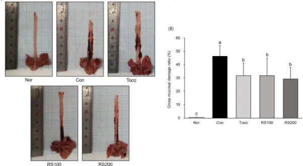
식도 점막의 손상 정도를 육안으로 관찰한 결과, 아무런 처 치를 하지 않은 Nor군의 식도 점막에서는 손상이 발견되지 않았으나, 급성 역류성 식도염 유발 후 증류수를 처리한 Con
군에서는 식도 전체에서 병변이 발견되었다. 반면에 Toco군 에서는 Con군 대비 식도의 병변이 31% 감소하였고, RS100 군에서 32%, RS200군 37% 병변이 감소하였다. RS100군에 서는 양성대조군인 Toco군과 비슷한 식도 보호효과를 나타냈 으며, RS200군에서는 Toco군 대비 6% 더 우수한 식도 점막 보호 효과를 나타냈다 (Fig. 1).
Fig. 1. Surgical induction of acute reflux esophagitis.
A representative gross image; (A), esophageal ulcer ratio; (B). All data are expressed means ± SD (n=8). Normal rats; Nor, acute reflux esophagitis rats; Con, tocopherol 30 ㎎/㎏ body weight/day-treated acute reflux esophagitis rats; Toco, Mixture of Rhei Rhizoma and Scutellariae Radix 100 ㎎/㎏ body weight/day-treated acute reflux esophagitis rats; RS100, Mixture of Rhei Rhizoma and Scutellariae Radix 200 ㎎/㎏ body weight/day-treated acute reflux esophagitis rats; RS200. Different letters above the bars indicate statistically significant differences at p < 0.05.
2. 조직학적 검사
아무런 처치를 하지 않은 Nor군의 식도 조직에서의 조직학적 변화가 관찰되지 않았으나, 급성 역류성 식도염 유발 후 증류 수를 처리한 Con군에서는 두꺼워진 조직과 식도 상피의 탈락
및 염증 세포의 침착을 관찰하였다. RS100군에서는 Con군에 비하여 상피층의 탈락 정도가 감소하였고, RS200군에서는 식도 상피의 탈락이 현저하게 감소하였으며 염증세포의 침윤 정도 또한 감소한 것을 확인하였다 (Fig. 2).
Fig. 2. Esophagus histological examination through H&E staining.
Normal rats; Nor, acute reflux esophagitis rats; Con, tocopherol 30 ㎎/㎏ body weight/day-treated acute reflux esophagitis rats; Toco, Mixture of Rhei Rhizoma and Scutellariae Radix 100 ㎎/㎏ body weight/day-treated acute reflux esophagitis rats; RS100, Mixture of Rhei Rhizoma and Scutellariae Radix 200 ㎎/㎏ body weight/day-treated acute reflux esophagitis rats; RS200.
3. 혈청 내 활성산소 및 간 손상 지표 측정
급성 역류성 식도염 유발 동물 모델에서 산화적 스트레스 바이오 마커인 ROS
21)와 간 손상 지표인 AST 및 ALT를 혈청을 이용하여 측정하였다. ROS 측정 결과, Nor군에 비하여 Con 군에서는 99% 증가하여 약 2배 높은 수치를 나타냈으며, Con군
대비 RS100군과 RS200군에서 각각 14%, 20% 감소하였다.
AST 수치를 확인한 결과, Nor군에 비하여 Con군에서는 51% 증가한 반면 Con군에 비하여 RS200군에서는 7% 감소 하였다. 또한, ALT 수치는 Nor군에 비하여 Con군에서 증가 하였으며, Con군 대비 RS100군에서 17%, RS200군에서는 27% 감소하였다 (Fig. 3).
Fig. 3. ROS, AST, and ALT levels in serum.
Reactive oxygen species (ROS) level; (A), aspartate aminotransferase (AST) level; (B), alanine aminotransferase (ALT) level; (C). All data are expressed means ± SD (n=8). Normal rats; Nor, acute reflux esophagitis rats; Con, tocopherol 30 ㎎/㎏ body weight/day-treated acute reflux esophagitis rats; Toco, Mixture of Rhei Rhizoma and Scutellariae Radix 100 ㎎/㎏ body weight/day-treated acute reflux esophagitis rats; RS100, Mixture of Rhei Rhizoma and Scutellariae Radix 200 ㎎/㎏ body weight/day-treated acute reflux esophagitis rats; RS200.
Different letters above the bars indicate statistically significant differences at p < 0.05.
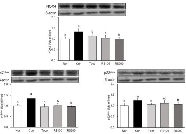
4. 식도 조직 내 NADPH 단백질 발현량 분석
식도 조직 내에서 NADPH 단백질인 NOX4, p47
phox, p22
phox단백질 발현을 확인하였다
22). Nor군에 대비 Con군에 서 NOX4 33%, p47
phox34%, p22
phox24% 증가하였고, 반면 Con군 대비 RS100군에서 NOX4 22%, p47
phox25% 감소하였 으며, RS200군에서 NOX4 26%, p47
phox25% 감소하였으며.
또한, p22
phox는 RS200군에서 감소하였다 (Fig. 4).
5. 식도 조직 내 MAPK 단백질 발현량 분석
식도 조직 내에서 MAPK 단백질인 c-Fos, c-Jun, p-p38, p-ERK 및 p-JNK 단백질 발현을 확인하였다
23, 24). Nor군 대비 Con군에서 MAPK 단백질의 발현이 증가하였으며, 특히 c-Jun의 발현이 52% 증가하였다. 반면에 Con군 대비 RS100
군에서 p-ERK는 감소하였으며, c-Fos, c-Jun, p-p38 및 p-JNK의 발현은 감소하였다. 또한, Con군과 대비하여 RS200군에서는 모든 MAPK 단백질의 발현이 감소하여 Nor 군과 비슷한 수치를 나타냈으며, 특히 발현이 50% 이상 증가 하였던 c-Jun의 수치를 25% 감소시켜 발현을 크게 억제한 것을 확인하였다 (Fig. 5).
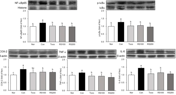
6. 식도 조직 내 염증성 단백질 발현량 분석
식도 조직 내에서 염증성 단백질인 NF-κBp65, p-IκBα,
COX-2, TNF-α 및 IL-6 단백질 발현을 확인하였다
23-25).
Nor군 대비 Con군에서 염증성 단백질의 발현이 증가하였으며,
Con군 대비 RS200군에서 NF-κBp65, p-IκBα, TNF-α
및 IL-6의 발현이 20% 이상 감소하였으며, COX-2 또한
Con군 대비 14% 감소하였다 (Fig. 6).
Fig. 4. Expression of NADPH oxidase proteins in esophagus.
All data are expressed means ± SD (n=8). Normal rats; Nor, acute reflux esophagitis rats; Con, tocopherol 30 ㎎/㎏ body weight/day- treated acute reflux esophagitis rats; Toco, Mixture of Rhei Rhizoma and Scutellariae Radix 100 ㎎/㎏ body weight/day-treated acute reflux esophagitis rats; RS100, Mixture of Rhei Rhizoma and Scutellariae Radix 200 ㎎/㎏ body weight/day-treated acute reflux esophagitis rats;
RS200. Different letters above the bars indicate statistically significant differences at p < 0.05.
Fig. 5. Expression of MAPK proteins in esophagus.
All data are expressed means ± SD (n=8). Normal rats; Nor, acute reflux esophagitis rats; Con, tocopherol 30 ㎎/㎏ body weight/day- treated acute reflux esophagitis rats; Toco, Mixture of Rhei Rhizoma and Scutellariae Radix 100 ㎎/㎏ body weight/day-treated acute reflux esophagitis rats; RS100, Mixture of Rhei Rhizoma and Scutellariae Radix 200 ㎎/㎏ body weight/day-treated acute reflux esophagitis rats;
RS200. Different letters above the bars indicate statistically significant differences at p < 0.05.
Fig. 6. Expression of inflammation related proteins in esophagus.
All data are expressed means ± SD (n=8). Normal rats; Nor, acute reflux esophagitis rats; Con, tocopherol 30 ㎎/㎏ body weight/day- treated acute reflux esophagitis rats; Toco, Mixture of Rhei Rhizoma and Scutellariae Radix 100 ㎎/㎏ body weight/day-treated acute reflux esophagitis rats; RS100, Mixture of Rhei Rhizoma and Scutellariae Radix 200 ㎎/㎏ body weight/day-treated acute reflux esophagitis rats;
RS200. Different letters above the bars indicate statistically significant differences at p < 0.05.
7. 식도 조직 내 항염증 단백질 발현량 분석
식도 조직 내에서 항염증 단백질인 IL-4와 IL-10의 발현 을 확인하였다
26, 27). Nor군 대비 Con군에서 항염증 단백질의
발현이 감소한 반면 Con군 대비 RS100군에서 IL-4 20%, IL-10 16% 증가하였고, RS200군에서 IL-4 29%, IL-10 20% 증가하였다 (Fig. 7).
Fig. 7. Expression of anti-inflammation related proteins in esophagus.
All data are expressed means ± SD (n=8). Normal rats; Nor, acute reflux esophagitis rats; Con, tocopherol 30 ㎎/㎏ body weight/day- treated acute reflux esophagitis rats; Toco, Mixture of Rhei Rhizoma and Scutellariae Radix 100 ㎎/㎏ body weight/day-treated acute reflux esophagitis rats; RS100, Mixture of Rhei Rhizoma and Scutellariae Radix 200 ㎎/㎏ body weight/day-treated acute reflux esophagitis rats;
RS200. Different letters above the bars indicate statistically significant differences at p < 0.05.
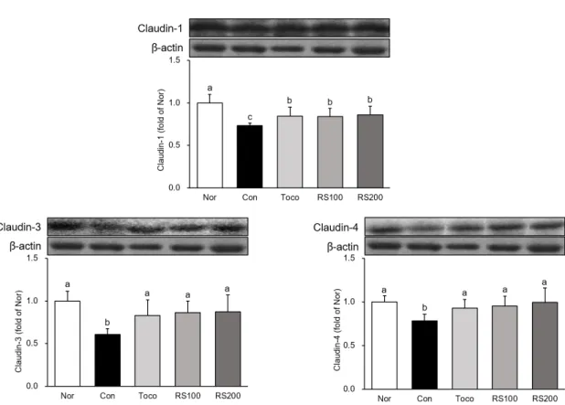
8. 식도 조직 내 tight junction 단백질 발현량 분석
식도 조직 내에서 tight junction 단백질인 Claudin-1, Claudin-3 및 Claudin-4의 발현을 확인하였다
28). Nor군 대
비 Con군에서 tight junction 단백질의 발현이 감소한 반면
Con군 대비 RS100군 및 RS200군에서 tight junction 단백
질의 발현이 증가하였으며, 특히 Claudin-3의 발현이
RS100군에서 43%, RS200군에서 44% 크게 증가한 것을 확
인하였다 (Fig. 8).
Fig. 8. Expression of tight junction proteins in esophagus.
All data are expressed means ± SD (n=8). Normal rats; Nor, acute reflux esophagitis rats; Con, tocopherol 30 ㎎/㎏ body weight/day- treated acute reflux esophagitis rats; Toco, Mixture of Rhei Rhizoma and Scutellariae Radix 100 ㎎/㎏ body weight/day-treated acute reflux esophagitis rats; RS100, Mixture of Rhei Rhizoma and Scutellariae Radix 200 ㎎/㎏ body weight/day-treated acute reflux esophagitis rats;
RS200. Different letters above the bars indicate statistically significant differences at p < 0.05.
Ⅳ. 고 찰
역류성 식도염은 하부 식도 괄약근의 불안정한 활동으로 위 안의 내용물이 역류하여 식도 조직에 손상을 일으키는 등 합병증이 유발된 상태를 말하며, 심한 경우 하부식도 점막에 염증이 생기면서 출혈 및 점막 괴사 박리 등이 일어나 섬유화 협착이 되는 질환이다. 1990년대 초반까지는 우리나라를 포 함한 아시아 국가에서 역류성 식도염의 유발율은 매우 낮게 나타났으나, 2000년대에 들어서면서 유발율의 증가함에 따라 관심 및 연구가 증가하는 추세이다
29-31). 현재 역류성 식도염 치료를 위해 사용되어지고 있는 치료제로는 제산제, 히스타민 H
2수용체 길항제 및 양성자 펌프 억제제 (PPIs) 등이 있으나, 장기간 사용 시 부작용이 뒤따른다고 보고가 되어있다
32). 이 러한 문제로 부작용이 없는 새로운 치료제 개발이 필요한 실 정이며, 본 실험에서는 새로운 치료 소재 개발을 위해 염증에 효과가 있다고 알려진 大黃과 黃芩을 이용하여 역류성 식도염 에 대한 효능을 평가하였으며, 양성 대조군으로는 천연 항산 화제로 알려진 tocopherol을 사용하였다.
약물의 효과를 확인할 수 있으면서 독성을 나타내지 않을 농도인 100 ㎎/㎏와 200 ㎎/㎏의 농도로 大黃과 黃芩 (1:1) 혼합물을 DW에 녹여 동물에게 경구투여한 후 수술을 통해 급 성 역류성 식도염을 유발하였으며, 동물의 식도 조직을 적출 하여 육안으로 관찰한 결과, 정상군에서는 식도 점막의 손상을 관찰할 수 없었으나 대조군에서는 식도 전체에서 손상 및 출
혈을 관찰할 수 있었으며, 大黃과 黃芩 혼합물 투여군에서는 식도 점막의 손상이 현저하게 감소된 것을 확인하였다 (Fig. 1).
부검을 통해 혈액에서 혈청을 분리하여 산화적 스트레스 바이오 마커인 ROS와 간 손상 지표인 AST 및 ALT를 측정하 였다. 활성산소종인 ROS는 체내 산화적 스트레스를 유도하여 단백질 및 DNA 손상을 일으키는 원인이 되며, 생성량이 과도 해지면 인체에 악영향을 미치게 된다
21). 또한, NADPH 산화효 소는 활성산소종의 원인이 되며, 세포막에 존재하는 p47
phox및 p22
phox등 세포질 인자는 NADPH 효소의 활성에 관여한 다고 알려져 있다
22). 본 실험에서 RS200군에서 ROS 및 AST, ALT 수치를 효과적으로 감소시켰으며 (Fig. 3), NADPH 단 백질인 NOX4, p47
phox및 p22
phox또한 유의하게 억제하였다 (Fig. 4).
염증반응은 많은 염증 매개인자의 유도 및 여러 사이토카 인의 상호작용에 의해 야기되는 복잡한 과정이다. TNF-α, IL-6을 포함한 사이토카인 및 COX-2와 같은 인자들은 급성 및 만성 염증을 일으키는 매개체로 작용한다
25). 염증성 사이 토카인 및 염증성 단백질의 발현은 NF-κB에 의해 조절된다.
NF-κB는 정상적인 조건에서 억제제인 IκB와 결합되어 비활성
상태로 존재하다가 외부로부터 염증 자극을 받게되면 IκB가
인산화되면서 NF-κB의 활성화를 일으키며, p38, ERK 및
JNK와 같은 MAPK 또한 NF-κB의 활성화를 조절하는 것으로
알려져 있다
23,24). 본 실험에서 大黃과 黃芩 혼합물의 투여는
전사조절인자인 c-Fos, c-Jun 및 MAPK 인자의 발현을 유
의하게 억제하였을 뿐만 아니라 NF-κB 및 관련되어진 염증성 단백질의 발현 또한 유의하게 억제하였으며, 항염증 단백질인 IL-4와 IL-10
26, 27)의 발현을 유의하게 증가시켰다. 이는 大 黃과 黃芩 혼합물이 급성 역류성 식도염 동물 모델에서 MAPK 및 NF-κB 경로를 억제함으로써 식도 점막의 손상을 감소시 키는 것으로 판단된다.
세포사이의 junction 중 tight junction은 세포-세포의 결 합을 연계하는 다중 단백질 복합체로써 상피의 전해질과 수분 의 이동 및 세포 내 신화전달 등 다양한 생물학적 기능을 가진 다
28). Tight junction의 중요한 구성요소인 Claudin 단백질 은 막단백질로써 세포의 극성이 형성과 유지에 중요한 역할을 하며, 다양한 신호 전달 단백질과의 상호작용을 통해 세포간 부착 및 물질이동을 조절한다
28, 33). 식도염 유발에 의해 식도 조직이 손상되면 상피 및 내피 결합조직에서는 tight junction 의 손상이 야기되며, 이는 Claudin 단백질의 감소를 유도한다
34)
. 본 실험에서 급성 역류성 식도염을 유발한 군에서는 Claudin 단백질 (Claudin-1, Claudin-3, Claudin-4)의 발현이 감소 하였으며, 大黃과 黃芩 혼합물 투여군에서는 Claudin 단백질의 발현이 유의하게 증가한 것을 확인하였다. 이는 大黃과 黃芩 혼합물 투여가 상피세포의 결합조직을 보호하였음을 나타낸다.
Ⅴ. 결 론
본 연구에서는 항염증 효과가 있다고 알려진 大黃과 黃芩의 혼합물이 급성 역류성 식도염 동물 모델에서 보이는 식도 점막 보호 효과에 대해 연구하였다. 大黃과 黃芩 혼합물을 농도별로 투여한 후 수술을 통해 역류성 식도염을 유발하였으며, 혈액 및 식도 조직을 채취하였다. 혈액을 이용하여 산화적 스트레스 바이오 마커, 간 손상 지표를 측정하였고, 식도조직을 이용하여 NADPH 단백질, MAPK 단백질, 염증성 단백질, 항염증 단백 질 및 tight junction 단백질의 발현을 분석하였으며, 다음과 같은 결과를 얻었다.
1. 급성 역류성 식도염 동물 모델에서 大黃과 黃芩의 혼합물 은 식도 점막 병변 및 궤양 범위를 유의하게 감소시켰다.
2. 大黃과 黃芩의 혼합물은 급성 역류성 식도염 동물 모델 에서 ROS를 효과적으로 감소시켰으며, 간 손상 지표인 AST 및 ALT 또한 효과적으로 감소시켰다.
3. 大黃과 黃芩의 혼합물은 NADPH 단백질인 NOX4, p47
phox및 p22
phox단백질 발현을 유의하게 억제하였다.
4. 大黃과 黃芩의 혼합물은 역류성 식도염 유발에 의해 증 가된 MAPK (c-Fos, c-Jun, p-p38, p-ERK, p- JNK)와 염증성 단백질 (NF-κBp65, p-IκBα, COX-2, TNF-α, IL-6)의 발현을 유의하게 억제하였다.
5. 大黃과 黃芩의 혼합물은 항염증 단백질인 IL-4와 IL-10 의 발현을 효과적으로 증가시켰다.
6. Tight junction 단백질 분석 결과, 大黃과 黃芩의 혼합 물은 Claudin-1, Claudin-3 및 Claudin-4 단백질 발 현을 증가시켜 식도 조직의 결합조직 내 손상을 억제하 였다.
이상의 결과로부터 급성 역류성 식도염 동물 모델에서 大黃 과 黃芩의 혼합물은 MAPK 및 NF-κB 경로를 억제하고 tight junction을 보호함으로써 식도 점막을 보호하여 손상을 억제 하였음을 알 수 있으며, 大黃과 黃芩의 혼합물이 역류성 식도염 치료를 위한 소재로서의 가능성이 있음을 알 수 있다.
감사의 글
본 연구는 한국연구재단 (No. 2017R1A2B2006858 and No. 2019R1I1A1A01064068)의 지원에 의해 수행되었습니다.
References