https://doi.org/10.5624/isd.2018.48.4.269
Introduction
Dental implants are considered to be an ideal option for the rehabilitation of edentulous jaws due to their sta- ble outcomes and high success rates.1 Despite the high predictability of dental implants, complications are still encountered. These complications may be associated with various factors, such as treatment planning, the anatomy
of the site, and the surgical technique and experience of the practitioner.2 A thorough clinical and radiological ex- amination plays an important role in avoiding complica- tions and enhancing the success of the treatment.
Intraoral radiography, panoramic radiography, com- puted tomography(CT), and cone-beam CT(CBCT) are frequently-used imaging modalities in treatment planning that improve long-term treatment success.3 Of these mo- dalities, panoramic radiography has been widely used, since it has the advantages of being cost-effective, readily available, and providing high-resolution images.4 Pan- oramic radiography is a unique tool in terms of offering
Comparing the precision of panoramic radiography and cone-beam computed tomography in avoiding anatomical structures critical to dental implant surgery: A retrospective study
Öznur Özalp 1,*, Hüseyin Alican Tezerişener 1, Burak Kocabalkan 1,
Ulviye Şebnem Büyükkaplan 2, Mehmet Mustafa Özarslan 2,
Göksel Şimşek Kaya
1,Mehmet Ali Altay
1, Alper Sindel
11Department of Oral and Maxillofacial Surgery, Faculty of Dentistry, Akdeniz University, Antalya, Turkey
2Department of Prosthodontics, Faculty of Dentistry, Akdeniz University, Antalya, Turkey
ABSTRACT
Purpose: The aim of this study was to evaluate the correlations between measurements made using panoramic radiography and cone-beam computed tomography(CBCT) based on certain anatomical landmarks of the jaws, with the goal of preventing complications due to inaccurate measurements in the pre-surgical planning phase of dental implant placement.
Materials and Methods: A total of 56 individuals who underwent panoramic radiography and a CBCT evaluation before dental implant surgery were enrolled in the study. Measurements were performed to identify the shortest vertical distance between the alveolar crest and neighboring anatomical structures, including the maxillary sinus, nasal floor, mandibular canal, and foramen mentale. The differences between the measurements on panoramic radiography and CBCT images were statistically analyzed.
Results: Statistically significant differences were observed between the measurements on panoramic radiography and CBCT for all anatomical structures(P<.05). The correlation coefficients(r) between the paired samples obtained from panoramic radiography and CBCT were closely correlated(P<.05), with r values varying from 0.921 and 0.979 for different anatomical regions.
Conclusion: The results of this study support the idea that panoramic radiography might provide sufficient information on bone height for preoperative implant planning in routine cases or when CBCT is unavailable.
However, an additional CBCT evaluation might be helpful in cases where a safety margin cannot be respected due to insufficient bone height.(Imaging Sci Dent 2018; 48: 269-75)
KEY WORDS: Cone-Beam Computed Tomography; Dental Implants; Radiography, Panoramic
Copyright ⓒ 2018 by Korean Academy of Oral and Maxillofacial Radiology
This is an Open Access article distributed under the terms of the Creative Commons Attribution Non-Commercial License(http://creativecommons.org/licenses/by-nc/3.0) which permits unrestricted non-commercial use, distribution, and reproduction in any medium, provided the original work is properly cited.
Imaging Science in Dentistry·pISSN 2233-7822 eISSN 2233-7830 Received July 23, 2018; Revised October 7, 2018; Accepted October 19, 2018
*Correspondence to : Dr. Öznur Özalp
Department of Oral and Maxillofacial Surgery, Faculty of Dentistry, Akdeniz University, Dumlupinar Boulevard, Campus, Antalya, 07058, Turkey
Tel) 90-507-577-1530, Fax) 90-242-310-6967, E-mail) [email protected]
a large amount of information about the dentition and the jaws with a small radiation dose.5 However, distortions in the horizontal plane and magnification in the vertical plane are inevitable consequences of the working princi- ple of panoramic radiography devices. In addition, incor- rect patient positioning and technical or processing errors have substantial effects on the accuracy of images.6 Fur- thermore, 2-dimensional(2D) radiographic examinations are incapable of displaying the available bone width and the exact relationships with neighboring anatomical struc- tures.7 Additionally, structures outside the focal trough are blurred and appear as shadows and artifacts on panoramic radiography.
The emergence of dentomaxillofacial software appli- cations for presurgical planning was a major milestone in oral and maxillofacial surgery.8 Since the introduction of CBCT in the late 1990s, 3-dimensional(3D) technology with a lower radiation dose has become widely used in oral and maxillofacial surgery practice.9 CBCT scans have several advantages over 2D images and CT, including re- al-size data, the potential for generating a comprehensive set of 2D images, the ability to perform vertical scanning with the patient in a natural seated position, isotropic vox- el size, less disturbance from metal artifacts, and Digital Imaging and Communications in Medicine compatibility.
Furthermore, CBCT obtains a large amount of data in a relatively short time period of exposure to X-ray radiation and provides high-resolution images in several orthogonal planes for accurate measurements.9 Hence, CBCT offers a smaller dose of radiation, lower costs, and better energy efficiency than CT. Moreover, it is suitable not only for preoperative diagnoses, but also for real-time intraopera- tive assessments.10
A precise radiographic assessment of the jaws is cru- cial for presurgical planning and implant placement. Pan- oramic radiography and CBCT are the most frequently used imaging modalities in dental implantology. Although
a considerable amount of research has been published on the use of panoramic radiography and CBCT in dental implantology, debate continues about the best imaging modality in presurgical implant planning. Some of these studies concluded that panoramic radiography was a re- liable and safe technique for determining bone height, while others claimed that presurgical planning using pan- oramic radiography might lead to an inaccurate designa- tion of the length of the implants, which could eventually result in complications such as nerve injury and sinus per- forations.9,11-13
Therefore, the aim of this study was to evaluate the cor- relations between measurements made using panoramic radiography and CBCT based on certain anatomical land- marks of the jaws, with the goal of preventing complica- tions due to inaccurate measurements in the pre-surgical planning phase of dental implant placement.
Materials and Methods
This study was approved(reference number: 631/01112 017) by the Clinical Research Ethics Committee of the University of Akdeniz, Antalya, Turkey, and written in- formed consent was obtained from each patient. A retro- spective study was conducted of 56 patients(30 male, 26 female; 32-79 years old, mean age of 58 years) who un- derwent preoperative panoramic radiography and CBCT evaluation for dental implant surgery between August 2016 and September 2017 at the Department of Oral and Maxillofacial Surgery.
The inclusion criteria were the availability of clear dig- ital panoramic radiography and CBCT images showing at least 1 edentulous region in the neighborhood of the maxillary sinus, nasal floor, mental foramen, and/or man- dibular canal in any quadrant. Images that demonstrated positioning errors, artifacts, unequal magnification or geometric distortion, and unclear anatomical structures
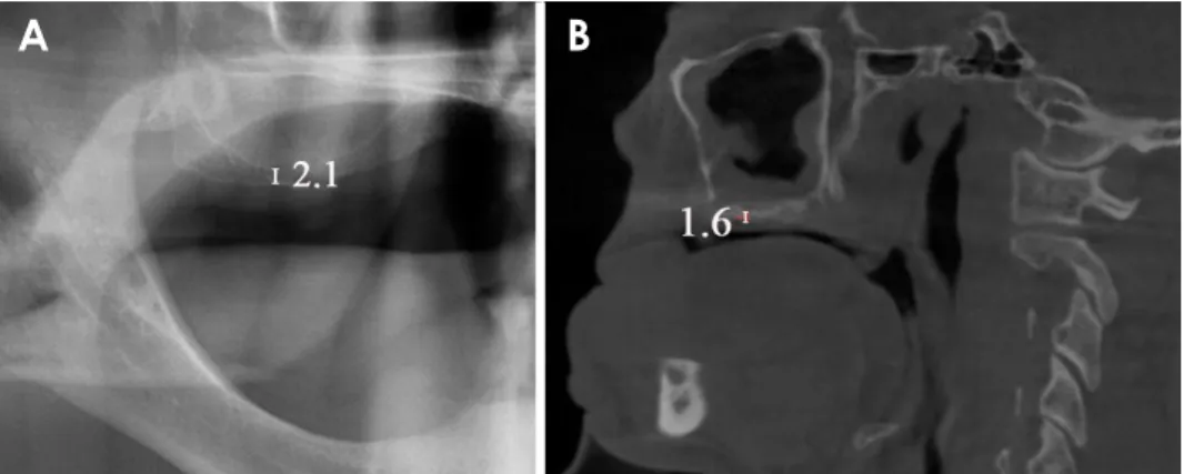
Fig. 1. Measurement of the short- est vertical distance between the alveolar crest and the bottom of the maxillary sinus on a panoramic im- age (A) and a cone-beam computed tomography image (B).
A B
were excluded from the study data.
The digital panoramic radiographs were obtained using the same device(Planmeca ProMax® 2D; Planmeca, Hel- sinki, Finland) with a 1.2 magnification ratio, a voltage of 60kV, a current of 5mA, and a minimum exposure time of 17 s. The CBCT images were taken using the Planme- ca Promax® 3D(Planmeca, Helsinki, Finland) at a voltage of 80kV, a current of 12mA, a scanning field of 16×5 cm, and a scanning time of 12 s. All measurements were performed using the Planmeca Romexis® Viewer v.3.8.1 (Planmeca, Helsinki, Finland) on panoramic images at 1 :1 magnification and on CBCT scans in the coronal and sagittal planes. The shortest vertical distances between the alveolar crest and neighboring anatomical structures were
measured, as follows: 1. The shortest distance between the bottom of the left and/or right maxillary sinus and the alveolar crest(Fig. 1). 2. The shortest distance between the bottom of the left and/or right nasal floor and the al- veolar crest(Fig. 2). 3. The shortest distance between the top of the mandibular canal and the alveolar crest in the area of the left and/or right mandibular second molar(Fig.
3). 4. The shortest distance between the top of the left and/or right foramen mentale and the alveolar crest(Fig.
4).Measurements were independently performed by 2 oral and maxillofacial surgeons, and the mean values were used for the analysis. The intraclass correlation coeffi- cient(ICC) was used to determine interobserver and in-
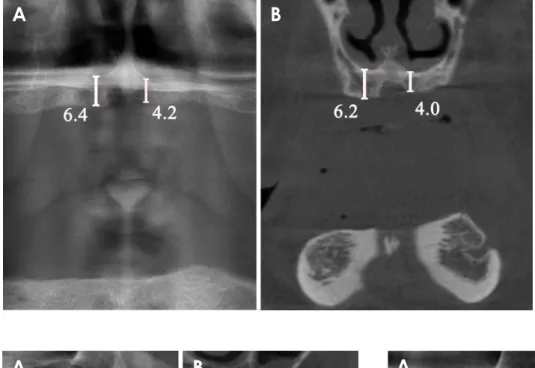
Fig. 2. Measurement of the shortest vertical distance between the alveo- lar crest and the bottom of the nasal floor on a panoramic image (A) and a cone-beam computed tomography image (B).
B A
Fig. 4. Measurement of the shortest vertical distance between the alveolar crest and the top of the foramen mentale on a panoramic image (A) and a cone-beam computed tomography image (B).
B A
Fig. 3. Measurement of the shortest vertical distance between the alveolar crest and the top of the mandibular canal on a panoramic image (A) and a cone-beam computed tomography image (B).
B
A
traobserver reliability.
Data were analyzed using SPSS version 22(IBM Corp., Armonk, NY, USA). The normality of the data was as- sessed by a visual inspection of histograms, QQ-plots, box plots, and the Shapiro-Wilk test. The paired t-test was utilized to compare the measured values on panoramic radiography and CBCT images. The level of statistical significance was set at P<.05. Pearson correlation analy- sis was used to analyze the relationships between the data obtained using the 2 methods. The correlation coefficient (r) between the paired samples was calculated, and the measures were considered to be closely correlated if r was between 0.5 and 1.
Results
After the eligibility criteria were assessed, the final sample was composed of 30 men(53.6%) and 26 women (46.4%), who ranged in age from 32 to 79 years(mean, 58 years). Statistically significant differences were not observed for sex or age(P>.05). The number of mea- surements of each anatomical region and the mean values of the distances between the anatomical structures and the alveolar crest on panoramic radiography and CBCT im- ages are shown in Table 1.
The ICC values for intraobserver reliability were 0.96 and 0.98, respectively, and the ICC value for interobserv- er reliability was 0.89. The ICC values indicated good to excellent reliability.14
Statistically significant differences were found between the measurements on panoramic and CBCT images for all anatomical structures(P<.05)(Table 2). The correlation coefficients(r) between the paired samples obtained from panoramic radiography and CBCT were closely correlat- ed(P<.05), with r values varying between 0.921 and
0.979 for the different anatomical structures(Table 3).
In the maxillary sinus, a statistically significant dif- ference was found between the measured values on panoramic radiography and CBCT images(t47=2.758, P<.05). On average, these measurements on panoramic images were 0.36mm higher than those made on CBCT images(95% CI, 0.097-0.620mm). The measured values on panoramic and CBCT images for the distance between the floor of the maxillary sinus and the alveolar crest were very closely and positively correlated(r=0.967, P<.05).
Similarly, panoramic radiography and CBCT showed statistically significant differences in measurements at the region of the nasal floor(t36=4.054, P<.05). On average, these measurements on panoramic images were 0.67mm higher than those on CBCT images(95% CI, 0.337-1.012 mm). The measured values on panoramic and CBCT im- ages for the distance between the nasal floor and the al- veolar crest were very closely and positively correlated (r=0.921, P<.05).
In the region of the mandibular canal, there was also a statistically significant difference between the measure- ments made on panoramic radiography and CBCT images (t49=6.723, P<.05). On average, these measurements on panoramic images were 0.76mm higher than those on CBCT images(95% CI, 0.533-0.988). The measured values on panoramic and CBCT images for the distance between the mandibular canal and the alveolar crest were very closely and positively correlated(r=0.979, P<.05).
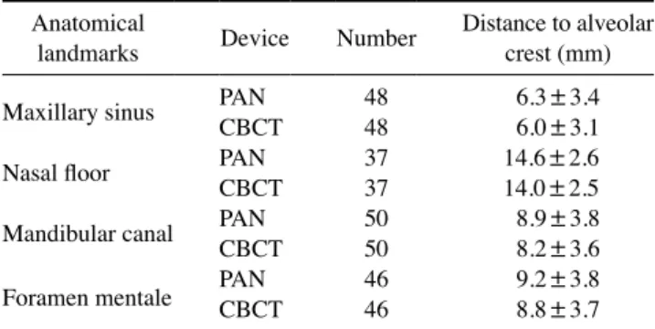
Table 1. The number and mean values of measurements of the distances between the alveolar crest and anatomical landmarks measured on panoramic radiography(PAN) and cone-beam com- puted tomography(CBCT)
Anatomical
landmarks Device Number Distance to alveolar crest (mm)
Maxillary sinus PAN 48 6.3±3.4
CBCT 48 6.0±3.1
Nasal floor PAN 37 14.6±2.6
CBCT 37 14.0±2.5
Mandibular canal PAN 50 8.9±3.8
CBCT 50 8.2±3.6
Foramen mentale PAN 46 9.2±3.8
CBCT 46 8.8±3.7
Table 2. Comparison of the differences in measurements of dis- tance from the anatomical landmarks to the alveolar crest between panoramic radiography and cone-beam computed tomography (CBCT)
Anatomical
landmarks Difference
(mm) Lower Upper Sig.
(2-tailed)
Maxillary sinus 0.36±0.9 0.1 0.6 .008*
Nasal floor 0.7±1.0 0.3 1.0 .000*
Mandibular canal 0.8±0.8 0.5 1.0 .000*
Foramen mentale 0.4±0.9 0.1 0.7 .003*
*P<.05
Table 3. Correlation coefficients between the paired samples obtained from panoramic radiography and cone-beam computed tomography at different anatomical landmarks
Anatomical region Correlation coefficient
Maxillary sinus 0.967*
Nasal floor 0.921*
Mandibular canal 0.979*
Foramen mentale 0.974*
*P<.05
Finally, the difference between the measured values on panoramic radiography and CBCT images was statis- tically significant for the foramen mentale(t45=3.197, P<.05). On average, these measurements on panoramic images were 0.4mm higher than those on CBCT images (95% CI, 0.149-0.655). The measured values on pan- oramic and CBCT images for the distance between the foramen mentale and the alveolar crest were very closely and positively correlated(r=0.974, P<.05).
Discussion
Various imaging modalities are currently available in oral and maxillofacial radiology, of which intraoral radio- graphs, panoramic radiography, CT, and CBCT are the most commonly preferred techniques in dental implan- tology. Intraoral radiographs have been widely used, and have the advantages of low cost and ready availability.
However, the inherent 2D nature of intraoral radiographs may lead to anatomical superimposition and geometric distortion.15 Isidor16 reported that due to superimposition, it was not possible to detect an unfavorable marginal bone level or the absence of osseointegration on 2D images.
Furthermore, several studies have shown that the restrict- ed preoperative diagnostic ability of 2D images in dental implant surgery may cause implant failure.17 Osseous de- struction as a result of periodontal disease or peri-implan- titis is an increasing problem in the long-term success of implant treatment.18 Three-dimensional images also show interproximal alveolar bone levels in the oro-vestibular direction, which is an essential criterion for follow-up of the peri-implant bone.19
Panoramic radiography provides a comprehensive 2D radiological examination of the jaws. The main advantag- es of panoramic radiography include a low radiation dose, relatively short exposure time, and comfort and simplicity of examination.20 However, the lower image quality com- pared to intraoral radiographs, geometric distortions such as unequal magnification and elongation, superimposition of the cervical spine, and the presence of ghost images are among the disadvantages of panoramic radiography.21 Laster et al.22 reported that horizontal measurements on panoramic radiography may be unreliable due to distor- tion and overlapping.
The outcomes of implant treatment have become highly predictable in recent years.23 Nonetheless, the relationship of implants with important vital structures can signifi- cantly affect the success of the surgical procedure. Thus, restricting the preoperative diagnostic examination to 2D
imaging methods may potentially cause implant failure.17 Tang et al.21 recommended 3D imaging in cases where implant surgery posed a risk of damaging vital structures.
Jacobs and Quirynen7 and Hassan and Jacobs24 also sug- gested using CT to evaluate extensive structures, such as the maxillary sinus. Similarly, Dreiseidler et al.25 reported that the image quality of CT and CBCT was superior to that of panoramic radiography, although CT and CBCT had the disadvantages of not being available in many lo- cal hospitals due to technical requirements and high cost.
In recent years, maxillofacial CBCT has been widely used in dental implantology.26 CBCT is advantageous due to its high spatial resolution, short scanning time, and rapid image acquisition.27 Monsour and Dudhia6 reported that patients undergoing CT examinations were exposed to a higher radiation dose than those who underwent ex- aminations using panoramic radiography and CBCT.
To date, the use of different imaging techniques for pre-implant evaluation has been analyzed in several studies. Kopecka et al.28 compared the use of panoram- ic radiography and CBCT in assessing the interantral bone height available for dental implant placement and reported that panoramic radiography was sufficient in the incisor region, but not in the canine region. In a cadaver- ic study, Hu et al.11 compared the measurement errors on panoramic radiography and CBCT images and found that the mean presurgical measurement error was significantly smaller for CBCT than for panoramic radiography in the maxillary region, whereas it did not differ significantly in the mandibular region.
In contrast, Renton et al.29 reported that a majority of idiopathic trigeminal neuropathies(90%) were found in patients who had undergone pre-surgical 2D radiographic evaluation, while only 10% of cases presented such neu- ropathies after pre-surgical CBCT had been taken. In ac- cordance with Renton et al.,29 Angelopoulos et al.30 also reported that CBCT images enabled a more precise evalu- ation of the mandibular canal.
In another study, Tang et al.21 compared the magnifica- tion rate of panoramic radiography in measuring differ- ent maxillofacial loci to that of CBCT and reported that the distances measured by panoramic radiography were closely correlated with those measured by CBCT. Similar- ly, the authors found strong positive correlations between panoramic radiography and CBCT in measurements of alveolar bone height in the regions of the maxillary sinus, nasal fossa, mandibular canal, and foramen mentale.
Vazquez et al.31 investigated the incidence of altered mental nerve sensation after implant placement in the
posterior segment of the mandible using only panoramic radiographs for the preoperative evaluation. They report- ed that when a safety margin of at least 2mm above the mandibular canal was respected, panoramic radiogra- phy was sufficient to evaluate the available bone height prior to the insertion of posterior mandibular implants.
Regarding the mandibular canal region, Gerlach et al.32 also recommended a vertical safety margin of at least 1.7mm when planning surgery using CBCT-based data.
In accordance with the recommendations of Vazquez et al.31 and Gerlach et al.,32 the present study revealed that the average difference between panoramic radiography and CBCT ranged from 0.36mm to 0.76mm. However, in cases where the available bone is not sufficient, it may be necessary to place an implant deeper than the safety measurement, meaning that it is not always possible to respect a safety margin of 2mm.33 In such cases, although the current study revealed that the average difference be- tween panoramic radiography and CBCT was less than 1 mm, the use of CBCT is recommended for more accurate planning.
In conclusion, the results of this study support the idea that panoramic radiography might provide sufficient in- formation on bone height for preoperative implant plan- ning in routine cases or when CBCT is not available.
However, an additional CBCT evaluation might be help- ful in cases where a safety margin cannot be respected due to insufficient bone height.
References
1. Ali SA, Karthigeyan S, Deivanai M, Kumar A. Implant reha- bilitation for atrophic maxilla: a review. J Indian Prosthodont Soc 2014; 14: 196-207.
2. Misch K, Wang HL. Implant surgery complications: etiology and treatment. Implant Dent 2008; 17: 159-68.
3. Jaju PP, Jaju SP. Clinical utility of dental cone-beam comput- ed tomography: current perspectives. Clin Cosmet Investig Dent 2014; 6: 29-43.
4. Ahlqwist M, Halling A, Hollender L. Rotational panoram- ic radiography in epidemiological studies of dental health.
Comparison between panoramic radiographs and intraoral full mouth surveys. Swed Dent J 1986; 10: 73-84.
5. Lecomber AR, Yoneyama Y, Lovelock DJ, Hosoi T, Adams AM. Comparison of patient dose from imaging protocols for dental implant planning using conventional radiography and computed tomography. Dentomaxillofac Radiol 2001; 30:
255-9.
6. Monsour PA, Dudhia R. Implant radiography and radiology.
Aust Dent J 2008; 53 Suppl 1: S11-25.
7. Jacobs R, Quirynen M. Dental cone beam computed tomogra- phy: justification for use in planning oral implant placement.
Periodontol 2000 2014; 66: 203-13.
8. Verstreken K, Van Cleynenbreugel J, Marchal G, Naert I, Suetens P, van Steenberghe D. Computer-assisted planning of oral implant surgery: a three-dimensional approach. Int J Oral Maxillofac Implants 1996; 11: 806-10.
9. Amarnath GS, Kumar U, Hilal M, Muddugangadhar BC, An- shuraj K, Shruthi CS. Comparison of cone beam computed tomography, orthopantomography with direct ridge mapping for pre-surgical planning to place implants in cadaveric man- dibles: an ex-vivo study. J Int Oral Health 2015; 7(Suppl 1):
38-42.
10. Klatt JC, Heiland M, Marx S, Hanken H, Schmelzle R, Pohlenz P. Clinical indication for intraoperative 3D imaging during open reduction of fractures of the mandibular angle. J Craniomaxillofac Surg 2013; 41: e87-90.
11. Hu KS, Choi DY, Lee WJ, Kim HJ, Jung UW, Kim S. Reli- ability of two different presurgical preparation methods for implant dentistry based on panoramic radiography and cone- beam computed tomography in cadavers. J Periodontal Im- plant Sci 2012; 42: 39-44.
12. Pertl L, Gashi-Cenkoglu B, Reichmann J, Jakse N, Pertl C.
Preoperative assessment of the mandibular canal in implant surgery: comparison of rotational panoramic radiography (OPG), computed tomography (CT) and cone beam computed tomography (CBCT) for preoperative assessment in implant surgery. Eur J Oral Implantol 2013; 6: 73-80.
13. Correa LR, Spin‐Neto R, Stavropoulos A, Schropp L, da Silveira HE, Wenzel A. Planning of dental implant size with digital panoramic radiographs, CBCT‐generated panoramic images, and CBCT cross‐sectional images. Clin Oral Im- plants Res 2014; 25: 690-5.
14. Koo TK, Li MY. A guideline of selecting and reporting intra- class correlation coefficients for reliability research. J Chiropr Med 2016; 15: 155-63.
15. Corpas Ldos S, Jacobs R, Quirynen M, Huang Y, Naert I, Duyck J. Peri‐implant bone tissue assessment by comparing the outcome of intra‐oral radiograph and cone beam computed tomography analyses to the histological standard. Clin Oral Implants Res 2011; 22: 492-9.
16. Isidor F. Clinical probing and radiographic assessment in rela- tion to the histologic bone level at oral implants in monkeys.
Clin Oral Implants Res 1997; 8: 255-64.
17. Greenstein G, Cavallaro J, Romanos G, Tarnow D. Clinical recommendations for avoiding and managing surgical com- plications associated with implant dentistry: a review. J Peri- odontol 2008; 79: 1317-29.
18. Charyeva O, Altynbekov K, Zhartybaev R, Sabdanaliev A.
Long-term dental implant success and survival--a clinical study after an observation period up to 6 years. Swed Dent J 2012; 36: 1-6.
19. Ritter L, Elger M, Rothamel D, Fienitz T, Zinser M, Schwarz F, et al. Accuracy of peri-implant bone evaluation using cone beam CT, digital intra-oral radiographs and histology. Dento- maxillofac Radiol 2014; 43: 20130088.
20. Suomalainen A, Pakbaznejad Esmaeili E, Robinson S. Dento- maxillofacial imaging with panoramic views and cone beam CT. Insights Imaging 2015; 6: 1-16.
21. Tang Z, Liu X, Chen K. Comparison of digital panoramic
radiography versus cone beam computerized tomography for measuring alveolar bone. Head Face Med 2017; 13: 2.
22. Laster WS, Ludlow JB, Bailey LJ, Hershey HG. Accuracy of measurements of mandibular anatomy and prediction of asymmetry in panoramic radiographic images. Dentomaxillo- fac Radiol 2005; 34: 343-9.
23. Choi JW. Assessment of panoramic radiography as a national oral examination tool: review of the literature. Imaging Sci Dent 2011; 41: 1-6.
24. Hassan B, Jacobs R. Cone beam computed tomography - 3D imaging in oral and maxillofacial surgery. Eur Med Imaging Rev 2008; 1: 38-40.
25. Dreiseidler T, Mischkowski RA, Neugebauer J, Ritter L, Zöller JE. Comparison of cone-beam imaging with orthopan- tomography and computerized tomography for assessment in presurgical implant dentistry. Int J Oral Maxillofac Implants 2009; 24: 216-25.
26. Sheikhi M, Dakhil-Alian M, Bahreinian Z. Accuracy and reli- ability of linear measurements using tangential projection and cone beam computed tomography. Dent Res J (Isfahan) 2015;
12: 271-7.
27. Dalessandri D, Laffranchi L, Tonni I, Zotti F, Piancino MG, Paganelli C, et al. Advantages of cone beam computed to- mography (CBCT) in the orthodontic treatment planning of cleidocranial dysplasia patients: a case report. Head Face Med 2011; 7: 6.
28. Kopecka D, Simunek A, Streblov J, Slezak R, Capek L. Mea- surement of the interantral bone in implant dentistry using panoramic radiography and cone beam computed tomogra- phy: a human radiographic study. West Indian Med J 2014;
63: 503-9.
29. Renton T, Dawood A, Shah A, Searson L, Yilmaz Z. Post-im- plant neuropathy of the trigeminal nerve. A case series. Br Dent J 2012; 212: E17.
30. Angelopoulos C, Thomas S, Hechler S, Parissis N, Hlavacek M.
Comparison between digital panoramic radiography and cone- beam computed tomography for the identification of the man- dibular canal as part of presurgical dental implant assessment.
J Oral Maxillofac Surg 2008; 66: 2130-5.
31. Vazquez L, Saulacic N, Belser U, Bernard JP. Efficacy of pan- oramic radiographs in the preoperative planning of posterior mandibular implants: a prospective clinical study of 1527 consecutively treated patients. Clin Oral Implants Res 2008;
19: 81-5.
32. Gerlach NL, Meijer GJ, Maal TJ, Mulder J, Rangel FA, Borst- lap WA, et al. Reproducibility of 3 different tracing methods based on cone beam computed tomography in determining the anatomical position of the mandibular canal. J Oral Maxillo- fac Surg 2010; 68: 811-7.
33. Greenstein G, Cavallaro J, Tarnow D. Practical application of anatomy for the dental implant surgeon. J Periodontol 2008;
79: 1833-46.