Osteoporosis is a systemic skeletal disease characterized by low bone mass and micro-architectural deterioration of bone tissue with a consequent increase in both bone fragility and susceptibility to fractures.1) In addition to ver- tebral, hip, and distal forearm fractures, humeral fractures are among the most common osteoporotic fractures, and the average lifetime risk in a 50-year-old Caucasian to ex-
perience this type of fracture has been estimated at 12.9%
for women and 4.1% for men.2) Osteoporotic fractures are associated with significant morbidity, and fractures of the hip, vertebrae, and humerus have been shown to be as- sociated with excess mortality.3,4) Fractures of the proximal humerus account for 5% of all fractures and are the third most common fracture pattern occurring in individuals over the age of 65.5,6)
The widely used bisphosphonate (BP) therapy de- creases bone resorption and hypercalcemia and reduces osteolysis of bone metastasis.7,8) A number of randomized clinical trials have shown that BPs increase the bone den- sity and reduce the risk of vertebral, non-vertebral, and hip fractures.8,9) Nevertheless, BPs are not recommended
Influence of Early Bisphosphonate Administration for Fracture Healing in Patients with Osteoporotic
Proximal Humerus Fractures
Joong-Bae Seo, MD, Jae-Sung Yoo, MD, Jee-Won Ryu, MD, Kun-Woong Yu, MD
Department of Orthopedic Surgery, Dankook University College of Medicine, Cheonan, Korea
Background: Bisphosphonates are generally known to adversely affect fracture healing because they inhibit osteoclastic bone resorption. However, some authors argue that bisphosphonates have no adverse effect on the restoration of the mechanical integ- rity of long bones after fractures. It is unclear whether bisphosphonates can be initiated safely in patients with acute proximal hu- merus fractures. The aim of this study was to determine whether the early use of a bisphosphonate affects healing and outcomes of osteoporotic proximal humerus fractures treated with a locking compression plate.
Methods: Between August 2004 and June 2013, a total of 82 osteoporotic patients who underwent locking plate fixation of proxi- mal humerus fractures were enrolled retrospectively. The patients were divided into two groups according to the timing of the commencement of treatment with alendronate after surgery: group A (n = 34, initiation of the bisphosphonate treatment within two weeks after surgery) and group B (n = 48, control group, initiation of the treatment three months after surgery). Patients were as- sessed for radiographic union at 2, 6, 10, and 16 weeks, 6 months, and 1 year after surgery. Clinical assessments were performed using the Constant score and American Shoulder and Elbow Surgeons (ASES) score at 1 year after surgery.
Results: No significant differences were observed between the two groups with respect to radiographic and clinical outcomes af- ter locking plate fixation. All patients obtained fracture union, and the mean time to radiographic union was similar in group A and group B (6.3 and 6.6 weeks, respectively; p = 0.67).
Conclusions: This study shows that the early initiation of bisphosphonate treatment does not affect bone union or clinical out- comes in patients with an osteoporotic proximal humerus fracture treated by locking compression plate fixation.
Keywords: Proximal humeral fracture, Osteoporosis, Bisphosphonate
Copyright © 2016 by The Korean Orthopaedic Association
This is an Open Access article distributed under the terms of the Creative Commons Attribution Non-Commercial License (http://creativecommons.org/licenses/by-nc/4.0) which permits unrestricted non-commercial use, distribution, and reproduction in any medium, provided the original work is properly cited.
Clinics in Orthopedic Surgery • pISSN 2005-291X eISSN 2005-4408 Received April 12, 2016; Accepted July 13, 2016
Correspondence to: Joong-Bae Seo, MD
Department of Orthopedic Surgery, Dankook University College of Medicine 119 Dandae-ro, Dongnam-gu, Cheonan 31116, Korea
Tel: +82-41-550-3060, Fax: +82-41-556-3238 E-mail: [email protected]
in patients with acute fractures because they are known to delay the process of fracture healing and suppress re- modeling of the callus.10,11) However, Kim et al.12) reported that the early administration of BPs does not appear to affect the rate of healing of an intertrochanteric fracture or the incidence of complications. Gong et al.13) argued that the early initiation of a BP treatment did not affect fracture healing or clinical outcomes. Cao et al.11) reported that after a bone fracture, BPs did not appear to interfere with the early phases of repair; rather, continuous BP use resulted in a larger, stronger callus. Recently, Li et al.14) reported that early administration of BPs after surgery did not appear to delay fracture healing time either radiologi- cally or clinically. Furthermore, according to the changes in bone mineral density (BMD) and bone turnover mark- ers, the anti-resorptive efficacy of BPs given immediately after surgical repair should positively affect the rate of sub- sequent fractures based on a meta-analysis of randomized controlled trials.
It is unclear whether BPs can be initiated safely in patients who have sustained an acute proximal humerus fracture. We hypothesized that the early initiation of a BP treatment for patients treated with a locking compression plate fixation system would not affect fracture healing or clinical outcomes. The aim of this study was to determine whether the early use of a BP affects healing and outcomes of osteoporotic proximal humerus fractures treated with a locking compression plate.
METHODS
Subjects
Following Dankook University Hospital Institutional Re- view Board exempt approval (IRB no.: DKUH IRB 2013- 10-011), a total of 127 patients over the age of 50 years who had undergone locking compression plate fixation of proximal humerus fractures and had been diagnosed with osteoporosis were enrolled retrospectively from August 2004 to June 2013. All patients were recommended BMD examinations preoperatively.
Fracture pattern grading was accomplished radio- graphically using the Neer classification system: a part/
segment was considered displaced if it was separated from its neighboring segment by more than 1 cm or angulated by more than 45°.15,16)
The inclusion criteria were: (1) preoperative BMD confirming osteoporosis with a T-score of < –2.5; (2) no BP therapy during two years preceding the fracture; and (3) 2-, 3-, or 4-part displaced fractures (including surgical neck fractures) of the proximal humerus treated with lock- ing compression plate fixation. The exclusion criteria were:
(1) refusal to take or a contraindication for BP medication;
(2) a condition capable of affecting BMD or bone metabo- lism, such as renal or adrenal insufficiency, diabetes, rheu- matoid arthritis, or thyroid disease; (3) fractures treated with another method (conservative treatment, Kirschner wire fixation, screw fixation, intramedullary nailing, or prosthesis); (4) open fractures; (5) stable fractures; (6) isolated tuberosity fractures; (7) head split fractures and fracture dislocations; and (8) fractures with significant ip- silateral injuries that could prevent early rehabilitation. All
Table 1. Demographic Data
Variable Group A (early oral bisphosphonate) Group B (late oral bisphosphonate) p-value
Gender (male:female) 10:24 15:33 0.277
Age (yr) 65.2 ± 9.9 67.7 ± 8.9 0.445
Bone mineral density (T-score) –2.8 ± 0.9 –3.0 ± 0.9 0.394
Body mass index (kg/m2) 23.5 ± 3.3 22.9 ± 3.9 0.324
ASA score 1.8 1.6 0.386
Fracture pattern (Neer classification) 0.172
2 Part 4 6
3 Part 13 18
4 Part 17 24
ASA: American Society of Anesthesiologists.
patients received an oral BP (alendronate, 70 mg) weekly.
In group A (34 patients), the BP treatment was started within 2 weeks after surgery, whereas in group B (48 pa- tients), it was initiated 3 months after surgery, as fractures usually heal within 3 months. For an effective comparison, patients who started to receive the BP treatment between 2 weeks and 3 months after surgery were excluded. A total of 45 patients were excluded, leaving a study sample of 82 patients.
The investigators examined the sex, age, type of fracture, BMD, body mass index, and American Society of Anesthesiologists (ASA) scores and found no statistically significant difference between the groups (Table 1).
Surgical Technique and Postoperative Care
All patients underwent surgery for proximal humerus fracture fixation using a proximal humeral internal lock- ing system (PHILOS) plate (Synthes; Stratec Medical Ltd., Mezzovico, Switzerland). All the operations were per- formed by the same surgeon (JBS) with the patient placed in the beach chair position on a radiolucent table under general anesthesia with the aid of intraoperative fluo- roscopy. An anterior deltopectoral approach was chosen for the exposure of all fractures. The biceps tendon was identified and retracted, and with the fracture exposed between the tuberosities and behind the bicipital groove, transosseous sutures were occasionally used to assist with the reduction of tuberosity fragments. The fracture was re- duced and held temporarily with K-wires. Reduction was checked fluoroscopically and the PHILOS plate was then
applied using a minimum of four proximal locking screws.
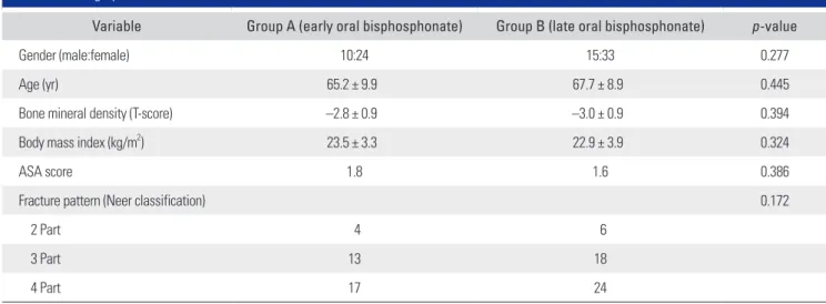
The plate was positioned with the aid of an aiming device 5 to 8 mm distal to the upper end of the greater tuberosity and 2 to 4 mm posterior to the bicipital groove. Five to six locking angularly stable screws were placed into the hu- meral head and three to four standard cortical screws were placed in the shaft. Final intraoperative images were taken to verify correct screw placement, and the range of motion (ROM) was assessed to minimize the risk of any postop- erative impingement (Fig. 1).
The wound was closed over a suction drain, which was removed 24 hours later. Postoperatively, the arm was supported in a sling. Pendular movements were started from the first postoperative day and the shoulder was mo- bilized with active assisted exercises, which was followed by active exercises three weeks later.
Clinical and Radiological Evaluation
The patients were routinely followed at 2, 6, 10, 16 weeks, 6 months, and 1 year after surgery. Standard anteroposte- rior and axillary radiographs were obtained at each visit.
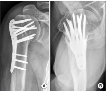
Radiogrpahic union was defined as the presence of bridg- ing of trabeculae or osseous bone in three of four (medial, lateral, anterior, and posterior) cortices (Fig. 2). In each evaluation of the union of proximal humerus fractures, the images were interpreted by two orthopedic surgeons (JSY and JWR). In cases where no agreement could be reached on the union by the two observers, the mean union times were used as a representative value.
A B
Fig. 1. Immediate postoperative radiographs. (A) Anteroposterior view.
(B) Axillary view.
A B
Fig. 2. Radiographs taken at 6 weeks after operation showing union of the proximal humerus fracture. (A) Anteroposterior view to confirm the medial and lateral cortices. (B) Axillary view to confirm the anterior and posterior cortices.
To evaluate loss of fixation, the cervicodiaphyseal angle (formed by the intersection between a line per- pendicular to the anatomical neck and a line parallel to the axis of the humeral diaphysis) was measured on true shoulder anteroposterior view radiographs by two ortho- pedic surgeons (JSY and JWR) immediately postoperative- ly and at the last follow-up (Fig. 3). To minimize interob- server bias, the mean difference of the cervicodiaphyseal angle between immediate postoperative and last follow-up measurements was used as a representative value. Fixation failure was defined as more than 10° of difference of the cervicodiaphyseal angle between immediate postoperative and last follow-up measurements.
Clinical outcomes were assessed at 1 year after sur- gery using the American Shoulder and Elbow Surgeons (ASES) scoring system, Constant score (CS),17) and the ROM of the shoulder. The ROMs of the shoulder assessed were the forward flexion, external rotation, and internal rotation.
Statistical Analysis
The reliability of agreement in determining radiological fracture healing between two observers who were blinded to the treatment was assessed using Kappa coefficients. Kappa coefficients were interpreted as follows: < 0 denoting poor agreement, 0 to 0.2 slight agreement, 0.2 to 0.4 fair agree- ment, 0.4 to 0.6 moderate agreement, 0.6 to 0.8 substantial agreement, and > 0.8 almost perfect agreement.18)
Group results were compared using the Pearson chi-square and the Student t-test for continuous variables.
Correlation analyses were conducted to identify the rela- tionships between the variables. Statistical analyses were performed with use of SPSS ver. 21.0 (IBM Co., Armonk, NY, USA), and p-values of < 0.05 were considered statisti- cally significant.
RESULTS
Radiologic Outcomes
All patients showed evidence of fracture healing. In group A, radiographic fracture union was obtained at six weeks in 28 patients (82%) and at ten weeks in 6 patients (18%).
In group B, radiographic fracture union was achieved at 6 weeks in 38 patients (79%) and at 10 weeks in 10 patients (21%). The average time to radiographic union was simi- lar (6.3 weeks; range, 6 to 10 weeks) between group A and group B (6.6 weeks; range, 6 to 10 weeks) (p = 0.65). There was no significant intergroup difference in the proportion of patients who obtained healing at 6 weeks after surgery (p
= 0.81) (Table 2). Interobserver reliability reached the level of “almost perfect agreement” with a weight kappa coef- ficient of 0.82.
Five patients in group A (14.7%) and 4 patients in group B (8.3%) showed fixation failure with more than 10°
of difference in the cervicodiaphyseal angle measured on radiographs immediately postoperatively and at the last follow-up. The mean difference of the cervicodiaphyseal angle was not significantly different between group A (av- erage, 3.7°; range, 0° to 24°; standard deviation, 6.1°) and group B (average, 2.2°; range, 0° to 17°; standard deviation, 7.3°) (p = 0.238) (Table 2).
Fig. 3. Measurement of the cervicodiaphyseal angle.
Table 2. Radiologic Outcomes
Variable Group A (early oral bisphosphonate) Group B (late oral bisphosphonate) p-value
Time to radiographic union (wk) 6.3 (6–10) 6.6 (6–10) 0.571
Patients with healing at 6 postoperative weeks 28 (82.4) 38 (77.2) 0.160
Difference of cervicodiaphyseal angle (°) 3.7 (0–24) 2.2 (0–17) 0.238
Values are presented as mean (range) or number (%).
Clinical Outcomes
At 1 year after surgery, the ASES score averaged 80.2 points (range, 62 to 92 points) in group A and 78.6 points (range, 58 to 94 points) in group B. No significant differ- ence was observed in the mean ASES score between the two groups (p = 0.61). In addition, the mean CS at 1 year after surgery was 71.6 points (range, 54 to 90 points) in group A and 73.1 points (range, 58 to 92 points) in group B, indicating no significant difference between the groups (p = 0.63). The respective mean values of the ROM of the shoulder were 130.5° (forward flexion), 44.2° (external ro- tation), and the eleventh thoracic level (internal rotation) in group A and 128.4°, 46.2°, and the tenth thoracic level in group B (p = 0.89) (Table 3).
DISCUSSION
In this study, all patients showed evidence of fracture healing without any statistically differences between two groups. Moreover, clinical results based on the ASES score were satisfactory in both groups.
Eriksen et al.19) reported that the administration of zoledronic acid for more than two weeks after surgical treatment for low-energy hip fractures increased hip BMD scores and significantly reduced the risks of subsequent vertebral and hip fractures, while also decreasing mortality.
Dirschl et al.20) reported that the loss of femoral neck BMD in patients with a hip fracture was five times greater than that found in a normal population. They recommended pharmacological or some other forms of intervention dur- ing the first critical year following a hip fracture to prevent accelerated bone loss and reduce the risk of subsequent fractures during this period.
Although the BP therapy reduces the risks of a secondary fracture and mortality,21,22) the optimal tim- ing of BP administration following a proximal humerus
fracture in osteoporotic patients remains unclear. Several animal studies have demonstrated that BPs can delay cal- lus remodeling.11,23) Clinically, Odvina et al.24) observed significantly delayed healing of fractures in patients on long-term alendronate. Rozental et al.25) compared 43 dis- tal radius fracture patients who were taking a BP with 153 controls: the time to union in the BP group was 55 days compared to 49 in the control group. However, these clini- cal studies did not evaluate the effect of BP medication on fracture healing in patients who had no history of taking BPs. In contrast, some authors have reported that BPs promote fracture healing. Amanat et al.26) demonstrated that a single dose of zoledronic acid significantly increased the callus volume and mechanical strength. Fleisch27) re- ported that the callus size was either not influenced or was increased by BPs, and it never decreased due to the slow- ing of callus resorption. Moreover, this resulted in a para- doxical increase in the mechanical strength.
In the present study, there was no delay in the heal- ing of proximal humerus fractures fixed by locking plate fixation. Recently, several authors have reported that the early initiation of BPs does not delay the healing of frac- tures fixed by a plate or by nailing.12,13) One possible ex- planation is the healing mechanism of proximal humerus fractures treated with locking plate fixation. Gong et al.13) noted that fractures treated by means of plate fixation in- volve primary bone healing between fragments rather than bridging due to external callus formation. The difference between cancellous and cortical bone could be another reason. In a fracture of compact long bones, where frac- ture bone debris must be absorbed to allow room for new bone formation,28) a resorption process is critical initially.
However, proximal humerus fractures involve cancellous bone, in which the space for new bone formation is larger than that in compact bones. Therefore, we speculate that Table 3. Clinical Outcomes at 24 Weeks after Operation
Variable Group A (early oral bisphosphonate) Group B (late oral bisphosphonate) p-value
ASES score, mean (range) 80.2 (62–92) 78.6 (58–94) 0.612
Constant score, mean (range) 71.6 (54–90) 73.1 (58–92) 0.631
Range of motion 0.892
Forward flexion (°) 130.5 128.4
External rotation (°) 44.2 46.2
Internal rotation 11th thoracic level 10th thoracic level
ASES: American Shoulder and Elbow Surgeons.
the healing of proximal humerus fractures stabilized by a locking plate may not be suppressed by a reduction in the resorption process by BPs owing to the spacious environ- ment offered by cancellous bone.
Our study had several limitations. First, we defined radiographic fracture union as bridging of trabeculae or osseous bone across fracture lines, but a quantitative as- sessment of this type of union may be necessary by, for instance, measuring the callus size on computed tomog- raphy scans. Second, in order to evaluate the influence of BP administration on fracture healing, it would have been better to compare the effects on the BMD and bone me- tabolism under the same condition. Third, the sample size was not large enough to evaluate the outcomes between two groups. Finally, the duration of follow-up was short;
a longer follow-up is needed to determine the prevalence
of complications and the long-term effects of an early os- teoporosis treatment. Nevertheless, the significance of this this lies in the fact that it attempted to analyze the influ- ence of early BP administration in osteoporotic proximal humerus fractures.
In conclusion, early initiation of BP treatment in patients with an osteoporotic proximal humerus fractures treated by locking compression plate fixation did not affect fracture healing or clinical outcomes, although our sample size was too small to detect a rare complication of non- union.
CONFLICT OF INTEREST
No potential conflict of interest relevant to this article was reported.
REFERENCES
1. Consensus development conference: prophylaxis and treat- ment of osteoporosis. Osteoporos Int. 1991;1(2):114-7.
2. Johnell O, Kanis J. Epidemiology of osteoporotic fractures.
Osteoporos Int. 2005;16 Suppl 2:S3-7.
3. Johnell O, Kanis JA, Oden A, et al. Mortality after osteopo- rotic fractures. Osteoporos Int. 2004;15(1):38-42.
4. Johnell O, Kanis JA. An estimate of the worldwide preva- lence and disability associated with osteoporotic fractures.
Osteoporos Int. 2006;17(12):1726-33.
5. Helmy N, Hintermann B. New trends in the treatment of proximal humerus fractures. Clin Orthop Relat Res. 2006;
442:100-8.
6. Baron JA, Barrett JA, Karagas MR. The epidemiology of pe- ripheral fractures. Bone. 1996;18(3 Suppl):209S-213S.
7. Silverman S, Christiansen C. Individualizing osteoporosis therapy. Osteoporos Int. 2012;23(3):797-809.
8. Reginster JY, Burlet N. Osteoporosis: a still increasing prev- alence. Bone. 2006;38(2 Suppl 1):S4-9.
9. Osugi K, Miwa S, Marukawa S, Marukawa K, Kawaguchi Y, Nakato S. Diaphyseal femoral fatigue fracture associated with bisphosphonate therapy: 3 more cases. Acta Orthop.
2011;82(1):112-3.
10. Li C, Mori S, Li J, et al. Long-term effect of incadronate disodium (YM-175) on fracture healing of femoral shaft in growing rats. J Bone Miner Res. 2001;16(3):429-36.
11. Cao Y, Mori S, Mashiba T, et al. Raloxifene, estrogen, and alendronate affect the processes of fracture repair differently
in ovariectomized rats. J Bone Miner Res. 2002;17(12):2237- 46.
12. Kim TY, Ha YC, Kang BJ, Lee YK, Koo KH. Does early administration of bisphosphonate affect fracture healing in patients with intertrochanteric fractures? J Bone Joint Surg Br. 2012;94(7):956-60.
13. Gong HS, Song CH, Lee YH, Rhee SH, Lee HJ, Baek GH.
Early initiation of bisphosphonate does not affect healing and outcomes of volar plate fixation of osteoporotic distal radial fractures. J Bone Joint Surg Am. 2012;94(19):1729-36.
14. Li YT, Cai HF, Zhang ZL. Timing of the initiation of bisphosphonates after surgery for fracture healing: a sys- tematic review and meta-analysis of randomized controlled trials. Osteoporos Int. 2015;26(2):431-41.
15. Neer CS 2nd. Displaced proximal humeral fractures: I. classifi- cation and evaluation. J Bone Joint Surg Am. 1970;52(6):1077- 89.
16. Neer CS 2nd. Displaced proximal humeral fractures: II.
treatment of three-part and four-part displacement. J Bone Joint Surg Am. 1970;52(6):1090-103.
17. Constant CR, Gerber C, Emery RJ, Sojbjerg JO, Gohlke F, Boileau P. A review of the Constant score: modifications and guidelines for its use. J Shoulder Elbow Surg. 2008;17(2):355- 61.
18. Landis JR, Koch GG. The measurement of observer agree- ment for categorical data. Biometrics. 1977;33(1):159-74.
19. Eriksen EF, Lyles KW, Colon-Emeric CS, et al. Antifracture efficacy and reduction of mortality in relation to timing of
the first dose of zoledronic acid after hip fracture. J Bone Miner Res. 2009;24(7):1308-13.
20. Dirschl DR, Henderson RC, Oakley WC. Accelerated bone mineral loss following a hip fracture: a prospective longitu- dinal study. Bone. 1997;21(1):79-82.
21. Beaupre LA, Morrish DW, Hanley DA, et al. Oral bisphos- phonates are associated with reduced mortality after hip fracture. Osteoporos Int. 2011;22(3):983-91.
22. Harrington JT, Ste-Marie LG, Brandi ML, et al. Risedro- nate rapidly reduces the risk for nonvertebral fractures in women with postmenopausal osteoporosis. Calcif Tissue Int. 2004;74(2):129-35.
23. Li J, Mori S, Kaji Y, Mashiba T, Kawanishi J, Norimatsu H.
Effect of bisphosphonate (incadronate) on fracture healing of long bones in rats. J Bone Miner Res. 1999;14(6):969-79.
24. Odvina CV, Zerwekh JE, Rao DS, Maalouf N, Gottschalk FA, Pak CY. Severely suppressed bone turnover: a potential
complication of alendronate therapy. J Clin Endocrinol Metab. 2005;90(3):1294-301.
25. Rozental TD, Vazquez MA, Chacko AT, Ayogu N, Bouxsein ML. Comparison of radiographic fracture healing in the distal radius for patients on and off bisphosphonate therapy.
J Hand Surg Am. 2009;34(4):595-602.
26. Amanat N, McDonald M, Godfrey C, Bilston L, Little D.
Optimal timing of a single dose of zoledronic acid to in- crease strength in rat fracture repair. J Bone Miner Res.
2007;22(6):867-76.
27. Fleisch H. Can bisphosphonates be given to patients with fractures? J Bone Miner Res. 2001;16(3):437-40.
28. Bindra RP. Basic pathology of the hand, wrist, and forearm:
bone and joint. In: Berger RA, Weiss AP, eds. Hand surgery.
Philadelphia, PA: Lippincott Williams & Wilkins; 2004.
1-21.