분절성 쇄골 골절의 수술적 치료와 비수술적 치료 - 증례 보고 -
하성식ㆍ홍기도ㆍ심재천ㆍ서이락ㆍ남태석
삼육서울병원 정형외과
The Different Treatment Methods for Segmental Fractures of the Clavicle
- Cases Report -
Sung-Sik Ha, M.D., Ki-Do Hong, M.D., Jae-Cheon Sim, M.D. Yi-Rak Seo, M.D., Tae-Seok Nam, M.D., Ph.D.
Department of Orthopedic Surgery, Sahmyook Medical Center, Seoul, Korea
Received January 16, 2017 Revised April 9, 2017 Accepted June 13, 2017 Correspondence to:
Tae-Seok Nam, M.D., Ph.D.
Department of Orthopedic Surgery, Sahmyook Medical Center, 82 Mangu- ro, Dongdaemun-gu, Seoul 02500, Korea
Tel: +82-2-2210-3580 Fax: +82-2-2210-2673 E-mail: [email protected] Financial support: None.
Conflict of interests: None.
Segmental fractures of the clavicle are very rare. Therefore, to date, there has not been a clear, stan- dardized method of management of segmental clavicle fractures. Herein, two patients with a segmen- tal fracture are described: One patient was treated conservatively, while another patient was treated operatively. Both patients showed excellent results. We discuss the various management options with a literature review.
Key Words: Clavicle, Conservative treatment, Treatment
Copyright © 2017 The Korean Fracture Society. All rights reserved.
This is an Open Access article distributed under the terms of the Creative Commons Attribution Non-Commercial License (http://creativecommons.org/licenses/by-nc/4.0) which permits unrestricted non-commercial use, distribution, and reproduction in any medium, provided the original work is properly cited.
Among clavicle fractures, 69% occur in the diaphysis, 28% in the distal and 3% in the proximal position.1) Seg- mental fractures involving the distal and the proximal ends of the clavicle are even rarer. The injury mechanism is often unclear and the management of these fractures remains controversial. We report two cases with a segmental frac- ture are described: One patient was treated conservatively and another patient was treated operatively and both of all,
excellent results were achieved. The management of seg- mental clavicle injuries is discussed.
Case Reports
1. Case 1
A 40-year-old man presented to the outpatient clinic
with pain over the right clavicle following a history of fall from height. There was no external wound. Physical ex- amination revealed tenderness on proximal and distal por- tion of the clavicle. Movements of the shoulder joint were restricted due to referred pain in the clavicle. There was no evidence of neurovascular deficit and no other significant combined injuries.
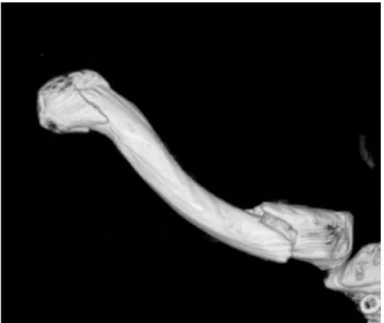
Radiographs showed a segmental fracture clavicle with lateral and medial end of clavicle (Fig. 1). Computed to- mography showed segmental proximal and distal fracture with displacement, especially proximal end (Fig. 2).
Initially, we planned operative treatment for the fracture.
But he refused operative treatment under anesthesia. So, we changed the initial plan. Within 2 weeks after trauma, if the degree of displacement is more progress than initial radiography, we plan to perform operative treatment. He was applied figure-eight-bandage to prevent shortening of clavicle for 8 weeks. We checked the radiography of the clavicle weekly. Fortunately, the degree of displacement was not more progress than initial radiography. He removed the brace at the time of 8 weeks after trauma. After 3 months later, he was able to move full range of motion. At the last follow-up (3 years after trauma), we checked the radiogra- phy (Fig. 3).
2. Case 2
A 62-year-old male, right hand dominant, presented to the emergency department with pain over the right clavicle following a history of bicycle accident. There was no exter- nal wound. The skin was not at risk.
Physical examination revealed tenderness on right mid shaft and distal portion of the clavicle. Movements of the shoulder joint were restricted due to pain. There was no evidence of neurovascular deficit and no other significant combined injuries.
Radiographs showed a segmental clavicle fracture with distal end and mid-shaft of clavicle (Fig. 4). Computed to- mography showed mid-shaft and distal fracture with dis- placement, especially distal end (Fig. 5).
A
B
Fig. 1. Both clavicle anteroposterior (A) and lordotic view (B) radiogra- phies showed segmental fracture of the clavicle (arrows).
Fig. 2. Computed tomography showed three-dimensional reconstruc- tion of the right clavicle.
A
B
Fig. 3. At 3 years after trauma, both clavicle anteroposterior (A) and lordotic view (B) radiographies showed complete union of the right clavicle.
We planned operative treatment for the fracture. Op- eration was performed under general anesthesia in a beach chair position. Closed reduction and percutaneous pinning with a Steinmann pin of mid-clavicle fracture was done.
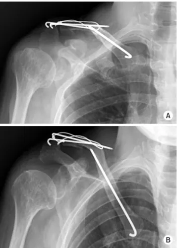
Open reduction with pinning and wiring of distal clavicle was done. Post-operative radiograph showed the final fixa- tion achieved (Fig. 6).
Postoperatively, the arm was supported in an arm-sling, with the patient instructed to perform regular hand grasping and elbow range of motion exercises, as well as gentle pen- dulum exercises of the shoulder. After then, passive stretch- ing exercise and strengthening exercise were followed. The patient had pain-free full range of movement of shoulder joint after 12 weeks.
Discussion
Clavicle fractures are common, comprising 2.6% of all adult fractures. Midshaft clavicular fractures are the most common, ranging from 69% and 82%, distal fractures comprise 21% to 28%, and proximal fractures occur be- tween 2% and 3%.1) The segmental clavicle fracture is an uncommon pattern. One study reported that 0.8% of clavi- cle fractures was segmental.2)
A
B
Fig. 4. Right clavicle anteroposterior (A) and lordotic view (B) radiogra- phies showed a segmental fracture of the clavicle (arrows).
Fig. 5. Computed tomography showed a three-dimensional recon- struction image of the right clavicle.
A
B Fig. 6. At 7 months after trauma, Right clavicle anteroposterior (A) and lordotic view (B) radiographies were shown.
Clavicle fractures are usually caused by a direct trauma to the shoulder, but the mechanism of injury leading to a segmental clavicle fracture is not well understood. Some reports described that this type fracture occur due to two separate concurrent forces.3) One is direct force on the point of the shoulder girdle due to fall. The other is an indirect force, which may give rise to clavicle fractures by the shear- ing force delivered from the humerus to the sternum.
Fractures of the proximal clavicle are difficult to visualize because overlying structures in the chest obscure the view of the proximal end. For rare proximal fractures, particularly those that extend into the sternoclavicular joint, a Hobb’s view or a serendipity view or computed tomography scan may be helpful to access the fractures and identify their relationship to the sternoclavicular joint. Recently, one re- ported a case of neglected segmental clavicle fracture.4) Ac- cording to the report, initially operation was performed on distal end of clavicle using hook plate. After operation, pa- tient had ongoing pain over the medial clavicular region. He underwent operative treatment for medial clavicle fracture on 6 weeks later after initial injury. We recommend that physician examine carefully including tenderness on ipsilat- eral sternoclavicular joint for the patient of clavicle fracture.
This simple step is very helpful for management of clavicular fracture and avoidance of misdiagnosis.
The management of segmental fractures has little stan- dard guidance in the literature, with some reports of suc- cessful operative3,5) and non-operative management.6) Miller et al.5) presented a report of fracture of the medial and lateral extremity of the clavicle with the use of a reconstruction plate and another with locking T-plate. Authors emphasized that due to the rarity of the case, there is no routine surgical technique described for the treatment of this type of fracture.
Recently reported cases were summarized (Table 1).4,6-10) According to previous reports, operative treatment over- weighted than conservative treatment. Operative technique was plate and screw on proximal lesion in all cases and K- wire fixation on or hook plate on distal lesion.
In this report, we compared two possible methods, one operatively and one conservatively treatment, for the rare
fracture pattern of clavicle. And the results showed excel- Table
1. Summary of Treatment for Segmental Clavicle Fracture Study (year)
Patient demogra- phyCause of injuryTreatmentClassificationOperative technique Remarks Age (yr)SexProximalDistal Pang et al.(2003)6) 19MaleFell off a pull-up barNon-operationBipolar--- 76MaleMotorcycle accidentNon-operationBipolar--- Sethi et al.(2012)8) 70FemaleFall down a flight of seven stairsNon-operationBipolar--- Grossi (2015)7)41MaleFell from a roofOperationSegmentalPlatingK-wire- Marjoram and Chakrabarti(2015)9)40MaleFell from a motorcycleOperationSegmentalPlatingHook plate- Varelas et al.(2015)10)68FemaleSlipped on iceOperationBipolarPlate & screwPlate & screw- Yalizis et al.(2016)4) 38MaleFell from a bikeOperationBipolarPlatingHook plateLate diagnosis for medial le- sion (2 stage operation) This report40MaleFell from a heightNon-operationBipolar--- 62MaleBike accidentOperationSegmentalK-wireTension band wiring-
lent outcomes for both treatments. The treatment should be based on the individual fracture pattern and patient charac- teristics.
요 약
분절성 쇄골 골절은 매우 드물며, 그 치료 방법에 대해 명 확히 정립되어 있지 않다. 분절성 쇄골 골절을 가진 환자에서 각각 보존적 치료를 시행한 예와 수술적 치료를 시행한 예를 통해 좋은 결과를 얻었던 2예를 보고자 한다. 또한 문헌 고찰 을 통해 유사한 경우의 치료에 대해 논하고자 한다.
색인 단어:
쇄골, 보존적 치료, 치료ORCID
하성식, http://orcid.org/0000-0002-8138-9489 홍기도, http://orcid.org/0000-0002-0861-6868 심재천, http://orcid.org/0000-0002-3451-4961 서이락, http://orcid.org/0000-0002-8683-4844 남태석, http://orcid.org/0000-0003-0589-5790
References
1. Robinson CM: Fractures of the clavicle in the adult. Epidemiol- ogy and classification. J Bone Joint Surg Br, 80: 476-484, 1998.
2. Jupiter JB, Leffert RD: Non-union of the clavicle. Associated complications and surgical management. J Bone Joint Surg Am, 69: 753-760, 1987.
3. Heywood R, Clasper J: An unusual case of segmental clavicle fracture. J R Army Med Corps, 151: 93-94, 2005.
4. Yalizis MA, Hoy GA, Ek ET: A rare case of bipolar clavicle fracture. Case Rep Orthop, 2016: 4309828, 2016.
5. Miller D, Smith KD, McClelland D: Bipolar segmental clavicle fracture. Eur J Orthop Surg Traumatol, 19: 337-339, 2009.
6. Pang KP, Yung SW, Lee TS, Pang CE: Bipolar clavicular injury.
Med J Malaysia, 58: 621-624, 2003.
7. Grossi EA. Segmental clavicle fracture. Rev Bras Ortop, 46:
733-735, 2015.
8. Sethi K, Newman SD, Bhattacharya R. An unusual case of bi- polar segmental clavicle fracture. Orthop Rev (Pavia), 4: e26, 2012.
9. Marjoram TP, Chakrabarti A. Segmental clavicle fracture and acromio-clavicular joint disruption: an unusual case report.
Shoulder Elbow, 7: 187-9, 2015.
10. Varelas N, Joosse P, Zermatten P. Operative Treatment of an atypical segmental bipolar fracture of the clavicle. Arch Trauma Res, 4: e29923, 2015.