- 27 -
Anti-obese effects of mulberry ( Morus alba L.) root bark through the inhibition of digestive enzymes and 3T3-L1 adipocyte differentiation
Yong-Xiang Wu1, You-Jeong Kim, Sha Li1, Myung-Chul Yun1, Jin-Mi Yoon1, Jin-Young Kim1, Sung-Il Cho1, Kun-Ho Son2, Taewan Kim1*
1
Department of Food Science and Biotechnology, Andong National University, Andong 760-749, Korea
2
Department of Food and Nutrition, Andong National University, Andong 760-749, Korea
소화효소 저해 및 지방세포 분화 억제활성에 의한 상백피의 항비만 효능
우용시앙1․김유정1․리샤1․윤명철1․윤진미1․김진영1․조성일1․손건호2․김태완1*
1안동대학교 식품생명공학과, 2안동대학교 식품영양학과
Abstract
Anti-obese effects of mulberry (Morus alba L.) root bark was investigated in vitro by measuring its inhibitory effect against 3T3-L1 preadipocyte differentiation and digestive enzymes such as α-amylase, α-glucosidase and pancreatic lipase. Ethanol extract of mulberry root bark (MRE) showed the potent inhibitory activities on α-amylase, α-glucosidase and pancreatic lipase with IC50 values of 7.86±0.36, 0.12±0.03 and 7.93±0.11 mg/mL, respectively.
Furthermore, MRE significantly suppressed cellular lipid accumulation in 3T3-L1 cells in a dose-dependent manner.
To elucidate the mechanism of MRE, we performed qRT-PCR and Western blotting for the expression of genes related with adipogenesis and lipogenesis. Treatment of MRE markedly suppressed the protein expression of PPARγ, C/EBPα and SREBP-1c, as well as FAS and ACC, which are the key transcription factors and metabolic enzymes in adipogenesis and lipogenesis. In addition, qRT-PCR analysis indicated that the anti-adipogenesis effect of MRE might be due to its inhibition at transcription levels. These results demonstrate that MRE can effectively suppress adipocyte differentiation and inhibit key enzymes related to obesity. Our findings suggest that mulberry root bark may have a potential benefit in preventing obesity.
Key words:mulberry root bark, adipogenesis, 3T3-L1 preadipocyte, obesity
Introduction
1)
Obesity has become a serious global health problem in recent decades. Obesity can lead to many chronic diseases such as dyslipidemia, type 2 diabetes mellitus (T2DM), cardiovascular diseases, cancer and hypertension (1-5).
Among them, T2DM is the most closely associated with obesity and they seem to share common causative factors,
*Corresponding author. E-mail:[email protected] Phone:82-54-820-6157, Fax:82-54-820-6264
Received 2 December 2014; Revised 23 December 2014;
Accepted 27 January 2015.
Copyright ⓒ The Korean Society of Food Preservation. All rights reserved.
chemical abnormalities and clinical complications (6).
Obesity is mainly caused by excessive food calorie intake and an abnormal accumulation of body fat (7). Consumption of a high-fat diet is a major risk factor that increases the chances of developing obesity and T2DM. Inhibition of digestive enzymes such as α-amylase, α-glucosidase and pancreatic lipase can decrease carbohydrate digestion and retard fat absorption, and therefore may be one of the most effective approaches to controlling obesity and related metabolic disorders.
Adipocyte, also known as fat cell, is associated with lipid homeostasis and energy balance (8). Therefore it has been widely used as an effectivein vitromodel for the evaluation of adipocyte differentiation. Adipocyte differentiation is the
process by which undifferentiated preadipocytes are concerted to differentiated adipocyte. Adipocyte differentiation is regulated by two key transcription factors, peroxisome proliferator-activated receptor γ (PPARγ) and CCATT/
enhancer binding protein α (C/EBPα) (9). C/EBPα and PPARγ are activated during the differentiation process and promote together the expression of downstream adipose specific genes involved in adipose phenotype as well as in glucose and lipid metabolism (10,11). Sterol regulatory element binding protein-1c (SREBP-1c) is a well-known upstream regulator of PPARγ in the adipogenesis pathway (12). Moreover, SREBP-1c could regulate the genes involved in lipogenesis such as fatty acid synthase (FAS) and acetyl-CoA carboxylase (ACC) (13). Thus, controlling adipocyte differentiation has become a major target to prevent and treat obesity and related metabolic disorders.
The medicinal use of the root bark of mulberry has been documented in the Pharmacopoeia of People’s Republic of China and British Herbal Pharmacopoeia for a long history.
Recently, it has been primarily used as antioxidant (14,15), antiasthmatic (16), antidepressant (17) and skin whitening ingredients (14). Besides, it was reported to have anti- inflammatory and antiviral effects in vitro (18,19) and a hypolipidemic activity in vivo (15,20,21). Moreover, plants of this genus were known to be rich in flavonoids and prenylated flavonoids. The prenylated flavonoids such as morusin, kuwanon C, sanggenon D and kuwanon G isolated from mulberry root bark were reported to possess antioxidant, antimicrobial and anti-inflammatory activities (14,22,23).
Recently, the phenolic compounds including maclurin, rutin, isoquercitrin, resveratrol and morin isolated from mulberry root bark showed potential antioxidant and whitening activities (24). It was reported that moracin M has a hypoglycemic effect on alloxan-diabetic mice (21). Additionally, morusalbanol A, a Diels-Alder type adduct with a new carbon skeleton, significantly attenuated the H2O2-induced cell damage and could be used as an euro-protective agent (25).
However, to date, there have been no previous studies on the anti-adipogenic activity of mulberry root bark using 3T3-L1 cells and on the investigation of its molecular mechanism.
This study was aimed to investigate the inhibitory effects of mulberry root bark on digestive enzymes and 3T3-L1 preadipocytes differentiation. MRE was further evaluated for the potential mechanism underlying the regulation of cell differentiation, in which the gene expression patterns involved in adipogenesis and lipogenesis of 3T3-L1 adipocyte cells were analyzed.
Materials and methods
Materials
3T3-L1 cells were purchased from American Type Culture Collection (ATCC). Dulbecco’s modified Eagle’s medium (DMEM), fetal bovine serum (FBS), calf bovine serum (Calf), phosphate-buffered saline (PBS, pH 7.4) and other tissue culture reagents were purchased from PAA Laboratories GmbH (Pasching, Austria). Type II crude porcine pancreatic lipase, p-nitrophenyl palmitate (PNP), α-amylase from porcine pancreas Type VI-B, α-glucosidase from saccharomyces cerevisiae, ρ-nitrophenyl-α-D-glucopyranoside (ρNPG) and all other chemicals were bought from Sigma-Aldrich Co (St.
Louis, MO, USA). Antibodies for target proteins (PPARγ, C/EBPα, SREBP-1c, FAS, ACC and β-actin) were acquired from Cell Signaling Technology (Beverly, MA, USA). For qRT-PCR test, the TRlzol Reagent was obtained from Invitrogen (Carlsbad, CA), PrimerScriptTM1st Strand cDNA Synthesis Kit from TaKaRa Bio Inc. (Otsu, Japan) and SYBR Green from PhileKorea Technology (Daejeon, Korea).
Preparation of mulberry root bark extracts Dried root barks of mulberry were purchased from Omni Herb Company and extracted with 70% ethanol (MRE) and hot water (MRW). The extracts were filtered and evaporated under reduced pressure using a vacuum rotary evaporator.
The extracts were then freeze-dried and stored at -70℃ until used. The extraction yield obtained from MRE and MRW were 16.71% and 17.23% (w/w), respectively.
Digestive enzymes inhibitory assays
The α-amylase inhibitory activities of samples (MRE and MRW) were measured according to the method described by Ali et al. (26) and their α-glucosidase inhibitory activities were determined based on the method of Kim et al. (27).
Pancreatic lipase inhibitory activities were determined according to the method of Kim et al. (28). In this study, acarbose and orlistat were used as positive controls. The percent inhibition of digestive enzymes was calculated as follows: Inhibition (%)=[(ODcontrol-ODsample)/ODcontrol]×100.
The IC50values were calculate by linear regression analysis.
Cell culture and adipocyte differentiation 3T3-L1 cells were cultured in T-flask with Dulbecco’s modified Eagle’s medium (DMEM) containing 10% Calf and 10 μg/mL of penicillin/streptomycin at 37℃ in 5% CO2
atmosphere. Then, cells were subcultured after cells grown
to more than 80-90% confluence. Two days after confluence (day 0), cells were stimulated to differentiate with differentiation medium containing DMEM with 10% FBS, 0.5 mM 3-isobutyl-1-methyl-xanthine, 5 μg/mL insulin and 1 μM dexamethasone for 2 days (day 2). Cells were then maintained in DMEM supplemented with 10% FBS and 5 μg/mL insulin for another 2 days (day 4), followed by culturing with DMEM with 10% FBS for an additional 4 days (day 8). Test samples were added in medium at various concentrations throughout the whole culture period (day 0~8).
Cell viability assay
The cytotoxicity of the sample was measured using MTT (3-(4,5-dimethylthiazol-2-yl)-2,5-diphenyltetrazolium bromide) method (29). Briefly, the cells were seeded at 104cells/well onto flat bottomed 96-well culture plates and treated with test samples at various concentrations. After incubation for 48 hr, the cells were incubated with MTT solution (5 mg/mL) at 37℃ for 3 hr. The MTT-containing media was removed and dimethyl sulfoxide was added to dissolve formazan precipitates, then the optical density was measured at 570 nm using a microplate reader (Molecular Devices, Sunnyvale, CA, USA).
Oil red O staining
Cells were washed three times with phosphate buffered saline (PBS) and fixed for 30 min with 4% formaldehyde.
The fixed cells were washed three times with distilled water.
Oil red O (0.5% in 60% isopropanol) was diluted with water (3:2), filtered through Whatman paper and incubated with the fixed cells for 1 hr at room temperature. Plates were rinsed three times with distilled water and the stained fat droplets in the adipocytes were visualized by inverted microscopy and photographed. Spectrophotometric analysis of the stain was performed by dissolving the stained lipid droplets with isopropanol and measuring at 520 nm.
Western blotting analysis
Extracted and quantified proteins (30 μg) from 3T3-L1 cells were separated with 10% SDS-PAGE gels and transferred onto PVDF membranes. The membranes were blocked in 5% skim milk containing TBST (Tris-buffered saline/Tween) for 3 hr. Then, the membranes were probed overnight with primary antibodies (PPARγ, C/EBPα, SREBP-1c, FAS and ACC). After being washed with TBST buffer, membranes were incubated with secondary antibodies for 1 hr. The expression levels of PPARγ, C/EBPα,
SREBP-1c, FAS and ACC were determined using Supersignal West Pico chemiluminescence detection reagents (Pierce Biotechnology, Rockford, IL) and Konica X-ray film (Konica Co., Tokyo, Japan). Incubation with monoclonal mouse β -actin antibody was performed for comparative control.
RNA extraction and qRT-PCR
Confluent cultures of 3T3-L1 cells in 60 mm dish were induced as previously described. Total RNA was isolated using TRlzol Reagent (Invitrogen, Carlsbad, CA) according to the manufacturer’s protocol. After cDNA was prepared from isolated RNA by using PrimerScriptTM1st Strand cDNA Synthesis Kit (TaKaRa), the cDNA was used as a template for real time-PCR with the illumine ECOTM qPCR system (PhileKorea Technology, Daejeon, Korea). The specific primers are given in Table 1. The mRNA level was normalized using β-actin as an internal control. Analysis was carried out in triplicates.
Table 1. Primers used for quantitative Real time-PCR mRNA Forward (F); Reverse (R) primers PPARγ F: 5’-TCG CTG ATG CAC TGC CTA TG-3’
R: 5’-GAG AGG TCC ACA GAG CTG ATT-3’
C/EBPα F: 5’-CCT TCA ACG ACG AGT TCC TG-3’
R: 5’-TGG CCT TCT CCT GCT GTC-3’
SREBP-1c F: 5’-GGA GAC ATC GCA AAC AAG CTG A-3’
R: 5’-CAG ACT GCA GGC CAG ATC CA-3’
FAS F: 5’-ATC CTG GAA CGA GAA CAC GAT CT-3’
R: 5’-AGA GAC GTG TCA CTC CTG GAC CT-3’
ACC F: 5’-GAG TGA CTG CCG AAA CAT CTC TG-3’
R: 5’-GCC TCT TCC TGA CAA ACG AGT-3’
β-actin F: 5’-GTT GGA CCT GAC AGA CTA CCT CA-3’
R: 5’-GTT GCC AAT AGT GAT GAC CT-3’
Statistical analysis
All values were expressed in terms of mean±standard deviation (SD) of at least three independent experiments, tested by analysis of variance (ANOVA) with Duncan’s multiple range tests. Differences were considered to be statistically significant when the p-value was less than 0.05.
Results and Discussion
Effects of mulberry root bark extracts on α-amylase and α-glucosidase activities
α-amylase and α-glucosidase, the two important starch- hydrolyzing enzymes, are essential during digestion of carbohydrate. Inhibitors of these enzymes can decrease
carbohydrate digestion and increase overall carbohydrate digestion time, causing a reduction in the rate of glucose absorption and consequently blunting the postprandial plasma glucose rise (30,31). In addition, inhibition of these enzymes may reduce excessive food calorie intake and retard fat accumulation. Therefore, development of α-amylase and α -glucosidase inhibitors may be one of the most effective approaches to controlling obesity and T2DM. As shown in Table 2, MRE showed dose-dependent inhibition on α -amylase activity with IC50value of 7.86±0.36 mg/mL. MRW also exhibited some α-amylase inhibitory activity with IC50
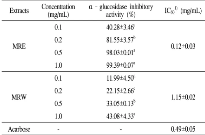
value of 20.10±0.86 mg/mL. Acarbose was used as a positive control in this study, with IC50 value of 0.09 mg/mL. In addition, both MRE and MRW had appreciable inhibitory activity against α-glucosidase with IC50 values of 0.12±0.03 mg/mL and 1.15±0.02 mg/mL, respectively (Table 3).
Especially, MRE inhibited α-glucosidase activity much stronger than positive control acarbose (IC50 value was 0.49±0.05 mg/mL). These results indicate that mulberry root bark extracts could be utilized as natural sources for potential anti-obesity and anti-diabetic application through inhibition of α-amylase and α-glucosidase activities.
Effect of mulberry root bark extracts on pancreatic lipase activity
Pancreatic lipase, also known as pancreatic triacylglycerol lipase, is very important in hydrolysis of triacyglycerols (32).
Pancreatic lipase inhibition is a valuable pathway to retard fat absorption and therefore attenuates obesity (33). The pancreatic lipase inhibitory activity of ethanol and water extracts from mulberry root bark is shown in Table 4. MRE Table 2. Effect of mulberry root bark extracts on α-amylase activity
Extracts Concentration
(mg/mL) α–amylase inhibitory
activity (%) IC501)mg/mL)
MRE
1 12.80±3.92d
7.86±0.36
2 21.34±0.87c
5 39.65±5.07b
10 58.85±0.89a
MRW
1 ND2)
20.10±0.86
2 0.50±1.65c
5 13.60±4.95b
10 22.29±2.36a
Acarbose - - 0.09±0.01
1)IC50 : Concentration required for 50% inhibition of α-amylase activity.
2)ND : Not detected.
Table 3. Effect of mulberry root bark extracts on α-glucosidase activity
Extracts Concentration
(mg/mL) α–glucosidase inhibitory
activity (%) IC501)(mg/mL)
MRE
0.1 40.28±3.46c
0.12±0.03 0.2 81.55±3.57b
0.5 98.03±0.01a 1.0 99.39±0.07a
MRW
0.1 11.99±4.50d
1.15±0.02 0.2 22.15±2.66c
0.5 33.05±0.13b 1.0 43.08±4.33a
Acarbose - - 0.49±0.05
1)IC50 : Concentration required for 50% inhibition of α-glucosidase activity.
exhibited pancreatic lipase inhibitory activity, with IC50 value of 7.93±0.11 mg/mL. And the IC50 value of MRW was 75.13±0.81 mg/mL. The reference compound of orlistat was used in this study, with IC50value of 0.01 mg/mL. Several studies also suggest that the extract of plants such as tea, soybean and ginseng inhibit pancreatic lipase activity (31,33).
These results demonstrate that mulberry root bark has potential as a pancreatic lipase inhibitor.
Table 4. Effect of mulberry root bark extracts on pancreatic lipase activity
Extracts Concentration
(mg/mL) Lipase inhibitory
activity (%) IC501)(mg/mL)
MRE
1 22.95±0.99d
7.93±0.11
5 35.63±4.32c
10 57.72±1.30b 20 68.16±2.01a
MRW
1 ND2)
75.13±0.81
5 ND
10 5.54±3.33
20 10.76±3.39
Acarbose - - 0.01
1)IC50 : Concentration required for 50% inhibition of pancreatic lipase activity.
2)ND : Not detected.
Effect of mulberry root bark extracts on adipogenic differentiation in 3T3-L1 cells
Many current understandings of adipogenesis are based on 3T3-L1 cells. 3T3-L1 preadipocyte cells have been served as a well-established in vitro model to search for new health benefit food or agents for the obesity and related metabolic
(A)
Ce ll v iab ilit y ( % of co nt ro l)
0 20 40 60 80 100 120
MRW MRE (mg/mL) CON 50 100 200 400 50 100 200 400
(B)
Re lat ive fa t a cc um ula tio n ( % of M DI )
0 20 40 60 80 100 120
a
d
CON MDI 50 100 200 50 100 200
MRW MRE (mg/mL)
ab ab ab a
b
c
(C)
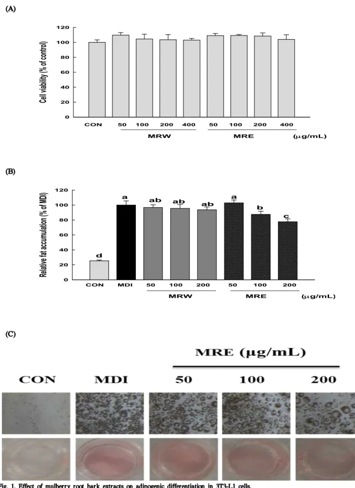
Fig. 1. Effect of mulberry root bark extracts on adipogenic differentiation in 3T3-L1 cells.
(A) Cell viability of 3T3-L1 cells exposed to increasing concentration of MRW and MRE. Cells were treated with different concentration of MRW and MRE for 48 h and cell viability was analyzed using an MTT assay. (B) 3T3-L1 preadipocyte were induced to differentiate into adipocytes by the MDI [0.5 mM 3-isobytyl-1 -methylxanthine, 1 μM dexamethasone, and 5 μg/mL insulin]. Following 8-d differentiation, differentiated adipocytes were fixed and stained with Oil Red O in order to visualize lipid droplets.
(C) Lipid accumulation was quantified by measuring the absorbance at 520 nm. Data are expressed in terms of mean±standard deviation (SD) of at least three independent experiments, tested by analysis of variance (ANOVA) with Duncan’s multiple range tests. Differences were considered statistically different at p<0.05.
disorders in numerous studies (34). In this study, first of all, the effects of mulberry root bark extracts (MRE and MRW) on cell viability and the differentiation of 3T3-L1 cells were evaluated by MTT assay and Oil Red O staining assay. As evidenced by the MTT assay, no significant cytotoxicity was observed at concentrations up to 400 μg/mL, as compared with the non-treated control (Fig. 1A). Treatment of 3T3-L1 cells with MRE decreased adipocyte differentiation, in a dose-dependent manner, as indicated by a decrease in Oil
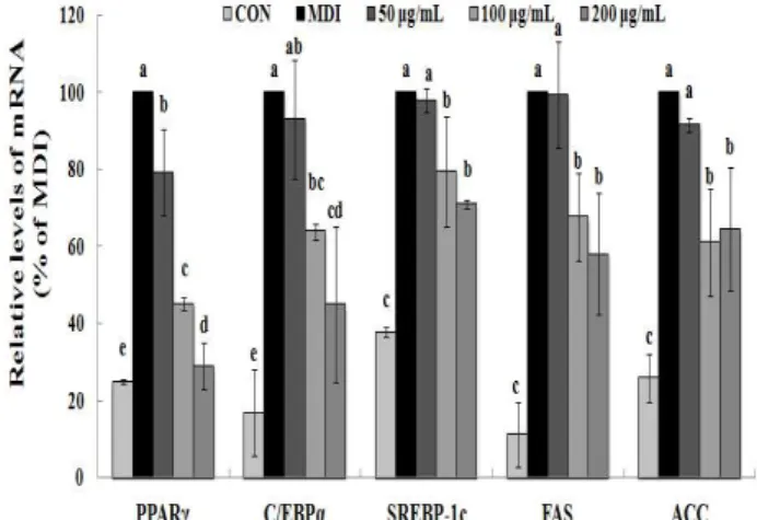
Fig. 2. Inhibitory effects of MRE on adipogenesis and lipogenesis mRNA expression in 3T3-L1 cells.
Total RNA was extracted and cDNA was prepared. Equivalent amounts of cDNA were amplified using primers specific for PPARγ, C/EBPα, SREBP-1c, FAS, ACC and β-actin.
Data are expressed in terms of mean±standard deviation (SD) of at least three independent experiments, tested by analysis of variance (ANOVA) with Duncan’s multiple range tests. Differences were considered statistically different at p<0.05.
Red O incorporation (Fig. 1C). The lipid accumulations were significantly decreased by 12.52% and 22.41% (p<0.05) at 100 μg/mL and 200 μg/mL of MRE, respectively (Fig. 1B).
However, the inhibition of relative fat accumulation was not detectable in MRW. Many plant extracts and their derivatives showed the potent inhibitory activities on adipocyte differentiation and have been used as anti-obesity dietary supplements (35). These results indicate that mulberry root bark extract can efficiently block adipocyte differentiation and may have a potential benefit in preventing obesity.
Effects of MRE on adipogenesis and lipogenesis gene expressions in 3T3-L1 cells
To gain a better understanding of the molecular mechanisms underlying the anti-adipogenic effect, we conducted qRT-PCR and Western blotting analysis to examine the effect of MRE on the expression of key transcriptional factors.
PPARγ and C/EBPα are necessary and should be sufficient for adipogenesis in vivo and in vitro (10,11). We observed
that PPARγ and C/EBPα were strongly inhibited by MRE at the transcriptional levels (Fig. 2). The mRNA level of PPARγ was reduced up to 70.97% and that of C/EBPα mRNA was decreased by up to 55.09%, respectively. Consistent with the mRNA expression, the protein expression levels of transcription factors (PPARγ and C/EBPα ) showed a similar alteration pattern (p<0.05, Fig. 3). We also determined the gene expression of SREBP-1c, a key transcription regulator which is well-known upstream regulators of PPARγ in the adipogenesis pathway (12). It showed that the SREBP-1c mRNA was suppressed significantly by MRE (Fig. 2).
Furthermore, treatment of MRE induced down-regulation of SREBP-1c protein expression (p<0.05, Fig. 3). These results indicate that MRE suppresses SREBP-1c as the upstream regulator of PPARγ at the transcriptional level. However, the deep investigation of molecular mechanism of MRE on PPARγ upstream signaling pathway need to be studied further. Consequently, these observations suggest that MRE inhibits aipogenesis during adipocyte differentiation.
FAS and ACC are the two key enzymes in lipogenesis, a fact that renders they are important targets of anti-obesity research (36). Therefore, we further studied whether MRE regulated the genes expression of FAS and ACC. Treatment with MRE inhibited the transcription of FAS in a dose-dependent manner (p<0.05, Fig. 2). It also induced down-regulation of ACC transcription compared with fully differentiated adipocytes. Moreover, the protein level of FAS was fallen dramatically up to 97.14% (p<0.05, Fig. 3). ACC protein expression was decreased by up to 57.08% upon MRE addition (p<0.05, Fig. 3). These results indicate that MRE inhibits lipogenesis during adipocyte differentiation.
Conclusion
Our investigations indicate that mulberry root bark is an effective inhibitor of α-amylase, α-glucosidase and pancreatic lipase. In addition, MRE has potential of inhibiting adipogenic differentiation in 3T3-L1 cells. These effects of MRE may work on multiple molecular targets and complex mechanisms modulating the expression of transcription factors such as PPARγ, C/EBPα and SREBP-1c, and suppressing the expression of lipogenesis related proteins such as FAS and ACC. Consequently, based on these findings, we suggest that mulberry root bark can be utilized as a functional resource having pharmacological and health-promoting effects for anti-obesity. Additional studies of active compounds responsible for the anti-obese effect of MRE are currently underway.
PPARg / b-actin (% of MDI)
0 20 40 60 80 100 120
CON MDI 50 100 200 MRE (mg/mL)
a
c
b a a
a
C/EBPa / b-actin (% of MDI)
0 20 40 60 80 100 120
a
ab ab
c
bc a
CON MDI 50 100 200
MRE (mg/mL) cDMf o% (nti-aS b /c-1PBERI)
0 20 40 60 80 100 120
a ab ab
c
b a
CON MDI 50 100 200 MRE (mg/mL)
FAS / b-actin (% of MDI)
0 20 40 60 80 100 120
a
a
ab
c
d bc
CON MDI 50 100 200 MRE (mg/mL)
ACC / b-actin (% of MDI)
0 20 40 60 80 100 120
a b
bc
d
c a
CON MDI 50 100 200 MRE (mg/mL) Fig. 3. Suppressive effects of MRE on adipogenesis and lipogenesis protein expression in 3T3-L1 cells.
Western blotting was performed using 30 μg of each sample. The loading control was assessed using β-actin antibody. The relative intensities of PPARγ, C/EBPα, SREBP-1c, FAS, ACC expression compared with the β-actin expression were determined using Quantity One software. Data are expressed in terms of mean±standard deviation (SD) of at least three independent experiments, tested by analysis of variance (ANOVA) with Duncan’s multiple range tests. Differences were considered statistically different at p<0.05.
요 약
본 연구에서는 상백피의 소화효소 저해활성과 3T3-L1 전지방세포의 분화 억제능을 기반으로 항비만 효능소재로
서의 활용가능성을 평가하였다. 상백피의 에탄올 추출물
(MRE)은 α-amylase와 α-glucosidase, pancreatic lipase를 활 용한 소화효소 저해활성 평가 실험에서 각각 7.86±0.36, 0.12±0.03, 7.93±0.11 mg/mL 의 IC50 값을 보이며 우수한 억제 활성을 나타냈다. 또한 3T3-L1 전지방세포를 활용한
세포분화억제효능실험에서 MRE 처리군의 세포내 지방 축 적율은 농도 의존적으로 감소되었다. 상백피의 항비만 작 용 기전을 구명하기 위하여 adipogenesis 및 lipogenesis와 관련된 유전자 발현양상을 분석한 결과, 상백피 추출물 처 리군에서는 생체내 지방대사 조절에 중요한 역할을 하는 FAS와 ACC 뿐 아니라 adipogenesis와 lipogenesis와 관련된 주요 전사요소인 PPARγ와 C/EBPα, SREBP-1c의 유전자 발현이 현저하게 억제되었다. qRT-PCR 분석 결과, 상백피 추출물의 anti-adipogenesis 효능은 전사단계에서의 관련 유 전자 발현억제에 기인한다고 판단되었다. 본 실험결과 상 백피 추출물은 전지방세포의 분화와 세포내 지질합성을 저해하고 비만과 관련 된 소화효소에 대한 저해활성을 나타 내었다. 이러한 결과를 기반으로 상백피의 비만 예방 소재 로서의 잠재적인 가능성을 확인하였다.
Acknowledgement
This research was partially supported by the ministry of trade, industry and energy through the R&D supporting program for reginal industry (R0003475).
References
1. Barr EL, Cameron AJ, Balkau B, Zimmet PZ, Welborn TA, Tonkin AM, Shaw JE (2010) HOMA insulin sensitivity index and the risk of all-cause mortality and cardiovascular disease events in the general population : the Australian diabetes, obesity and lifestyle study.
Diabetologia, 53, 79-88
2. Liberopoulos EU, Mikhailidis DP, Elisaf MS (2005) Diagnosis and management of the metabolic syndrome in obesity. Obes Rev, 6, 283-296
3. Mokdad AH, Ford ES, Bowman BA, Dietz WH, Vinicor F, Bales VS, Marks JS (2003) Prevalence of obesity, diabetes, and obesityrelated health risk factors. JAMA, 289, 76-79
4. Nammi S, Koka S, Chinnala KM, Boini KM (2004) Obesity : an overview on its current perspectives and treatment options. Nutr J, 3, 3-10
5. Pischon T, Nothlings U, Boeing H (2008) Obesity and cancer. Pro Nutr Soc, 67, 128-145
6. Shamseddeen H, Getty JZ, Hamdallah IN, Ali MR (2011) Epidemiology and economic impact of obesity and type 2 diabetes. Surg Clin N Am, 91, 1163-1172
7. Evans RM, Barish GD, Wang YX (2004) PPARs and
the complex journey to obesity. Nat Med, 10, 355-361 8. An Y, Zhang Y, Li C, Qian Q, He W, Wang T (2011) Inhibitory effects of flavonoids from Abelmoschus manihot flowers on triglyceride accumulation in 3T3-L1 adipocytes. Fitoterapia, 82, 595-600
9. Spiegelman BM, Choy L, Hotamisligil GS, Graves RA, Tontonoz P (1993) Regulation of adipocyte gene expression in differentiation and syndromes of obesity/diabetes. J Biol Chem, 268, 6823-6826 10. Farmer SR (2006) Transcriptional control of adipocyte
formation. Cell Metab, 4, 263-273
11. Gregoire FM, Smas CM, Sul HS (1998) Understanding adipocyte differentiation. Physiol Rev, 78, 783-809 12. Rosen ED, Walkey CJ, Puigserver P, Spiegelman BM
(2000) Transcriptional regulation of adipogenesis. Genes Dev, 14, 1293-1307
13. Tabor DE, Kim JB, Spiegelman BM, Edwards PA (1999) Identification of conserved cis-elements and transcription factors required for sterol-regulated transcription of stearoyl-CoA desaturase 1 and 2. J Biol Chem, 274, 20603-20610
14. Chang LW, Juang LJ, Wang BS, Wang MY, Tai HM, Hung WJ, Chen YJ, Huang MH (2011) Antioxidant and antityrosinase activity of mulberry (Morus alba L.) twigs and root bark. Food Chem Toxicol, 49, 785-790 15. Yang XL, Yang L, Zheng HY (2010) Hypolipidemic and
antioxidant effects of mulberry (Morus alba L.) fruit in hyperlipidaemia rats. Food Chem Toxicol, 48, 2374-2379 16. Kim HJ, Lee HJ, Jeong SJ, Lee HJ, Kim SH, Park EJ (2011) Cortex Mori Radicis extract exerts antiasthmatic effects via enhancement of CD4+CD25+Foxp3+ regulatory T cells and inhibition of Th2 cytokines in a mouse asthma model. J Ethnopharmacol, 138, 40-46
17. Lee MS, Park WS, Kim YH, Kwon SH, Jang YJ, Han DS, Morita K, Her S (2013) Antidepressant-like effects of Cortex Mori Radicis extract via bidirectional phosphorylation of glucocorticoid receptors in the hippocampus. Behav Brain Res, 236, 56-61
18. Chao WW, Kuo YH, Li WC, Lin BF (2009) The production of nitric oxide and prostaglanding E2 in peritoneal macrophages is inhibited by Andrographis paniculata, Angelica sinensis and Morus alba ethyl acetate fractions. J Ethnopharmacol, 122, 68-75 19. Du J, He ZD, Jiang RW, Ye WC, Xu HX, But PPH
(2003) Antiviral flavonoids from the root bark ofMorus alba L.. Phytochemistry, 62, 1235-1238
20. EI-Beshbishy HA, Singab ANB, Sinkkonen J, Pihlaja
K (2006) Hypolipidemic and antioxidant effects ofMorus alba L. (Egyptian mulberry) root bark fractions supplementation in cholesterol-fedrats. Life Sci, 78, 2724-2733
21. Zhang M, Chen M, Zhang HQ, Sun S, Xia B, Wu FH (2009) In vivo hypoglycemic effects of phenolics from the root bark of Morus alba. Fitoterapia, 80, 475-477 22. Chi YS, Jong HG, Son KH, Chang HW, Kang SS, Kim HP (2001) Effects of naturally occurring prenylated flavonoids on enzymes metabolizing arachidonic acid:
cyclooxygenases and lipoxygenases. Biochem Pharmacol, 62, 1185-1191
23. Sohn HY, Son KH, Kwon CS, Kwon GS, Kang SS (2004) Antimicrobial and cytotoxic activity of 18 prenylated flavonoids isolated from medicinal plants :Morus alba L.,Morus mongolicaSchneider, Broussnetia papyrifera(L.) Vent, Sophora flavescens Ait and Echinosophora koreensis Nakai. Phytomedicine, 11, 666-672
24. Zheng ZP, Tan HY, Wang MF (2012) Tyrosinase inhibition constituents from the roots of Morus australis.
Fitoterapia, 83, 1008-1013
25. Chen HD, Ding YQ, Yang SP, Li XC, Wang XJ, Zhang HY, Ferreira D, Yue JM (2012) Morusalbanol A, a neuro-protective Dielse Alder adduct with an unprecedented architecture from Morus alba. Tetrahedron, 68,
6054-6058
26. Ali H, Houghton PJ, Soumyanath A (2006) Alpha- amylase inhibitory activity of some Malaysian plants used to treat diabetes : with particular reference to Phyllanthus amarus. J Ethnopharmacol, 107, 449-455
27. Kim YM, Jeong YK, Wang MH, Lee YH, Rhee HI (2005) Inhibitory effect of pine extract on alpha-glucosidase activity and postprandial hyperglycaemia. Nutrition, 21, 756-761
28. Kim JH, Kim HJ, Park HW, Youn SH, Choi DY, Shin CS (2007) Development of inhibitors against lipase and
α-glucosidase from derivatives of monascus pigment.
FEMS Microbiol Lett, 276, 93-98
29. Mosmann T (1983) Rapid colorimetric assay for cellular growth and survival : application to proliferation and cytotoxicity assays. J Immunol Methods, 65, 55-63 30. Bhandari MR, Nilubon JA, Hong G, Kawabata J (2008)
α-Glucosidase and α-amylase inhibitory activities of Nepalese medicinal herb Pakhanbhed (Bergenia ciliata, Haw.). Food Chem, 106, 247-252.
31. Liu S, Li D, Huang B, Chen Y, Lu X, Wang Y (2013) Inhibition of pancreatic lipase, α-glucosidase, α-amylase, and hypolipidemic effects of the total flavonoids from Nelumbo nucifera leaves. J Ethnopharmacol, 149, 263-269
32. Lowe ME (1994) Pancreatic triglyceride lipase and colipase : insights into dietary fat digestion. Gastroenterology, 107, 1524-1536
33. de la Garza AL, Milagro FI, Boque N, Campion J, Martinez JA (2011) Natural inhibitors of pancreatic lipase as new players in obesity treatment. Planta Med, 77, 773-785
34. Cho EJ, Rahman A, Kim SW, Baek YM, Hwang HJ, Oh JY, Hwang HS, Lee SH, Yun JW (2008) Chitosan oligosaccharides inhibit adipogenesis in 3T3-L1 adipocytes. J Microbiol Biotech, 18, 80-87
35. Hasani-Ranjbar S, Nayebi N, Larijani B, Abdollahi M (2009) A systematic review of the efficacy and safety of herbal medicines used in the treatment of obesity.
World J Gastroenterol, 15, 3073-3085
36. Kwon TH, Wu YX, Kim JS, Woo, JH, Park KT, Kwon OJ, Seo HJ, Park NH (2014) 6,6′-Bieckol inhibits adipocyte differentiation through downregulation of adipogenesis and lipogenesis in 3T3-L1 cells. J Sci Food Agric, Epub. 2014 Aug 21