Received: March 14, 2016, Revised: March 30, 2016, Accepted: April 8, 2016, Published online: October 5, 2016
Corresponding author: Chun Soo Park, Division of Pediatric Cardiac Surgery, Asan Medical Center, University of Ulsan College of Medicine, 88 Olympic-ro 43-gil, Songpa-gu, Seoul 05505, Korea
(Tel) 82-2-3010-3583 (Fax) 82-2-3010-6811 (E-mail) [email protected]
© The Korean Society for Thoracic and Cardiovascular Surgery. 2016. All right reserved.
This is an open access article distributed under the terms of the Creative Commons Attribution Non-Commercial License (http://creativecommons.org/
licenses/by-nc/4.0) which permits unrestricted non-commercial use, distribution, and reproduction in any medium, provided the original work is properly
cited.
Cardiology, and Department of Radiology, Asan Medical Center, University of Ulsan College of Medicine
A multistage plan and multidisciplinary approach are the keys to successful repair in patients with pulmo- nary atresia (PA) with ventricular septal defect (VSD) and major aortopulmonary collateral arteries (MAPCAs).
In this article, we present a multidisciplinary approach adopted to treat a patient with PA with VSD and MAPCAs associated with left pulmonary artery interruption.
Key words: 1. CHD, pulmonary atresia 2. CHD, septal defect
3. Major aortopulmonary collateral arteries
Case report
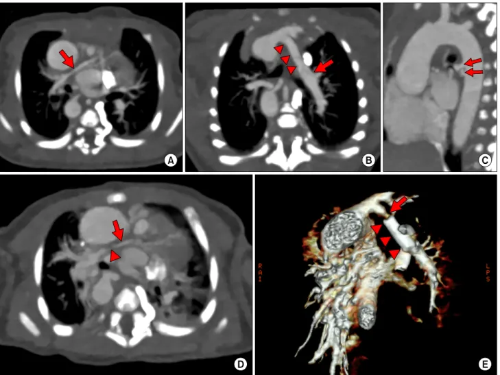
An infant weighing 2.91 kg was born at a gesta- tional age of 38+0 weeks. Prenatal echocardiography revealed pulmonary atresia (PA) with ventricular sep- tal defect (VSD) and major aortopulmonary collateral arteries (MAPCAs). Echocardiography performed im- med iately after birth revealed a d isconnected left pul- monary artery supplied by the left patent ductus ar- teriosus (PDA), originating from the left innominate artery, in addition to the abovementioned findings.
On the computed tomography (CT) performed 1 d ay after birth, a diminutive central right pulmonary ar- tery (RPA), a disconnected left pulmonary artery sup- plied by the left PDA, and 2 MAPCAs from the prox- imal descending thoracic aorta were observed (Fig.
1A–C).
We decided to allow the diminutive RPA to grow and delay the reconstruction of the central pulmo-
nary artery until the RPA was large enough to be re- liably enlarged surgically. As the first intervention, a stent was placed in the left PDA at 26 days of age.
On follow-up CT scan, severe narrowing of the PDA just proximal to the stent was observed and the RPA was found to be still diminutive (diameter <1.5 mm) (Fig. 1D, E). At 41 days of age, a central shunt was placed through a mid line sternotomy with a method introduced by Gates et al. [1] in 1998. Subsequently, a stent was reinserted into the proximal PDA with the stent-in-stent approach in the catheterization labo- ratory. The hospital course was uncomplicated, and the patient was discharged with prescriptions for war- farin, captopril, and diuretics at 65 days of age. The level of brain natriuretic peptide at discharge was 510 pg/mL.
At 5 months of age, a follow-up angiography and
CT scan revealed that the left pulmonary artery had
grown to a sufficient size with normal arborization