ABSTRACT
BACKGROUND: We evaluated long-term prognosis according to improvement of pulmonary hypertension (PH) and left ventricular ejection fraction (LVEF) in patients with heart failure with reduced ejection fraction (HFrEF) and PH.
METHODS: We included all consecutive patients with HFrEF and PH who had a baseline and follow-up echocardiographic examinations from September 2011 to March 2017. PH was defined as maximal velocity of tricuspid regurgitation (TR Vmax) over 3.0 m/s, and LVEF improvement was defined as LVEF change ≥ 15% from the baseline echocardiography.
Primary outcome was 5-year major adverse cardio-cerebrovascular events (MACCE).
RESULTS: We analyzed 271 patients. Mean LVEF was 28±8% and TR Vmax was 3.4±0.4 m/s. On follow-up, 183 (68%) showed improvement of LVEF, and 165 (61%) demonstrated improvement of PH. We classified patients into 4 groups according to improvement of PH and LVEF; group 1 (both improvement, 134 patients), group 2 (PH improvement only, 31 patients), group 3 (LVEF improvement only, 49 patients) and group 4 (no improvement, 57 patients). Group 4 had older age, higher incidence of myocardial infarction and aggravation of pre-existing HF. During the follow-up (31±20 months), 27% died and 40.8% experienced MACCE. Group 4 had the worst survival (HR=4.332, 95% CI=2.396-7.833, p<0.001), and group 3 had increased MACCE rate (HR=2.030, 95% CI=1.060-3.888, p=0.033) compared with group 1. Group 2 had similar long-term clinical events (HR=1.085, 95% CI=0.458-2.571, p=0.853) to group 1.
CONCLUSIONS: In patients with HFrEF and PH, persistence of PH and no LVEF improvement was associated with the worst long-term outcome.
Keywords: Pulmonary hypertension; Heart failure with reduced ejection fraction;
Follow-up echocardiography; Improvement outcomes
Original Article
Received: Mar 19, 2019 Revised: May 20, 2019 Accepted: Jun 5, 2019 Address for Correspondence:
Jae-Hyeong Park, MD, PhD
Department of Cardiology in Internal Medicine, Chungnam National University Hospital, Chungnam National University College of Medicine, 282 Munhwa-ro, Jung-gu, Daejeon 35015, Korea.
E-mail: [email protected] Copyright © 2019 Korean Society of Echocardiography
This is an Open Access article distributed under the terms of the Creative Commons Attribution Non-Commercial License (https://
creativecommons.org/licenses/by-nc/4.0/) which permits unrestricted non-commercial use, distribution, and reproduction in any medium, provided the original work is properly cited.
ORCID iDs Hee-Jin Kwon
https://orcid.org/0000-0002-4281-5211 Jae-Hyeong Park
https://orcid.org/0000-0001-7035-286X Jin Joo Park
https://orcid.org/0000-0001-9611-1490 Jae-Hwan Lee
https://orcid.org/0000-0002-6561-7760 In-Whan Seong
https://orcid.org/0000-0003-4628-0258 Conflict of Interest
The authors have no financial conflicts of interest.
Hee-Jin Kwon , MD1, Jae-Hyeong Park , MD, PhD1, Jin Joo Park , MD, PhD2, Jae-Hwan Lee , MD, PhD1, and In-Whan Seong , MD, PhD1
1 Department of Cardiology in Internal Medicine, Chungnam National University Hospital, Chungnam National University College of Medicine, Daejeon, Korea
2 Department of Cardiology, Cardiovascular Center, Seoul National University Bundang Hospital, Seongnam, Korea
Improvement of Left Ventricular Ejection Fraction and Pulmonary
Hypertension Are Significant Prognostic
Factors in Heart Failure with Reduced
Ejection Fraction Patients
INTRODUCTION
Pulmonary hypertension (PH) is common and is associated with high mortality and morbidity.1)2) Among various etiologies for PH, left ventricular (LV) dysfunction is the most common one and accounts for 65%–80% of all PH.3-8) A combination of elevated LV filling pressures and reactive pulmonary vascular remodeling results in PH secondary to left heart disease.3)6) Many patients with heart failure with reduced ejection fraction (HFrEF) have PH, and the presence of PH is associated with increased mortality and morbidity regardless of LV ejection fraction (LVEF) and stage of heart failure (HF).8-13) LVEF and degree of PH are not static and can change with treatment. A substantial number of patients with HFrEF recover LVEF in response to guideline directed medical therapy.14) Moreover, the presence and severity of PH can vary with treatment and it depend on the compensation status.15) Data on long-term prognosis of patients who experience improvement of LVEF and/or PH are scarce.
Hence, we evaluated the long-term clinical outcome of patients with HFrEF and PH according to the improvement of PH and/or LVEF.
METHODS
Study population
We screened all consecutive patients with HFrEF and PH in Chungnam National University Hospital from September 2011 to March 2017. We collected baseline clinical profiles from their medical records and echocardiographic data from digitally stored echocardiographic images. HFrEF was defined when patients had symptoms and signs of HF and LVEF < 40%.
Of them, we included only patients who underwent follow-up echocardiographic study within 12 months from the baseline echocardiography. Patients with other concomitant diseases including malignancy which can affect survival were excluded. We classified our study population into 4 groups according to the improvement of PH and/or LVEF based on the follow-up echocardiographic findings. Group 1 included patients with both improvements, group 2 with an improvement of PH only, group 3 with an improvement of LVEF only, and group 4 persistence of PH and no LVEF improvement. The primary endpoint was the development of major adverse cardio-cerebrovascular event (MACCE) including all- cause death, myocardial infarction, stroke or transient ischemic attack (TIA) and admission for HF for 5 years. HF hospitalizations were defined as unplanned hospitalizations of at least 24 hours due to worsening HF and were identified from their medical records. The study complied with the Declaration of Helsinki principles. The study protocol was approved by the institutional review board (IRB). The IRB waived the need for written informed consent from the study patients.
Echocardiographic measurement
We reanalyzed all echocardiographic images from baseline and follow-up echocardiographic studies. LVEF was calculated with the biplane modified Simpson's method with apical 4 chamber and apical 2 chamber views. The presence and degree of tricuspid regurgitation (TR) was found in the focused right ventricular view with color flow mapping. TR velocity was measured with the application of continuous wave Doppler to TR signal. Pulmonary artery systolic pressure (PASP) was estimated with the maximal velocity of TR (TR Vmax) and size of the inferior vena cava with respiratory change. In the follow-up echocardiographic examination, we checked the improvement of LVEF and change of PH based on baseline LVEF and TR Vmax levels. PH was defined TR Vmax ≥ 3.0 m/s by Doppler-derived velocity of
optimal TR jet. The improvement of PH was defined when TR Vmax decreased to < 3.0 m/s in the follow-up echocardiography. Improvement of LVEF was defined as LVEF ≥ 15% from baseline to follow-up echocardiography.
Statistical analysis
Data are presented as numbers and frequencies for categorical variables and as mean ± standard deviations for continuous variables. For comparisons among the groups, the χ2 test (or Fisher exact test when any expected count was < 5 for a 2 × 2 table) was performed for categorical variables, and the one-way analysis of variance was used for continuous variables.
We plotted survival curves according to our study groups using the Kaplan-Meier method with comparison using the log rank test. The time to first clinical event was analyzed using the multivariate Cox-proportional hazard analysis to determine the independent predictors of MACCE. In the multivariable analysis, we included all variables as covariates found to be statistically significant (p < 0.05) in the univariate analysis or variables known to be clinically important, excluding those with multicollinearity with others.
A two-sided p-value of < 0.05 was considered statistically significant. Statistical analyses were performed using SPSS version 22.0 (IBM Co., Chicago, IL, USA)
RESULTS
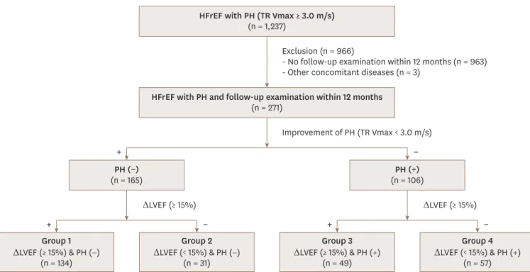
We screened 1237 consecutive patients with HFrEF and PH from September 2011 to March 2017.
Of them, 963 patients had no follow-up echocardiographic examination and 3 patients had malignancies affecting survival, so that total of 271 patients were included in this final analysis.
The mean age was 65 ± 14 years, 167 (62%) were males, 219 (81%) had de novo onset of HF.
Baseline, echocardiographic characteristics and clinical outcomes were summarized in the Table 1. Hypertension and dyslipidemia were common in our study population (48% and 47%, respectively). At baseline, the mean LVEF was 28.1 ± 8.3% and that of TR Vmax was 3.4 ± 0.4 m/s. Regarding the medical therapy, 91% received beta-blockers and 86% received renin- angiotensin system inhibitors. Follow-up echocardiographic examinations were performed at an average of 5.5 ± 3.6 months from the baseline echocardiography. One-hundred and eighty-three patients (68%) showed improvement of LVEF, and 165 (61%) demonstrated improvement of PH. The improvement of LVEF (74.4% vs. 38.5%, p < 0.001) and improvement of PH (67.1% vs. 34.6%, p < 0.001) were more frequent in patients with de novo HF group.
According to the improvement of PH and LVEF based on the follow up echocardiogram, we classified the study population into 4 groups as follows: 134 patients (49%), 31 patients (11%), 49 patients (18%) and 57 patients (21%) were classified as group 1, 2, 3, and 4, respectively (Figure 1). Overall, patients in group 4 were older, less likely to have de novo onset of HF. The use of guideline-directed medical therapy did not differ between the groups. In follow-up echocardiography, the mean LVEF were 45.3 ± 11.1% in patients with improved LVEF (group 1 and 3) and 29.8 ± 8.5% in patients without LVEF improvement (group 2 and 4). The TR Vmax was significantly lower in patients with improved PH (group 1 and 2) than patients with sustained PH (group 3 and 4) (2.4 ± 0.3 m/s vs. 3.4 ± 0.4 m/s, p < 0.001).
Adverse clinical outcomes
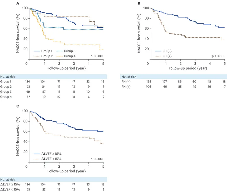
During the follow-up period (31 ± 20 months) from the follow-up echocardiography, 52 patients (19%) died, 65 (24%) had hospital admission for HF more than 1 episode and 17
(6%) admitted for stroke or TIA. Thus, 97 patients (36%) experience MACCEs. Group 4 had the lowest 5-year MACCE-free survival rate (19.0 ± 9.2%) than other groups (p < 0.001, Figure 2A).
Patients with improvement of PH (62.7 ± 5.4% vs 38.2 ± 6.7%, p < 0.001) and improved LVEF (60.5 ± 4.8% vs 36.6 ± 7.9%, p < 0.001) had significantly higher 5-year MACCE-free survival rate than the other group (Figure 2B and 2C).
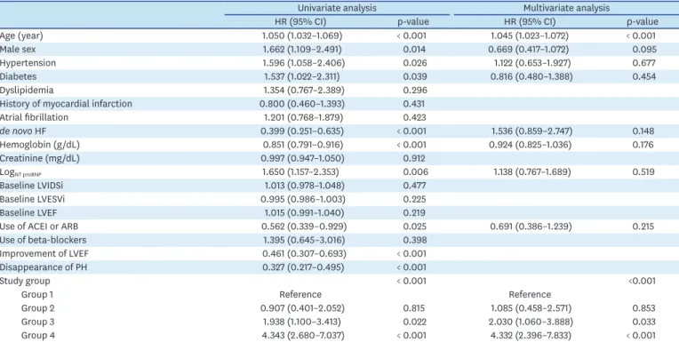
In the univariate analysis, age, male sex, hypertension, diabetes and NT proBNP concentration were associated with higher incidence of MACCE. Also, de novo HF, hemoglobin concentration, Table 1. Baseline and follow-up echocardiographic findings and clinical outcomes according to the study groups
Characteristics Total (n = 271) Group 1 (n = 134) Group 2 (n = 31) Group 3 (n = 49) Group 4 (n = 57) p-value
Age (year) 65 ± 14 62.7 ± 14.4 65.1 ± 11.9 63.7 ± 15.3 68.9 ± 12.9 0.043
Male gender 167 (62%) 82 (61%) 25 (81%) 27 (55%) 33 (58%) 0.113
Cardiovascular risk factors
Hypertension 131 (48%) 64 (48%) 15 (48%) 22 (45%) 30 (53%) 0.880
Diabetes mellitus 88 (33%) 37 (28%) 10 (32%) 17 (35%) 24 (42%) 0.265
Dyslipidemia 126 (47%) 43 (10%) 15 (48%) 45 (8%) 32 (56%) 0.301
Active smoker 64 (23%) 22 (16%) 5 (16%) 2 (4%) 7 (12%) 0.492
ESRD on regular hemodialysis 35 (13%) 13 (10%) 6 (20%) 13 (10%) 9 (16%) 0.411
Myocardial infarction 46 (17%) 15 (11%) 9 (29%) 6 (12%) 16 (28%) 0.007
Ischemic heart disease 73 (27%) 28 (21%) 9 (29%) 15 (31%) 21 (37%) 0.313
Type of heart failure < 0.001
de novo HF 219 (81%) 123 (92%) 24 (77%) 40 (82%) 32 (56%)
Aggravation of pre-existing HF 52 (19%) 11(8%) 7 (23%) 9 (18%) 25 (44%)
Medical treatment
Beta-blocker 246 (91%) 120 (90%) 29 (94%) 42 (92%) 52 (91%) 0.896
ACEI/ARB 234 (86%) 111 (88%) 27 (87%) 41 (84%) 48 (84%) 0.834
Spironolactone 154 (57%) 75 (56%) 15 (48%) 35 (71%) 29 (51%) 0.111
Diuretics 217 (80%) 108 (81%) 21 (68%) 41 (84%) 47 (83%) 0.311
Laboratory findings
Hemoglobin (g/dL) 12.7 ± 2.5 13.0 ± 2.5 13.2 ± 2.7 12.3 ± 2.8 12.1 ± 2.0 0.069
Creatinine (mg/dL) 1.6 ± 1.9 1.4 ± 1.3 1.6 ± 1.5 2.0 ± 2.8 1.7 ± 2.3 0.204
eGFR (mL/min/1.73 m2) 71.5 ± 36.3 75.2 ± 35.7 69.1 ± 33.6 65.6 ± 34.4 69.7 ± 40.7 0.434
LogNT-proBNP 3.6 ± 0.7 3.6 ± 0.6 3.2 ± 1.1 3.8 ± 0.5 3.7 ± 0.7 0.003
Echocardiography
Baseline LVIDSi (mm) 28.2 ± 6.3 27.9 ± 5.9 30.8 ± 7.8 26.5 ± 5.1 29.0 ± 6.9 0.023
Baseline LVIDDi (mm) 33.8 ± 5.7 33.3 ± 5.1 35.5 ± 7.7 32.2 ± 4.8 35.3 ± 6.2 0.016
Follow-up LVIDSi (mm) 25.4 ± 6.9 22.8 ± 5.3 29.7 ± 7.9 24.0 ± 6.2 30.2 ± 6.5 < 0.001
Follow-up LVIDDi (mm) 32.3 ± 5.9 30.5 ± 4.9 35.3 ± 7.5 31.7 ± 5. 35.6 ± 5.9 < 0.001
Baseline LVEDVi (mL) 89.5 ± 32.6 87.7 ± 31.7 99.8 ± 43.6 89.3 ± 26.5 88.0 ± 32.5 0.342
Baseline LVESVi (mL) 65.5 ± 28.0 65.8 ± 27.1 69.6 ± 37.1 66.1 ± 23.5 62.2 ± 28.4 0.711
Follow-up LVEDVi (mL) 77.1 ± 34.1 66.6 ± 25.4 99.0 ± 50.2 72.9 ± 26.2 93.7 ± 36.5 < 0.001
Follow-up LVESVi (mL) 48.1 ± 29.8 37.1 ± 19.6 68.5 ± 41.1 42.1 ± 21.7 68.7 ± 33.1 < 0.001
Baseline LVEF (%) 28.1 ± 8.3 26.2 ± 7.9 31.9 ± 8.7 27.2 ± 7.6 31.0 ± 8.0 < 0.001
Follow-up LVEF (%) 40.3 ± 12.6 45.6 ± 10.5 32.0 ± 9.3 44.6 ± 12.7 28.6 ± 7.9 < 0.001
ΔLVEF% 12.1 ± 12.7 19.4 ± 8.8 0.2 ± 3.7 17.4 ± 10.4 -2.8 ± 5.5 < 0.001
Baseline TR Vmax (m/sec) 3.6 ± 0.7 3.33 ± 0.3 3.36 ± 0.41 3.47 ± 0.41 3.45 ± 0.39 0.048
Follow-up TR Vmax (m/sec) 2.8 ± 0.6 2.5 ± 0.28 2.4 ± 0.24 3.3 ± 0.36 3.5 ± 0.40 < 0.001
E/e′ 21.4 ± 8.7 20.7 ± 8.4 18.9 ± 6.8 21.5 ± 8.0 24.2 ± 10.2 0.088
Clinical outcome
All cause death 52 22 (16%) 4 (13%) 9 (18%) 17 (30%) 0.131
Admission for HF 65 18 (13%) 6 (19%) 14 (29%) 27 (47%) < 0.001
Admission for stroke or TIA 17 6 (5%) 0 (0%) 4 (8%) 7 (12%) 0.086
MACCE 97 34 (25%) 7 (23%) 21 (43%) 35 (62%) < 0.001
ACEI: angiotensin converting enzyme inhibitor, ARB: angiotensin receptor blocker, eGFR: estimated glomerular filtration rate, ESRD: end-stage regnal disease, HF: heart failure, LVEDVi: body surface area indexed left ventricular end-diastolic volume, LVEF: left ventricular ejection fraction, LVESVi: body surface area indexed left ventricular end-systolic volume, LVIDDi: body surface area indexed left ventricular internal dimension, end-diastole, LVIDSi: body surface area indexed left ventricular internal dimension, end-systole, MACCE: major adverse cardio-cerebrovascular event, NT proBNP: N terminal pro B type natriuretic peptide, TIA: transient ischemic attack TR Vmax: maximal velocity of tricuspid regurgitation.
use of angiotensin converting enzyme inhibitor or angiotensin receptor blocker, improvement of LVEF and disappearance of PH were associated with lower MACCE (Table 2). Study group showed statistical significance, and group 4 had significantly higher MACCE rate. In the multivariate analysis, older age (hazard ratio [HR] = 1.045, 95% confidence interval [CI] = 1.023- 1.072, p < 0.001) and study group remained statistical significance. Group 4 had statistically significantly lower MACCE free survival (HR = 4.332, 95% CI = 2.396-7.833, p < 0.001, Table 2).
DISCUSSION
In this study, we evaluated changes in LVEF and PH from baseline to follow-up echocardiography in patients with HFrEF and PH, and showed that different long-term prognosis in patients with PH and HFrEF. Patients with improvement of LVEF and/or PH had better prognosis that those with persistent PH and LV systolic dysfunction.
The PH secondary to left-sided heart disease is classified as group 2 in the WHO classification.1)16) Within Group 2 PH, there are subtypes characterized by the presence or absence of pulmonary vascular disease.16) Left heart disease, including HFrEF, HF with preserved ejection fraction (HFpEF) and valvular heart disease results in an increase in left atrial pressure due to mainly diastolic dysfunction. The elevation of the left atrial pressure eventually increases the hydrostatic pressure in the pulmonary capillaries. Increased pulmonary artery pressure in patients with HF often represents a combination of increased left-sided filling pressures (passive component) and elevated pulmonary vascular resistance attributable to functional and structural abnormalities of the pulmonary vascular bed (reactive component).17)
HFrEF with PH and follow-up examination within 12 months (n = 271)
Group 1 ΔLVEF (≥ 15%) & PH (−)
(n = 134)
Group 2 ΔLVEF (< 15%) & PH (−)
(n = 31)
Group 3 ΔLVEF (≥ 15%) & PH (+)
(n = 49)
Group 4 ΔLVEF (< 15%) & PH (+)
(n = 57) PH (−)
(n = 165) PH (+)
(n = 106) HFrEF with PH (TR Vmax ≥ 3.0 m/s)
(n = 1,237)
Exclusion (n = 966)
- No follow-up examination within 12 months (n = 963) - Other concomitant diseases (n = 3)
Improvement of PH (TR Vmax < 3.0 m/s)
ΔLVEF (≥ 15%) ΔLVEF (≥ 15%)
−
−
− +
+ +
Figure 1. Scheme of study population. ΔLVEF: change of left ventricular ejection fraction, HFrEF: heart failure with reduced ejection fraction, PH: pulmonary hypertension, TR Vmax: maximal velocity of tricuspid regurgitation.
PH due to left heart disease is common in HFrEF patients and has been shown to be associated with worse clinical outcome.4)5)8)9)11-13)18) Aronson et al.9) first reported that TR Vmax > 2.5 m/s was associated higher mortality in patients with dilated cardiomyopathy.
Since then, several studies have reported a strong association between PH and mortality and morbidity in HF patients. However, few studies have reported long-term prognosis according to the improvement of LVEF and/or PH. We have shown through this study that the improvement in PH was indicative of a better long-term prognosis in HFrEF patients with PH.
Similar to our findings, Shalaby et al.19) reported that higher PASP at baseline was associated with worse survival in a study of cardiac resynchronization therapy recipients. They found that patients with reductions in PASP on the follow-up had better outcomes. In some patients with PH and HFrEF, acute phase with increased left atrial pressure may be reversible with standard HF treatment. However, chronic exposure to elevated pulmonary capillary wedge
p < 0.001 100
0
MACCE-free survival (%)
20
60 60
5 4 3 2 1
A
Follow-up period (year) Group 3 Group 4 Group 1
Group 2 80
40
No. at risk Group 2
Group 1 134
31
104 24
71 17
47 13
33 9
16 5 Group 4
Group 3 49
57
27 19
15 10
11 8
10 6
6 2
p < 0.001 100
0
MACCE-free survival (%)
20
5 4 3 2 1
B
Follow-up period (year) PH (−)
PH (+) 80
40
No. at risk PH (+)
PH (−) 165
106
127 46
86 25
60 19
42 16
18 7
p < 0.001 100
0
MACCE-free survival (%)
20 60
5 4 3 2 1
C
Follow-up period (year) ΔLVEF ≥ 15%
ΔLVEF < 15%
80
40
No. at risk ΔLVEF < 15%
ΔLVEF ≥ 15% 134 31
104 23
71 15
47 13
33 9
13 5
Figure 2. MACCE free survival according to the group by Kaplan-Meier survival analysis. Group 4 has the lowest 5- year MACCE-free survival rate (19.0 ± 9.2%) than other groups (A, p < 0.001 by Log-rank test). The group with improved PH (B, 62.7 ± 5.4% vs 38.2 ± 6.7%, p < 0.001 by Log-rank test) and improvement of left ventricular ejection fraction (C, 60.5 ± 4.8% vs 36.6 ± 7.9%, p < 0.001 by Log-rank test) have better 5-year MACCE-free survival rate than the other group.
ΔLVEF: change of left ventricular ejection fraction, MACCE: major adverse cardio-cerebrovascular event, PH: pulmonary hypertension.
pressure may lead to permanent vascular remodeling, irreversibly.3)20) Although there may be differences depending on the timing of follow up echocardiography and patient status, we thought that reactive PH was the main mechanism in patients with persistent PH regardless of LVEF. Reactive PH means chronic PH and irreversible vascular remodeling and may have caused a bad prognosis. Aronson et al.9) reported similar results. In a subgroup analysis of 242 patients from the Vasodilation in the Management of Acute Congestive Heart Failure trial, reactive PH patients had higher mortality rate than passive PH patients.
Limitations
There are several limitations in our study. First, this is a retrospective observational study in single-center. A large number of patients were excluded because a follow-up echocardiography was not performed. Second, patients were not in the same states at the time of taking follow- up echocardiographic examinations. Some patients underwent follow-up examination at a stable state when heart failure was well controlled, while others were followed up for acute exacerbations such as worsening of symptoms, hospitalization, or other problems. Third, we used echocardiography in the diagnosis of PH with HFrEF. It may be less accurate than the invasive method. Right heart catheterization (RHC) is a gold standard in the diagnosis of PH and accurate method for the measurement of PASP.3) However, RHC is an invasive procedure with associated risks, complications and cost.21) Thus, RHC is not indicated all PH patients with HFrEF and is recommended in patients considered heart transplantation.1) Echocardiography is a useful non-invasive imaging modality for estimating PASP using the Doppler-derived technique and also provides valuable information of the cardiac structure and performance. It is important for identification of the cause of PH.22) Because of its non-invasiveness, echocardiographic examinations can be performed repeatedly without giving no harm to patients. Further prospective studies with standardized protocols are needed to solve these limitations.
Table 2. Univariate and multivariate analysis in the prediction of major adverse cardio-cerebrovascular event during five year
Univariate analysis Multivariate analysis
HR (95% CI) p-value HR (95% CI) p-value
Age (year) 1.050 (1.032–1.069) < 0.001 1.045 (1.023–1.072) < 0.001
Male sex 1.662 (1.109–2.491) 0.014 0.669 (0.417–1.072) 0.095
Hypertension 1.596 (1.058–2.406) 0.026 1.122 (0.653–1.927) 0.677
Diabetes 1.537 (1.022–2.311) 0.039 0.816 (0.480–1.388) 0.454
Dyslipidemia 1.354 (0.767–2.389) 0.296
History of myocardial infarction 0.800 (0.460–1.393) 0.431
Atrial fibrillation 1.201 (0.768–1.879) 0.423
de novo HF 0.399 (0.251–0.635) < 0.001 1.536 (0.859–2.747) 0.148
Hemoglobin (g/dL) 0.851 (0.791–0.916) < 0.001 0.924 (0.825–1.036) 0.176
Creatinine (mg/dL) 0.997 (0.947–1.050) 0.912
LogNT proBNP 1.650 (1.157–2.353) 0.006 1.138 (0.767–1.689) 0.519
Baseline LVIDSi 1.013 (0.978–1.048) 0.477
Baseline LVESVi 0.995 (0.986–1.003) 0.225
Baseline LVEF 1.015 (0.991–1.040) 0.219
Use of ACEI or ARB 0.562 (0.339–0.929) 0.025 0.691 (0.386–1.239) 0.215
Use of beta-blockers 1.395 (0.645–3.016) 0.398
Improvement of LVEF 0.461 (0.307–0.693) < 0.001
Disappearance of PH 0.327 (0.217–0.495) < 0.001
Study group < 0.001 <0.001
Group 1 Reference Reference
Group 2 0.907 (0.401–2.052) 0.815 1.085 (0.458–2.571) 0.853
Group 3 1.938 (1.100–3.413) 0.022 2.030 (1.060–3.888) 0.033
Group 4 4.343 (2.680–7.037) < 0.001 4.332 (2.396–7.833) < 0.001
ACEI: angiotensin converting enzyme inhibitor, ARB: angiotensin receptor blocker, CI: confidence interval, HF: heart failure, HR: hazard ratio, LVEF: left ventricular ejection fraction, LVESVi: body surface area indexed left ventricular end-systolic volume, LVIDSi: body surface area indexed left ventricular internal dimension at end-systole, NT proBNP: N terminal pro B type natriuretic peptide, PH: pulmonary hypertension.
Conclusion
In patients with HFrEF and PH associated with HFrEF, the improvement of PH and LVEF were associated with favorable outcomes. The follow-up echocardiographic studies may help to determine prognosis in these patients by confirming the improvement of LVEF and PH compared with the baseline echocardiography.
REFERENCES
1. Galiè N, Humbert M, Vachiery JL, et al.. 2015 ESC/ERS guidelines for the diagnosis and treatment of pulmonary hypertension: the Joint Task Force for the Diagnosis and Treatment of Pulmonary Hypertension of the European Society of Cardiology (ESC) and the European Respiratory Society (ERS):
endorsed by: Association for European Paediatric and Congenital Cardiology (AEPC), International Society for Heart and Lung Transplantation (ISHLT). Eur Heart J 2016;37:67-119.
PUBMED | CROSSREF
2. Chung K, Strange G, Codde J, Celermajer D, Scalia GM, Playford D. Left heart disease and pulmonary hypertension: are we seeing the full picture? Heart Lung Circ 2018;27:301-9.
PUBMED | CROSSREF
3. Georgiopoulou VV, Kalogeropoulos AP, Borlaug BA, Gheorghiade M, Butler J. Left ventricular dysfunction with pulmonary hypertension: part 1: epidemiology, pathophysiology, and definitions. Circ Heart Fail 2013;6:344-54.
PUBMED | CROSSREF
4. Kjaergaard J, Akkan D, Iversen KK, et al. Prognostic importance of pulmonary hypertension in patients with heart failure. Am J Cardiol 2007;99:1146-50.
PUBMED | CROSSREF
5. Grigioni F, Potena L, Galiè N, et al. Prognostic implications of serial assessments of pulmonary hypertension in severe chronic heart failure. J Heart Lung Transplant 2006;25:1241-6.
PUBMED | CROSSREF
6. Rosenkranz S, Gibbs JS, Wachter R, De Marco T, Vonk-Noordegraaf A, Vachiéry JL. Left ventricular heart failure and pulmonary hypertension. Eur Heart J 2016;37:942-54.
PUBMED | CROSSREF
7. Simonneau G, Gatzoulis MA, Adatia I, et al. Updated clinical classification of pulmonary hypertension. J Am Coll Cardiol 2013;62:D34-41.
PUBMED | CROSSREF
8. Bursi F, McNallan SM, Redfield MM, et al. Pulmonary pressures and death in heart failure: a community study. J Am Coll Cardiol 2012;59:222-31.
PUBMED | CROSSREF
9. Aronson D, Eitan A, Dragu R, Burger AJ. Relationship between reactive pulmonary hypertension and mortality in patients with acute decompensated heart failure. Circ Heart Fail 2011;4:644-50.
PUBMED | CROSSREF
10. Cappola TP, Felker GM, Kao WH, Hare JM, Baughman KL, Kasper EK. Pulmonary hypertension and risk of death in cardiomyopathy: patients with myocarditis are at higher risk. Circulation 2002;105:1663-8.
PUBMED | CROSSREF
11. Kalogeropoulos AP, Siwamogsatham S, Hayek S, et al. Echocardiographic assessment of pulmonary artery systolic pressure and outcomes in ambulatory heart failure patients. J Am Heart Assoc 2014;3:e000363.
PUBMED | CROSSREF
12. Merlos P, Núñez J, Sanchis J, et al. Echocardiographic estimation of pulmonary arterial systolic pressure in acute heart failure. Prognostic implications. Eur J Intern Med 2013;24:562-7.
PUBMED | CROSSREF
13. Santas E, de la Espriella-Juan R, Mollar A, et al. Echocardiographic pulmonary artery pressure estimation and heart failure rehospitalization burden in patients with acute heart failure. Int J Cardiol 2017;241:407-10.
PUBMED | CROSSREF
14. Lupón J, Díez-López C, de Antonio M, et al. Recovered heart failure with reduced ejection fraction and outcomes: a prospective study. Eur J Heart Fail 2017;19:1615-23.
PUBMED | CROSSREF
15. Le Jemtel TH, Alt EU. Are hemodynamic goals viable in tailoring heart failure therapy? Hemodynamic goals are outdated. Circulation 2006;113:1027-32; discussion 1033.
PUBMED
16. Kalogeropoulos AP, Georgiopoulou VV, Borlaug BA, Gheorghiade M, Butler J. Left ventricular dysfunction with pulmonary hypertension: part 2: prognosis, noninvasive evaluation, treatment, and future research.
Circ Heart Fail 2013;6:584-93.
PUBMED | CROSSREF
17. Damy T, Goode KM, Kallvikbacka-Bennett A, et al. Determinants and prognostic value of pulmonary arterial pressure in patients with chronic heart failure. Eur Heart J 2010;31:2280-90.
PUBMED | CROSSREF
18. Santas E, Chorro FJ, Miñana G, et al. Tricuspid regurgitation and mortality risk across left ventricular systolic function in acute heart failure. Circ J 2015;79:1526-33.
PUBMED | CROSSREF
19. Shalaby A, Voigt A, El-Saed A, Saba S. Usefulness of pulmonary artery pressure by echocardiography to predict outcome in patients receiving cardiac resynchronization therapy heart failure. Am J Cardiol 2008;101:238-41.
PUBMED | CROSSREF
20. Delgado JF, Conde E, Sánchez V, et al. Pulmonary vascular remodeling in pulmonary hypertension due to chronic heart failure. Eur J Heart Fail 2005;7:1011-6.
PUBMED | CROSSREF
21. Hoeper MM, Lee SH, Voswinckel R, et al. Complications of right heart catheterization procedures in patients with pulmonary hypertension in experienced centers. J Am Coll Cardiol 2006;48:2546-52.
PUBMED | CROSSREF
22. Sciomer S, Magrì D, Badagliacca R. Non-invasive assessment of pulmonary hypertension: Doppler- echocardiography. Pulm Pharmacol Ther 2007;20:135-40.
PUBMED | CROSSREF