INTRODUCTION
Heterotopic brain tissue is defined as a mass-like lesion com- posed of mature brain tissue isolated from the cranial cavity or spinal cord (1). It usually occurs in the brain and extracra- nial midline structures, including the nose (2), nasopharynx, oropharynx, soft palate (3), lip, and tongue (4). Rarely, hetero- topic brain tissue has been reported to occur in the extracra- nial non-midline locations such as scalp, orbit, eye, lung, di- aphragm, peritoneum, skin, temporal bone, uterine cervix, and endometrium. Although many choristomas of the salivary gland origin have been reported in the middle ear and mas- toid (5, 6), heterotopic brain tissue is very uncommon in this region (1, 7-10). We herein describe a case of glial choristoma involving the middle ear and mastoid bone with a review of the literature.
CASE REPORT
A 50-yr-old man presented with progressive hearing loss and otorrhea of the left ear. Previously, he had frequently com- plained of ear fullness and hearing disturbance for 8 yr. Inter- mittent otorrhea was developed about 4 months ago. There was no history of congenital anomalies, trauma, or ear surgery.
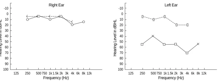
Physical examination revealed otorrhea in the external audi- tory canal of the left ear. The right ear was normal. Pure tone audiometry showed conductive hearing loss on the left ear
(Fig. 1). The right ear was normal. Otorrhea was improved after treatment of otitis externa. The patient had suffered from progressive hearing disturbance. Tympanic membrane showed attic retraction. Computed tomography revealed a mass-like lesion with soft tissue density in the middle ear cavity and mastoid antrum. Bony erosions were suspected in tegmen tympani and long process of incus of the left ear (Fig. 2). Pre- operative diagnosis was attic cholesteatoma. At operation, a graywhite fibrotic mass was detected in the epitympanic area.
Mesotympanum was intact. There was no bony erosions in tegmen and incus. Malleus and stapes were unremarkable.
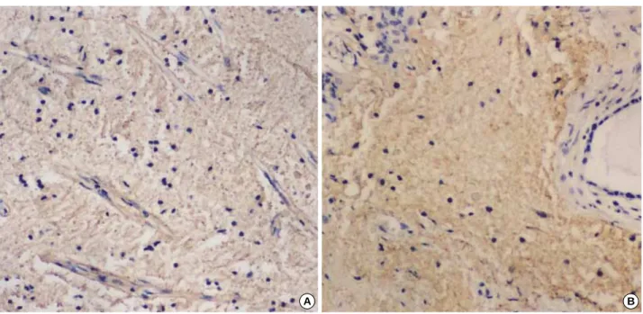
After the surgical removal of the epitympanic mass, subsequent examination about ossicular movement was normal. The pa- tient underwent left simple mastoidectomy with type I tym- panoplasty. During operation, there was no evidence of cranial bone defect or leakage of cerebrospinal fluid. Microscopic exami- nation of the hematoxylin-eosin stained section revealed a poor- ly circumscribed pale staining lesion with low cellularity. The lesion consisted of exclusively mature, disorganized glial tis- sue with fibrovascular tissue in a rather loose fibrillary back- ground (Fig. 3A). No mitotic figures or cellular atypia were noted. Scant lymphocytes were scattered within the glial tis- sue. Dystrophic calcifications were noted in degenerative foci.
Small distended glands, lined by ciliated, flat or cuboidal ep- ithelium, were present at the periphery of the lesion (Fig. 3B).
Other brain components, such as neurons, oligodendroglia, and meningeal, ependymal, or choroid elements were not de- tected. No clearly defined endodermal or mesodermal tissue
Jong Im Lee, Ki Kwon Kim, Yoon Keun Park*, Kyung Yoon Eah�, Jung Ran Kim
Departments of Pathology, Otolaryngology*, and Neurology�, College of Medicine, Dongguk University, Gyeongju, Korea
Address for correspondence Jong Im Lee, M.D.
Department of Pathology, College of Medicine, Dongguk University, 707 Sukjang-dong, Gyeongju 780-714, Korea
Tel : +82.54-770-2411, Fax : +82.54-770-2431 E-mail : [email protected]
155 J Korean Med Sci 2004; 19: 155-8
ISSN 1011-8934
Copyright � The Korean Academy of Medical Sciences
Glial Choristoma in the Middle Ear and Mastoid Bone : A Case Report
Heterotopic brain tissue usually involves extracranial midline structures of the head and neck such as nose, nasopharynx, and oral cavity. Its occurrence in the non-mid- line structures, including middle ear, is rare. We described a 50-yr-old-man with het- erotopic glial tissue in the middle ear and mastoid bone. The patient presented with progressive hearing loss for 8 yr. There was no history of congenital anomalies, trau- ma, or ear surgery. Computed tomography revealed a mass-like lesion with soft tis- sue density occupying the middle ear cavity and mastoid antrum. At the operation, a graywhite fibrotic mass was detected in the epitympanic area. Mesotympanum and ossicles were intact. The patient underwent left simple mastoidectomy with type I tympanoplasty. During operation, definite cranial bone defect or cerebrospinal fluid leakage was not found. Histologically, the lesion was composed of exclusively mature, disorganized glial tissue with fibrovascular elements in a rather loose fibrillary back- ground. Glial tissue showed diffuse positive reaction for glial fibrillar acidic protein and S100 protein on immunohistochemical study.
Key Words : Choristoma; Encephalocele; Brain; Neuroglia; Ear, Middle
Received : 11 November 2002 Accepted : 14 March 2003
could be identified. Additional sections were stained for glial fibrillar acidic protein (GFAP), S100 protein, cytokeratin (CK), and microtubule associated protein (MAP-2). The glial tis- sue revealed diffuse positive reaction for GFAP (Fig. 4A) and S100 protein (Fig. 4B), but negative reaction for MAP-2 and CK.
DISCUSSION
Since the initial description of heterotopic glial tissue over the dorsal surface of the cervical spinal cord in 1907 by Wol- bach (11), heterotopic brain or glial tissue have been report- ed in various sites. These lesions may be classified based on the
156 J.I. Lee, K.K. Kim, Y.K. Park, et al.
Fig. 1.Preoperative pure tone audiometry shows conductive hearing loss on the left ear (10/70 dB). Right ear is normal (7/10 dB).
Hearing Level in dBHL
Frequency (Hz) Right Ear -10
0 10 20 30 40 50 60 70 80 90 100
125 250 500 750 1k 1.5k 2k 3k 4k 6k 8k 12k
Hearing Level in dBHL
Frequency (Hz) Left Ear -10
0 10 20 30 40 50 60 70 80 90 100
125 250 500 750 1k 1.5k 2k 3k 4k 6k 8k 12k
C D
A B
Fig. 2. Coronal (A, B) and axial (C, D) computed tomography demonstrate a mass-like lesion with soft tissue density occupying the middle ear and mastoid cavity of the left ear (B, D).
location and possible pathogenetic mechanisms as follows (7, 12): 1) intraparenchymal central nervous system lesions, 2) dural and leptomeningeal lesions, 3) intracranial extracerebral lesions, 4) midline lesions, 5) distal lesions of the lung and uterus, and 6) extracranial non-midline lesions. Most report- ed cases have involved extracranial midline structures includ- ing the nose and nasopharynx (so-called nasal glioma) as well as the lips, oral cavity, oropharynx, palate (3), and tongue (4).
These lesions are most often considered to represent displaced
brain tissue during development and are therefore thought to be pathologically related to encephaloceles. Non-midline heterotopic brain tissue is rare. Cases have been reported to involve the scalp, eye and orbit, lung, peritoneum, and mid- dle ear (8). A review of the literature reveals that the most frequently reported choristomas of the middle ear is salivary gland tissue (5, 6), but heterotopic brain or glial tissue is very rare in this region (1, 7-10).
Unlike their midline counterparts, most middle ear hetero-
Choristoma in the Middle Ear 157
Fig. 3.Histologic features of the lesion. (A) The lesion is composed of exclusively mature disorganized glial tissue and fibrovascular elements in a rather loose fibrillary background. (B) Distended glandular structures are noted at the periphery of the glial tissue (H&E, ×100).
A B
Fig. 4.Immunohistochemical features. The lesion shows diffuse positive reaction for GFAP (A) and S100 protein (B) (×100).
A B
158 J.I. Lee, K.K. Kim, Y.K. Park, et al.
topic brain tissues are diagnosed in adult patients. Some au- thors have described chronic infection or inflammation, pre- vious trauma, or surgical procedures as predisposing factors for the development of middle ear heterotopia or encephalo- cele (7, 8). These clinical findings support the concept that heterotopic brain tissue in the middle ear region represents acquired encephalocele (7). However, our case had neither apparent predisposing factors nor evidence of connection be- tween the lesion and the central nervous system. Similar cases have been reported in the literature (7). In these cases, and also in our case, a possible explanation for pathogenesis is that the lesion might represent a true neuroglial heterotopia. Other possibilities, such as prior tiny congenital bone defect or remote trauma unrecognized at the time of presentation, can also be considered.
Histologically, heterotopic brain tissue is characterized by varying proportions of neurons and glia with associated chronic inflammation, choroid plexus, and ependymal or leptomenin- geal components. Rarely, heterotopic brain tissue of the middle ear was associated with choleastoma (8). Heterotopic brain tissue must be distinguished from true neoplasms such as glioma, ganglioglioma, meningioma, neuroma, and schwan- noma. The patient’s clinical presentation, the location of the lesion, its relation to surrounding structures as well as careful histologic examinations are helpful in differential diagnosis.
In the present case, the heterotopic glial tissue was admixed with distended glands lined by bland-looking flattened to cuboidal epithelial components at the periphery of the lesion, most likely representing entrapped tympanic cavity or Eusta- chian tube epithelium. Absence of elements unrelated to nor- mal anatomic structures is helpful to avoid the misdiagnosis as teratoma (13). The lesion of our case was exclusively com- posed of glial tissue lacking neuronal or other brain compo- nents. The relative lack of neuronal elements in some hetero- topic brain tissue has been attributed to the poor blood supply, resulting in neuronal ischemia and gliosis (14). Genut et al.
(15) have noticed the fact that neuronal precursors appeared in the developing brain at the 10th week and suggested that there would be no neuronal elements in the heterotopic brain, if the embryonic tissue has been separated prior to the 10th week.
It should be noted that there are no significant histologic differences between heterotopic brain tissue and encephalo- cele. It is important to distinguish heterotopic brain or glial tissue from more common middle ear encephalocele because of the risk of infection in the latter. The accurate diagnosis
of heterotopia versus encephalocele requires the knowledge of radiologic and operative findings of the patient.
REFERENCES
1. Klein MV, Schwaighofer BW, Sobel DF, Fantozzi RD, Hesselink JR.
Heterotopic brain in the middle ear: CT findings. J Comput Assist Tomogr 1989; 13: 1058-60.
2. Yeoh GP, Bale PM, de Silva M. Nasal cerebral heterotopia: the so- called nasal glioma or sequestered encephalocele and its variants.
Pediatr Pathol 1989; 9: 531-49.
3. Choi HJ, Lee YS, Kim YS, Kim KY, Kang CS, Shim SI. Heterotopic brain tissue in the soft palate. Korean J Pathol 1998; 32: 1039-41.
4. Abdelsayed RA, Wetherington RW, Bent JP 3rd, Sharpe DE. Glial choristoma of the tongue: a case report and review of the literature.
Oral Surg Oral Med Oral Pathol Oral Radiol Endod 1999; 87: 215-22.
5. Ha SL, Shin JE, Yoon TH. Salivary gland choristoma of the middle ear: a case report. Am J Otolaryngol 2000; 21: 127-30.
6. Buckmiller LM, Brodie HA, Doyle KJ, Nemzek W. Choristoma of the middle ear: a component of a new syndrome? Otol Neurotol 2001;
22: 363-8.
7. Gyure KA, Thompson LD, Morrison AL. A clinicopathological study of 15 patients with neuroglial heterotopias and encephaloceles of the middle ear and mastoid region. Laryngoscope 2000; 110: 1731-5.
8. McGregor DH, Cherian R, Kepes JJ, Kepes M. Case reports: hetero- topic brain tissue of middle ear associated with cholesteatoma. Am J Med Sci 1994; 308: 180-3.
9. Gulya AJ, Glasscock ME 3rd, Pensak ML. Neural choristoma of the middle ear. Otolaryngol Head Neck Surg 1987; 97: 52-6.
10. Slater DN, Timperley WR, Smith CM. Heterotopic middle ear glio- matosis. Histopathology 1988; 12: 230-1.
11. Wolbach SB. Congenital rhabdomyoma of the heart. Report of a case associated with multiple nests of neuroglia tissue in the meninges of the spinal cord. J Med Res 1907; 16: 495-520.
12. Harris CP, Townsend JJ, Klatt EC. Accessory brains (extracerebral heterotopias): unusual prenatal intracranial mass lesions. J Child Neurol 1994; 9: 386-9.
13. Gyure KA, Morrison AL, Jones RV. Intracranial extracerebral neu- roglial heterotopia: a case report and review of the literature. Ann Diagn Pathol 1999; 3: 182-6.
14. Whittet HB, Barker S, Anslow P, Leighton S. Heterotopic brain tissue:
a rare cause of adult recurrent meningitis. J Laryngol Otol 1990; 104:
328-30.
15. Genut AA, Miranda FG, Garcia JH. Organized cerebral heterotopia in the ethmoid sinus. A case report. J Neurol Sci 1976; 28: 339-44.