ABSTRACT
The Surgery Treatment Modality Committee of the Korean Gynecologic Oncologic Group (KGOG) has determined to develop a surgical manual to facilitate clinical trials and to improve communication between investigators by standardizing and precisely describing operating procedures. The literature on anatomic terminology, identification of surgical components, and surgical techniques were reviewed and discussed in depth to develop a surgical manual for gynecologic oncology. The surgical procedures provided here represent the minimum requirements for participating in a clinical trial. These procedures should be described in the operation record form, and the pathologic findings obtained from the procedures should be recorded in the pathologic report form. Here, we focused on radical hysterectomy and lymphadenectomy, and we developed a KGOG classification for those conditions.
Keywords: Manuals as Topic; Gynecologic Surgical Procedures; Hysterectomy, Lymph Node Excision
Surgical Manual
Received: Jul 26, 2016 Accepted: Aug 5, 2016 Correspondence to Ju-Won Roh
Department of Obstetrics and Gynecology, Dongguk University Ilsan Hospital, Dongguk University College of Medicine, 27 Dongguk-ro, Ilsandong-gu, Goyang 10326, Korea.
E-mail: [email protected] Copyright © 2017. Asian Society of Gynecologic Oncology, Korean Society of Gynecologic Oncology
This is an Open Access article distributed under the terms of the Creative Commons Attribution Non-Commercial License (http://
creativecommons.org/licenses/by-nc/4.0/) which permits unrestricted non-commercial use, distribution, and reproduction in any medium, provided the original work is properly cited.
ORCID Maria Lee
http://orcid.org/0000-0002-8017-3176 Yun Hwan Kim
http://orcid.org/0000-0001-9498-2938 Kwang Beom Lee
http://orcid.org/0000-0003-4634-176X Shin-Wha Lee
http://orcid.org/0000-0002-5088-1905 Seung-Hyuk Shim
http://orcid.org/0000-0001-8043-2257 Yong-Jung Song
http://orcid.org/0000-0002-6103-2466 Ju-Won Roh
http://orcid.org/0000-0002-2449-8742
Maria Lee,1 Chel Hun Choi,2 Yi Kyeong Chun,3 Yun Hwan Kim,4 Kwang Beom Lee,5 Shin-Wha Lee,6 Seung-Hyuk Shim,7 Yong-Jung Song,8 Ju-Won Roh,9
Suk-Joon Chang,10 Jong-Min Lee11
1Department of Obstetrics and Gynaecology, Seoul National University College of Medicine, Seoul, Korea
2 Department of Obstetrics and Gynecology, Samsung Medical Center, Sungkyunkwan University School of Medicine, Seoul, Korea
3 Department of Pathology, Cheil General Hospital & Women’s Healthcare Center, Dankook University College of Medicine, Seoul, Korea
4 Department of Obstetrics and Gynecology, Ewha Womans University Mokdong Hospital, Ewha Womans University School of Medicine, Seoul, Korea
5 Department of Obstetrics and Gynecology, Gachon University Gil Medical Center, Gachon University College of Medicine, Incheon, Korea
6 Department of Obstetrics and Gynecology, Asan Medical Center, University of Ulsan College of Medicine, Seoul, Korea
7 Department of Obstetrics and Gynecology, Research Institute of Medical Science, Konkuk University School of Medicine, Seoul, Korea
8 Department of Obstetrics and Gynecology, Pusan National University Yangsan Hospital, Pusan National University School of Medicine, Yangsan, Korea
9 Department of Obstetrics and Gynecology, Dongguk University Ilsan Hospital, Dongguk University College of Medicine, Goyang, Korea
10Departments of Obstetrics and Gynecology, Ajou University School of Medicine, Suwon, Korea
11 Department of Obstetrics and Gynecology, Kyung Hee University Hospital at Gangdong, Kyung Hee University School of Medicine, Seoul, Korea
Surgical manual of the Korean Gynecologic Oncology Group:
classification of hysterectomy and
lymphadenectomy
INTRODUCTION
To date, no surgical manual or standardized anatomical description of gynecologic oncology has been developed by the Korean Gynecologic Oncology Group (KGOG). The members of the Surgery Treatment Modality Committee (Surgery TMC) of the KGOG identified a need for a surgical manual to facilitate clinical trials and to improve communication between investigators by standardizing and precisely describing operating procedures.
We reviewed the current literature on anatomic terminology, identification of surgical components, and surgical techniques, and discussed them in depth in order to create a surgical manual for gynecologic oncology. Here, we focused on radical hysterectomy and lymphadenectomy, and we developed a KGOG classification for those conditions.
ANATOMICAL NOMENCLATURE
To simplify the anatomical terms for classification, we will consistently use the terminology
‘paracervix,’ ‘vesicouterine ligament,’ and ‘uterosacral ligament’ for the structures usually referred to by surgeons as the cardinal ligament (or lateral parametrium), anterior parametrium, and posterior parametrium, respectively [1].
The term ‘paracervix’ (cardinal ligament, Mackenrodt’s ligament, or parametrium) refers to the dorsolateral attachment of the cervix, tissues that surround the uterine artery between the uterine corpus and pelvic sidewall cranial to the ureter, connective tissue, and lymph channels.
The term ‘vesicouterine ligament’ (ventral parametrium) includes the anterior and posterior leaflets. The connective tissue of the anterior leaflet of the vesicouterine ligament; that is, the anterior portion of the so-called ureteral tunnel, can be identified following the complete separation of the uterine artery and superficial uterine vein from the ureter. The posterior leaflet of the vesicouterine ligament is the tissue that resides under the ureter and connects the posterior wall of the bladder and the lateral cervix/upper lateral vagina [2].
The term ‘uterosacral ligament’ (dorsal parametrium) refers to the fibrous tissue and non-striated muscular fibres that are attached to the front of the sacrum and travel from the uterus to the anterior aspect of the sacrum.
Throughout this classification, the term ‘nerve preservation’ refers to identify the hypogastric nerves, the inferior hypogastric nerve plexus (pelvic plexus), and its bladder branches, and indicates allowing the resection of oncologically relevant pericervical structures while preserving the sympathetic and parasympathetic innervations of the pelvic organs [3].
KGOG CLASSIFICATION OF HYSTERECTOMY
The Piver-Rutlege-Smith classification published in 1974 has achieved popularity [4,5].
However, several limitations, including inconsistencies in terminology, drawbacks associated with a uterocentric concept, exclusion of nerve-sparing and fertility-preserving techniques, and difficulties adapting the guidelines to advances in vaginal, laparoscopic, or Suk-Joon Chang
http://orcid.org/0000-0002-0558-0038 Jong-Min Lee
http://orcid.org/0000-0002-0562-5443 Conflict of Interest
No potential conflict of interest relevant to this article was reported.
Funding
This work was supported in part by the National Cancer Center Grant (NCC1310311, NCC1610070).
robotic surgery, makes continued use of the classification challenging. To overcome these limitations, Querleu and Morrow [1] proposed a new classification consisting of four types of radical hysterectomy, the Kyoto classification. We decided to create a KGOG classification of hysterectomy and lymphadenectomy based on the Kyoto classification, because it is considered contemporary and adequate for worldwide communication.
The classification has been modified and adapted to Korean circumstances by the Surgery TMC of the KGOG. There are two major changes. One is the deletion of the type B subclassification (B1 and B2), and the other is the inclusion of a fertility-preserving procedure, trachelectomy (cervicectomy), as a subclassification of types C and D.
The reasons for the elimination of the type B subclassification are as follows: (1) It is not easy and is sometimes impossible to differentiate the paracervical lymph nodes from the pelvic lymph nodes. (2) The basic reason for developing this classification is to provide separate descriptions of hysterectomy and lymphadenectomy. (3) Separate clinical indications for each operation have not been identified. (4) The clinical significance of the differentiation within type B is considered minimal. Table 1 summarises the four types of KGOG classification of hysterectomy.
1. Type A: minimum resection of the paracervix
This is an extrafascial hysterectomy. The paracervix is transected medial to the ureter and lateral to the cervix. The ureter does not need to be unroofed. The uterosacral and vesicouterine ligaments are transected close to the uterus. The length of the vaginal resection is generally less than 10 mm, and the vaginal part of the paracervix is not removed.
Table 1. Korean Gynecologic Oncologic Group classification of hysterectomy*
Extent of resection Ureter
A: Minimum resection
of the paracervix† Paracervix: transected medial to the ureter but lateral to the cervix Palpation or direct visualization without freeing from its bed Uterosacral and vesicouterine ligaments: transected closely to the uterus
Vaginal resection: generally less than 10 mm, without removal of the paracervix A (T)‡ Simple trachelectomy (cervicectomy)
Surgical removal of the uterine cervix without removal of the paracervix or vagina, very large conization
B: Transection of paracervix
at the ureter§ Paracervix: transected at the level of the ureteral tunnel Unroofed and rolled
laterally Uterosacral and vesicouterine ligaments: partial resection
Neural component of the paracervix: no resection
Vaginal resection: at least 10 mm of the vagina from the cervix or tumor B (T)‡ Radical trachelectomy (cervicectomy)
Surgical removal of the uterine cervix with the paracervix and vagina C: Transection of the paracervix
at its junction with the internal iliac vascular systemǁ
Transection of the uterosacral ligaments at the rectum Completely mobilized
Transection of the vesicouterine ligaments at the bladder
Resection: 15–20 mm of the vagina from the tumor or cervix and corresponding paracervix
C1 With autonomic nerve sparing/preservation
C2 Without autonomic nerve sparing/preservation
D: Entire resection of paracervix
with vessels Ultra-radical procedures, mostly indicated at the time of pelvic exenteration Completely mobilized D1 Resection of the paracervix at the pelvic side, with vessels arising from the internal iliac system,
exposing the roots of the sciatic nerve
D2 Resection of the paracervix at the pelvic side, with the internal iliac vessels plus the adjacent fascial or muscular structures
*Modification of the new classification of radical hysterectomy by Querleu et al. [1]. †It is similar to type I of the “Piver-Rutledge-Smith (PRS) classification” [4,5].
‡(T) means trachelectomy (cervicectomy). §This is similar to type II under the PRS classification. ǁThis is similar to type III under the PRS classification.
2. Type B: transection of the paracervix at the ureter
Partial resection of the uterosacral and vesicouterine ligaments is the key element of this category. The ureter is unroofed and rolled laterally, permitting transection of the paracervix at the level of the ureteral tunnel. The neural component of the paracervix caudal to the deep uterine vein is not resected. At least 10 mm of the vagina from the cervix or tumor is resected.
3. Type C: transection of the paracervix at the junction with the internal iliac vascular system
Following complete mobilization of the ureter, transection of the uterosacral ligament at the rectum and transection of the vesicouterine ligament at the bladder are characteristics of type C. In addition, 15 to 20 mm of the vagina from the tumor or cervix and the corresponding paracolpos is resected, depending on the extent of vaginal and paracervical involvement and the surgeon’s preference. Type C is divided into two types: C1, with autonomic nerve preservation; C2, without autonomic nerve preservation.
In type C1, the uterosacral ligament is transected after separation of the hypogastric nerves.
The bladder branches of the pelvic plexus are preserved in the lateral ligament of the bladder (i.e., the lateral part of the bladder pillar). If the caudal part of the paracervix is transected, careful identification of bladder nerves is subsequently required. In type C2, the paracervix is transected completely, including the part caudal to the deep uterine vein.
4. Type D: entire resection of paracervix with vessels
This rare type of surgery is characterised by additional ultraradical procedures, primarily indicated at the time of pelvic exenteration. In this type of surgery, the entire paracervix is resected. Type D is divided into two types: D1, resection of the entire paracervix along with the internal iliac vessels; D2, resection of the entire paracervix, with the internal iliac vessels and adjacent fascial or muscular structures.
Type D1 is a resection of the entire paracervix at the pelvic sidewall along with the internal iliac vessels, exposing the roots of the sciatic nerve. The procedure involves a total resection of the vessels of the lateral part of the paracervix. These vessels (i.e., inferior gluteal, internal pudendal, and obturator vessels) arise from the internal iliac vessel system. Type D2 is the same as D1 plus resection of the entire paracervix with the internal iliac vessels and adjacent fascial or muscular structures (i.e., pubococcygeus, iliococcygeus, coccygeus, and obturator muscles).
LYMPHADENECTOMY
We classified lymphadenectomy by its level and radicality. Anatomically, arteries are the most stable landmarks for lymphadenectomy. Four areas or levels are defined according to the corresponding arterial anatomy: (1) level 1, external and internal iliac (including obturator), (2) level 2, common iliac (including presacral), (3) level 3, para-aortic infra- inferior mesenteric artery (IMA); and (4) level 4, para-aortic infrarenal. If other lymph nodes are resected, specification of the procedure is necessary.
Although lymph nodes can cross borders, the limits between levels 1 and 2, levels 2 and 3, and levels 3 and 4 are the bifurcation of the common iliac artery, the bifurcation of the aorta, and the IMA, respectively.
We also defined types of lymphadenectomy by radicality, lymph node sampling (LNS), systematic lymph node dissection (LND), and debulking. LNS is defined as sampling of a sentinel node, suspicious nodes, or random sampling [6]. In a systematic pelvic LND (PLND), all lymph nodes and fatty tissues between the external and internal iliac arteries, from the bifurcation of the common iliac artery up to the circumflex vein and above the obturator nerve, are removed. A systematic para-aortic LND includes resection of all lymph nodes and fatty tissue surrounding the aorta, inferior vena cava, and renal vessels from the renal vein cranially to the midpoint of the common iliac vessels caudally, and extending laterally to the edge of the psoas major muscle. The range of the minimum number of lymph nodes for an adequate systematic PLND has been previously found to be 10 to 25 [6-11]. The number of LNs required can be modified according to the characteristics of a clinical trial.
Debulking of lymph nodes is defined as resection of bulky nodes [9,12].
OPERATION RECORD FORM
An operation record form (ORF) for cervical cancer has been established on the basis of the Synoptic Operative Template for Ovarian Cancer of the National Cancer Center of Korea.
A standardized ORF will encourage keeping of full and accurate records with all required information and surgical procedures for clinical trials on cervical cancer. The ORF for cervical cancer includes the following information (Fig. 1, Supplementary Fig. 1).
□CIN 1 (mild dysplasia) □CIN 2 (moderate dysplasia) □CIN 3 (severe dysplasia & CIS)
□Squamous cell carcinoma □Adenocarcinoma □Adenosquamous cell carcinoma
Operation record form for cervical cancer
Generalinformation Patient ID Name Operation date Operator Assistant FIGOstaging
□Ia1 □Ia2 □Ib1 □Ib2 □IIa1 □IIa2 □IIb □IIIa □IIIb □IVa □IVb Preoperative histologic diagnosis
□Neuroendocrine carcinoma □Others ( )
Disease status
□Primary disease □After neoadjuvant chemotherapy
□After chemoradiation □Recurrent disease
□Others ( )
Preoperative tumor marker
□SCC-Ag ( ) □CA-125 ( ) □CEA ( )
Anesthesia
□General □Spinal □Epidural □Local □Others
Patient's position
□Supine □Lithotomy □Others
Approach Laparotomy
□Lower midline incision □Extended lower midline incision
□Pfannenstiel’s incision □Maylard incision □Others ( )
Minimally invasive surgery
□Laparoscopic □Port numbers ( )
□Robotic □Port numbers ( )
Conversion □No
□Yes from ( ) to ( )
Reason □Bleeding □Adhesion □Organ injury
□Other organ invasion□Others ( )
Operation type: Hysterectomy (KGOG classification)
□Conization □LLETZ (LEEP) □Cold knife conization
□Trachelectomy (cervicectomy)
□Type A(T) Minimum resection of paracervix (simple trachelectomy; simple cervicectomy)
□Type B(T) Transection of the paracervix at the ureter (radical trachelectomy; radical cervicectomy)
□Type A Minimum resection of paracervix (extrafascial hysterectomy)
□Type B Transection of the paracervix at the ureter (modified radical hysterectomy)
Pelvic LN/Level 1 □RtLNS □RtLND □Lt LNS □Lt LND
Common iliac LN/Level 2 □Rt LNS □RtLND □Lt LNS □Lt LND
Para-aortic LN (infra-IMA)/Level 3 □LNS □LND Para-aortic LN (infra-renal)/Level 4 □LNS □LND
□Hysterectomy
□Right □Left
□Type C1 Transection of paracervix at the junction with the internal iliac vascular system with nerve preservation (nerve-sparing radical hysterectomy)
□Right □Left
□Type C2 Transection of paracervix at the junction with the internal iliac vascular system without nerve preservation (conventional radical hysterectomy)
□Right □Left
□Type D1 Resection of the entire paracervix along with the internal iliac vessels
□Bladder □Rectum □ Inferior gluteal vessel
□Internal pudendal vessel □Obturator vessel
□Others ( )
□Type D2 Resection of the entire paracervix, with the internal iliac vessels and adjacent fascial or muscular structure
(specify site: )
□Aborted (specify the reason: )
□Others ( )
Operation type: Lymphadenectomy (KGOG classification)
□None
□Debulking (specify site: )
□Others ( )
Combined procedures
Oophorectomy □Right □Left □Bilateral
Salpingectomy □Right □Left □Bilateral
Ovarian cystectomy □Right □Left □Bilateral
Ovarian transposition □Right □Left □Bilateral
□Other operation 1 (surgeon: ) (procedure: )
□Other operation 2 (surgeon: ) (procedure: )
Intraoperative findings Frozen biopsy Ascites Adhesion
□No
□No
□No
□Yes
□Yes
□Yes
(specify, if yes: )
( mL)
(specify, if yes: )
Suspicious invasion to adjacent organ
□No □Yes □Vagina ( )
□Paracervix ( )
□Vesicouterine ligament ( )
□Uterosacral ligament ( )
Lymph node enlargement
□No □Yes (specify, if yes: )
Nerve preservation procedure □No □Yes
identify nerve, if yes □ Superior hypogastric plexus
□Right hypogastric nerve □Left hypogastric nerve
□Right pelvic plexus □Left pelvic plexus
□Right bladder branch □Left bladder branch Specimen examination during surgery
Size of primary tumor ( cm of largest diameter)
Right paracervix width ( cm) length ( cm)
Left paracervix width ( cm) length ( cm)
Vaginal length ( cm)
Anti-adhesive used □No □Yes ( )
Intraoperative injury
□Ureter (specify, if yes: )
□Vessel (specify, if yes: )
□Nerve (specify, if yes: )
□Others (specify, if yes: )
Estimated blood loss ( mL)
Transfusion □No □Yes
(p-RBC pint, Plt conc pint, FFP pint, WB pint) Drain
Gauze count
□No Location
□Checked
□Yes
□LLQ□RLQ□LUQ□RUQ□Others ( )
□Not checked Wound closure
Peritoneum Fascia Subcutaneous Skin
□No
□No
□No
□No
□Yes
□Yes
□Yes
□Yes Remarks
Fig. 1. Operation record form for cervical cancer. CA-125, cancer antigen 125; CEA, carcinoembryonic antigen; CIN, cervical intraepithelial neoplasia; CIS, cervical carcinoma in situ; FIGO, International Federation of Gynecology and Obstetrics; FFP, fresh frozen plasma; IMA, inferior mesenteric artery; KGOG, Korean Gynecologic Oncology Group; LEEP, loop electrosurgical excision procedure; LLETZ, large loop excision of the transformation zone; LLQ, left lower quadrant; LN, lymph node; LND, lymph node dissection; LNS, lymph node sampling; Lt, left; LUQ, left upper quadrant; Plt conc, platelet concentration; p-RBC, packed-red blood cell; RLQ, right lower quadrant; Rt, right; RUQ, right upper quadrant; SCC-Ag, squamous cell carcinoma antigen; WB, whole blood.
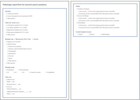
PATHOLOGIC REPORT FORM
The surgery TMC of the KGOG, in conjunction with the Gynecological Pathology Study Group, reviewed and analyzed several pathologic report forms (PRFs) for cervical cancer from domestic and international institutes. Finally, two kinds of PRF for cervical cancer were developed; one is for excision, and the other is for trachelectomy, hysterectomy, and pelvic exenteration. PRFs for cervical cancer include the following information (Figs. 2, 3, Supplementary Figs. 2, 3).
Pathologic report form for cervical cancer (excision)
Operation:
□Cold knife conization
□Loop electrosurgical excision procedure (LEEP)
□Other (specify) Tumor site: uterine cervix
□Left superior quadrant (12 to 3 o’clock),
□Left inferior quadrant (3 to 6 o’clock)
□Right inferior quadrant (6 to 9 o’clock)
□Right superior quadrant (9 to 12 o’clock)
□Other (specify)
Histologic type:□Microinvasive (T1a1, T1a2) □Invasive
□Squamous cell carcinoma
□Adenocarcinoma
□Endocervical,usualtype
□Mucinous (gastric/intestinal/signet-ring cell)
□Villoglandular
□Endometrioid
□Clear cell
□Serous
□Other (specify)
□Other (specify) Histologic grade:
□Keratinizing □Non-keratinizing
□G1 □G2 □G3 □Cannot be assessed □Not applicable
Tumor size:
□Microinvasive
Depth: mm
Horizontal extent: mm
□Invasive Greatest dimension: cm Additional dimensions (optional): × cm
Depth: mm
Margin:
(1) Endocervical Margin:
□Not involved: mm free from margin (specify location, if possible)
□Involved by invasive carcinoma/HSIL/LSIL/AIS (specify location, if possible) (2) Exocervical Margin:
□Not involved: mm free from margin (specify location, if possible)
□Involved by invasive carcinoma/HSIL/LSIL/AIS (specify location, if possible) (3) Deep Margin:
□Not involved: mm free from margin (specify location, if possible)
□Involved by invasive carcinoma/HSIL/LSIL/AIS (specify location, if possible) Vascular/lymphatic invasion:
□Absent □Present □Indeterminate
Fig. 2. Pathologic report form for cervical cancer managed by local excision. AIS, adenocarcinoma in situ; HSIL, high grade squamous intraepithelial lesion; LSIL, low grade squamous intraepithelial lesion.
Pathologicreportformforcervicalcancer(trachelectomy,hysterectomy,pelvicexenteration)
Operation:
□Simple trachelectomy □Radical trachelectomy
□Total hysterectomy □Radical hysterectomy
□Pelvic extenteration □Salpingectomy (right/left/bilateral)
□Salpingo-oophorectomy (right/left/bilateral) □Lymph node sampling/dissection (specify)
□Other (specify) Tumor site: uterine cervix
□Left superior quadrant (12 to 3 o’clock) □Left inferior quadrant (3 to 6 o’clock)
□Right inferior quadrant (6 to 9 o’clock) □Right superior quadrant (9 to 12 o’clock)
□Other (specify):
Histologic type:□Microinvasive (T1a1, T1a2) □Invasive
□Squamous cell carcinoma
□Adenocarcinoma
□Endocervical, usual type □Mucinous (gastric/intestinal/signet-ring cell)
□Villoglandular □Endometrioid
□Clear cell □Serous
□Other (specify)
□Other (specify) Histologic grade:
□Keratinizing □Non-keratinizing
□G1 □G2 □G3 □Cannot be assessed □Not applicable
Tumor size:
□Microinvasive
Depth: mm
Horizontal extent: mm
□Invasive Greatest dimension: cm Additional dimensions (optional): × cm
Depth: mm
Distal margin:
□Not involved: mm free from margin (specify location, if possible)
□Involved by invasive carcinoma/HSIL/LSIL/AIS (specify location, if possible)
Level 3, para-aortic infra-IMA: ( / ) Level 4, para-aortic infra-renal: ( / ) Other (specify)
Endocervical margin: (required in trachelectomy)
□Not involved: mm free from margin (specify location, if possible)
□Involved by invasive carcinoma/HSIL/LSIL/AIS (specify location, if possible) Parametrial invasion:
□Absent
□Present (right/left/bilateral): mm free from margin (optional) Vascular/lymphatic invasion:
□Absent □Present □Indeterminate
Other site involvement:
□Absent
□Present: Uterine corpus/Right ovary/Left ovary/Right salpinx/Left salpinx/Vagina/Urinary bladder/Rectum
□Other (specify)
Lymph node metastasis:□Absent □Present Greatest metastatic tumor dimension: mm Extranodal extent:□Absent,□Present ( mm)
Level 1, external and internal iliac (including obturator): Right ( / ), Left ( / ) Level 2, common iliac (including presacral): Right ( / ), Left ( / )
Fig. 3. Pathologic report form for cervical cancer managed by trachelectomy, hysterectomy, or pelvic exenteration. AIS, adenocarcinoma in situ; HSIL, high grade squamous intraepithelial lesion; LSIL, low grade
CONCLUSIONS
The purpose of this surgical manual is to facilitate future clinical trials and to improve communication between investigators by standardizing and describing operative procedures. The surgical procedures provided here represent the minimum requirements for participating in a clinical trial. These procedures should be systematically and properly described in the ORF, and the pathologic findings obtained from the procedures should be recorded in the PRF. This manual will be updated as necessary to include various clinical trials and to reflect the latest trends.
ACKNOWLEDGMENTS
For a clearer understanding of this KGOG classification of hysterectomy and
lymphadenectomy, a medical animation was produced by the HealthBreeze Corp. and is available on the KGOG website (http://goo.gl/aSuRo1).
SUPPLEMENTARY MATERIALS
Supplementary Fig. 1
Operation record form for cervical cancer Click here to view
Supplementary Fig. 2
Pathologic report form for cervical cancer (excision) Click here to view
Supplementary Fig. 3
Pathologic report form for cervical cancer (trachelectomy, hysterectomy, pelvic exenteration) Click here to view
REFERENCES
1. Querleu D, Morrow CP. Classification of radical hysterectomy. Lancet Oncol 2008;9:297-303.
PUBMED | CROSSREF
2. Fujii S, Takakura K, Matsumura N, Higuchi T, Yura S, Mandai M, et al. Precise anatomy of the vesico- uterine ligament for radical hysterectomy. Gynecol Oncol 2007;104:186-91.
PUBMED | CROSSREF
3. Roh JW, Lee DO, Suh DH, Lim MC, Seo SS, Chung J, et al. Efficacy and oncologic safety of nerve- sparing radical hysterectomy for cervical cancer: a randomized controlled trial. J Gynecol Oncol 2015;26:90-9.
PUBMED | CROSSREF
4. Piver MS, Rutledge F, Smith JP. Five classes of extended hysterectomy for women with cervical cancer.
Obstet Gynecol 1974;44:265-72.
PUBMED
5. Piver MS, Ghomi A. The twenty-first century role of Piver-Rutledge type III radical hysterectomy and FIGO stage IA, IB1, and IB2 cervical cancer in the era of robotic surgery: a personal perspective. J Gynecol Oncol 2010;21:219-24.
PUBMED | CROSSREF
6. Chan JK, Cheung MK, Huh WK, Osann K, Husain A, Teng NN, et al. Therapeutic role of lymph node resection in endometrioid corpus cancer: a study of 12,333 patients. Cancer 2006;107:1823-30.
PUBMED | CROSSREF
7. Panici PB, Scambia G, Baiocchi G, Matonti G, Capelli A, Mancuso S. Anatomical study of para-aortic and pelvic lymph nodes in gynecologic malignancies. Obstet Gynecol 1992;79:498-502.
PUBMED
8. Panici PB, Maggioni A, Hacker N, Landoni F, Ackermann S, Campagnutta E, et al. Systematic aortic and pelvic lymphadenectomy versus resection of bulky nodes only in optimally debulked advanced ovarian cancer: a randomized clinical trial. J Natl Cancer Inst 2005;97:560-6.
PUBMED | CROSSREF
9. Benedetti Panici P, Basile S, Maneschi F, Alberto Lissoni A, Signorelli M, Scambia G, et al. Systematic pelvic lymphadenectomy vs. no lymphadenectomy in early-stage endometrial carcinoma: randomized clinical trial. J Natl Cancer Inst 2008;100:1707-16.
PUBMED | CROSSREF
10. Chan JK, Kapp DS. Role of complete lymphadenectomy in endometrioid uterine cancer. Lancet Oncol 2007;8:831-41.
PUBMED | CROSSREF
11. Maggioni A, Benedetti Panici P, Dell’Anna T, Landoni F, Lissoni A, Pellegrino A, et al. Randomised study of systematic lymphadenectomy in patients with epithelial ovarian cancer macroscopically confined to the pelvis. Br J Cancer 2006;95:699-704.
PUBMED | CROSSREF
12. Hacker NF, Wain GV, Nicklin JL. Resection of bulky positive lymph nodes in patients with cervical carcinoma. Int J Gynecol Cancer 1995;5:250-6.
PUBMED | CROSSREF