Abdominal wall bleeding occurs in traumatized pa- tients or in patients after anticoagulation therapy with
the use of heparin or warfarin, and it also occurs as a complication of paracentesis, a femoral artery puncture for vascular intervention, or laparoscopic surgery (1-6).
Even though conservative management is the treatment of choice unless bleeding is severe or the course is com- plicated by infection, abdominal wall bleeding often re- quires emergency or urgent treatment that includes manual compression, surgery, or interventional em-
Embolotherapy Using N-butyl Cyanoacrylate for Abdominal Wall Bleeding
1Young Ho Choi, M.D., Young Hwan Koh, M.D., Daehee Han, M.D., Ji-hoon Kim, M.D., Joo Hee Cha, M.D., Eun Hye Lee, M.D., Chi Sung Song, M.D.
1Department of Radiology, Seoul National University Boramae Hospital Received June 24, 2007 ; Accepted November 17, 2007
Address reprint requests to : Young Ho Choi, M.D., Department of Radiology, Seoul National University Boramae Hospital, 425 Shindaebang-2-dong, Dongjak-gu, Seoul 156-707, Korea.
Tel. 82-2-840-2671 Fax. 82-2-831-2826 E-mail: [email protected]
Purpose: We describe our experience with the use of N-butyl cyanoacrylate (NBCA) embolization of abdominal wall bleeding and we evaluate the clinical effectiveness of the procedure.
Materials and Methods: Embolization was performed in nine patients with abdominal wall bleeding. The sites of embolization were the left first lumbar (n = 1), left second lumbar (n = 1), right inferior epigastric (n = 2), left inferior epigastric (n = 3), right cir- cumflex iliac (n = 1), and left circumflex iliac artery (n = 1). A coil was used with NBCA in one patient due to difficulty in selecting only a bleeding focus and anticipat- ed reflux. NBCA was mixed with Lipiodol at the ratio of 1:1 to 1:4. Blood pressure and heart rate were measured before and after the embolization procedure, and the serial hemoglobin and hematocrit levels and transfusion requirements were reviewed to evaluate hemostasis and rebleeding.
Results: Hemostasis was obtained in six out of the nine patients and technical success was achieved in all patients. There were no procedure-related complications. Four out of the nine patients died due to rebleeding of a subarachnoid hemorrhage (n = 1), mul- tiorgan failure (n = 1), and hepatic failure (n = 2) that occurred two to nine days after the embolization procedure. One patient had rebleeding. The five surviving patients had no rebleeding, and the patients continue to visit the clinic on an outpatient basis.
Conclusion: NBCA embolization is a clinically safe procedure and is effective for ab- dominal wall bleeding.
Index words :Abdominal wall Arteries
Hemorrhage
Embolization, therapeutic
bolotherapy (7-11). Manual compression is a noninva- sive procedure, and is considered as the first choice for abdominal wall bleeding. However, the use of manual compression is limited as it may be difficult to achieve compression with sufficient pressure exactly on the bleeding foci, even though ultrasonography can assist the physicians to localize the bleeding foci (12, 13).
Surgery is an invasive procedure, and surgeons prefer not to directly operate on patients without first attempt- ing to use other methods, especially immediately after
patients have undergone surgery for some other reason.
Recently, embolotherapy has been used in patients with bleeding at various sites with good outcomes (14-16).
Embolotherapy for abdominal wall bleeding can be a good substitution procedure in place of manual com- pression and surgery as it is less invasive than surgery and is more precise than the use of manual compres- sion.
Various embolic agents such as coils, gelfoam, polyvinyl alcohol particles or n-butyl cyanoacrylate
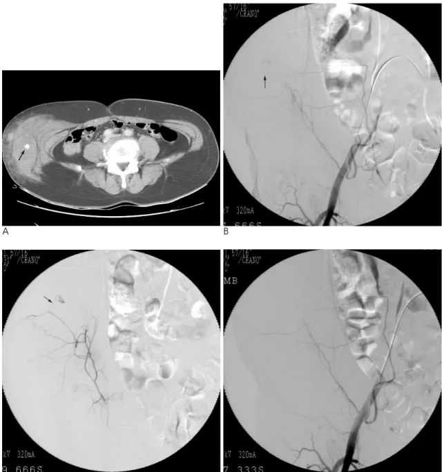
A B
C D
Fig. 1. NBCA embolization performed for abdominal wall bleeding (Patient No. 2).
A. A computed tomography scan demonstrates a bleeding focus (arrow) with a large hematoma at the right lateral abdominal wall.
B, C. A right external arteriogram and a selective right circumflex iliac arteriogram show a feeding artery (arrow) of the bleeding fo- cus that originates from a branch of the right circumflex iliac artery.
D. An arteriogram after embolization demonstrates no further bleeding.
(NBCA; B. Braun, Melsungen, Germany) are used to control bleeding (17-20). Liquid embolic agents, that can cast bleeding foci might be more effective for ab- dominal wall bleeding as the vascular system in the ab- dominal wall is in the form of a network and may per- mit adjacent collateral channels to feed bleeding foci, even when a feeding artery is occluded. We have used NBCA for abdominal wall bleeding and have evaluated its clinical effectiveness in this retrospective study.
Materials and Methods
NBCA embolization was performed in nine out of 11 patients with abdominal wall bleeding between September 2003 and July 2007 (Fig. 1). We used gelfoam or a coil for two patients as it was not possible to ap- proach the feeding branches sufficiently close to use NBCA. The nine patients that received NBCA emboliza- tion ranged in age from 18 to 78 years and the mean age was 50 years. The patient population included five men and four women. Embolization was performed in pa- tients with hematoma at the right flank (one patient), left flank (one patient), right anterior abdominal wall (two patients), left anterior abdominal wall (four patients), and left retroperitoneum (one patient). A coil was used along with NBCA in one patient with a hematoma at the
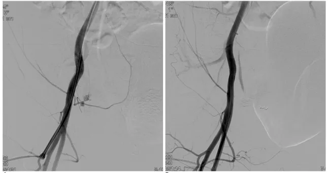
right anterior abdominal wall due to difficulty in select- ing only a bleeding focus, and due to anticipated reflux (Fig. 2).
Bleeding foci were located in the left first lumbar artery (one patient), left second lumbar artery (one pa- tient), right inferior epigastric artery (two patients), left inferior epigastric artery (three patients), right circum- flex iliac artery (one patient), and left circumflex iliac artery (one patient). The presence of a bleeding focus was confirmed on angiography. Underlying diseases are listed in Table 1.
This study was performed retrospectively with data obtained from medical records and telephone interview with the patients. The study received institutional re- view board approval and informed consent was ob- tained from patients and from patient families prior to undertaking the embolization procedure. Computed to- mography images obtained prior to embolization were reviewed to determine the type of underlying disease, the presence of arterial bleeding foci and the location of the foci.
Blood pressure and heart rate were measured before and after the embolization procedure, and serial hemo- globin and hematocrit levels and transfusion require- ments were reviewed to evaluate hemostasis and re- bleeding. A sudden stop of bleeding causes a change in
A B
Fig. 2. NBCA embolization performed in combination with a coil (Patient No. 6).
A. A right external iliac arteriogram demonstrates contrast extravasation from the right inferior epigastric artery.
B. Due to difficulty in selecting only a bleeding focus and anticipated reflux, we embolized the distal part of the mother vessel with a coil and then injected the NBCA mixture at the proximal part of the vessel. A post-embolization angiogram shows no further ex- travasation.
blood pressure and heart rate that can be an indicator of whether embolization was effective. Technical success was defined as bleeding foci no longer seen by the use of angiography after embolization, hemostasis was defined as bleeding that disappeared following angiographic and clinical evaluation and rebleeding was defined as bleed- ing that appeared again at angiographic or clinical evalu- ation.
Embolization Techniques
Angiography with the digital subtraction technique, abdominal aortography and selective arteriography were performed to detect bleeding foci. Arteriography in the lumbar artery, external iliac artery, inferior epi- gastric artery, and circumflex iliac artery were per- formed. A 5 Fr RH catheter or Davis catheter (Cook, Bloomington, IN U.S.A.) were used to select the lumbar artery and external iliac artery, and Microferret catheters and wires (Cook) or a Progreat catheter and
0.018 inch GT wire (Terumo, Tokyo, Japan) were used to select the inferior epigastric artery and circumflex ili- ac artery. The approach to the bleeding foci was per- formed cautiously to avoid triggering a vascular spasm where it impossible to inject the NBCA mixture or cause a delay in the procedure.
NBCA was prepared carefully without contact with ionic fluid or blood that causes liquid NBCA to polymer- ize and become solid. NBCA was mixed with iodized oil (Lipiodol; Laboratoire Guerbet, Roissy, France) at a ratio of 1:1 to 1:4, and the mixture provides radiopacity under fluoroscopy. A lower ratio (the use of additional Lipiodol) prolongs the time for NBCA to be solidify and is useful for embolizing bleeding foci that are difficult to approach and are located far from a microcatheter tip.
The NBCA mixture was injected into the bleeding foci after several test injections of liquid contrast agent to de- termine the injection power and to prevent a reflux to a Table 1. Clinical Findings in Nine Patients that Underwent NBCA Embolization
Patient Age/ Underlying Hematoma Bleeding Embolic Ratio of
Clinical No. Sex condition location foci agents NBCA Complications Hemostasis Rebleeding
follow-up used and Lipiodol
1 59/F Subarachnoid Left Left NBCA 1:1 No Yes No 2 days: death,
hemorrhage, flank second rebleeding of
disseminated lumbar subarachnoid
intravascular artery hemorrhage
coagulation
2 34/M Fall Right Right NBCA 1:3 No Yes No 10 months:
flank circumflex out
iliac artery patient visit
3 53/M Infectious Left Left first NBCA 1:2 No No No 2 days: death,
spondylitis, retroperi- lumbar multiorgan
LC toneum artery failure
4 50/M LC, ascites Left anterior Left inferior NBCA 1:2 No No No 2 days: death,
tapping abdominal epigastric hepatic
wall artery failure
5 49/F LC, ascites Left anterior Left inferior NBCA 1:4 No No Yes 9 days: death,
tapping abdominal epigastric hepatic
wall artery failure,
coagulopathy
6 59/F Angina, Right Right NBCA, coil 1:3 No Yes No 16 months:
coronary anterior inferior outpatient
angiography abdominal epigastric visit
wall artery
7 49/M Abdominal Right anterior Right inferior NBCA 1:3 No Yes No 11 months:
wall pain abdominal epigastric outpatient
after exercise wall artery visit
8 78/F Angina, Left anterior Left NBCA 1:3 No Yes No 10 months:
coronary abdominal circumflex outpatient
angiography wall iliac artery visit
9 18/M Repair of Left anterior Left inferior NBCA 1:3 No Yes No 8 months:
colostomy abdominal epigastric outpatient
wall artery visit
Note─ LC, liver cirrhosis; NBCA, N-butyl cyanoacrylate
proximal feeding artery or an embolism to non-target or- gans. The use of test injections was useful even when the viscosity of the contrast agent and NBCA mixture were different and there was a difference in the injec- tion power for the contrast agent and the NBCA mix- ture. Microcatheters were flushed with 5% or 10% glu- cose fluid before injection of the NBCA mixture.
Microcatheters were removed out of the body 1 to 2 seconds after injection of the NBCA mixture to prevent a cast of the NBCA mixture at the bleeding foci from be- ing attached to the tips of the microcatheters. Follow-up angiography was performed after embolization.
Results
Technical success with the use of NBCA for abdomi- nal wall bleeding was achieved in all nine patients even though a coil was added along with NBCA in one pa- tient. Hemostasis was obtained in six out of nine pa- tients and three patients did not show rise of a blood pressure and decline of heart rate before and after the embolization procedure. Procedure-related complica- tions did not occur in any patients.
Patients underwent clinical follow-up for 2 days to 16 months with a mean duration of 6 months. Four out of nine patients died two to nine days after embolization.
One male patient (Patient No. 3) had liver cirrhosis and underwent an L1 partial corpectomy and anterior fusion with the use of a mesh graft (T12-L2) due to infectious spondylitis (T11-L3) with a compression fracture (T11- L2). The patient was diagnosed with a large hematoma at the left retroperitoneum one day after surgery and the patient died of a multiorgan failure even though the bleeding focus, a branch of the left first lumbar artery, was embolized with the use of NBCA. Another female patient (Patient No. 1) had a hematoma at the left flank fed by a branch of the left second lumbar artery one day after a diagnosis of a subarachnoid hemorrhage and dis- seminated intravascular coagulation. The patient died due to rebleeding of a subarachnoid hemorrhage. The other two patients (Patient No. 4 and 5) had liver cirrho- sis with a large amount of ascites, and a hematoma at the left anterior abdominal wall that occurred after as- cites tapping. The patients died of a hepatic failure.
Patient No. 5 had rebleeding at the left inferior epigas- tric artery, but the focus was different from the initial bleeding focus. The remaining five patients had no re- bleeding and continue to be seen as outpatients.
Discussion
In this study, we used NBCA as an embolic agent for abdominal wall bleeding as we expected that the use of NBCA would provide better clinical effectiveness for hemostasis. The use of liquid embolic agents may be more favorable for a vascular system that is in the form of a network and may permit adjacent collateral chan- nels to feed bleeding foci, even when a feeding artery is occluded (17).
NBCA is a liquid agent, and it is polymerized if ex- posed to ionic materials (21-24). NBCA polymerization can be predictably prolonged by the addition of Lipiodol. The prolongation may help NBCA reach bleeding foci far from a microangiocather tip as early polymerization causes NBCA to form a cast before its ar- rival at the bleeding foci. We mixed NBCA with Lipiodol in the ratio of 1:1 to 1:4, and ratios of 1:1 or 1:2 were adopted when the catheter closely approached the bleeding foci and ratios of 1:3 or 1:4 were adopted when it was not possible to reach the bleeding foci closely.
Microcatheters were retracted from the body 1 to 2 sec- onds after the injection of the NBCA mixture. A quick removal was performed to prevent an NBCA cast at the bleeding foci from being attached to the microcatheter tip.
NBCA embolization can be performed effectively for various forms of bleeding. Kish et al. (17) reported that NBCA embolization was feasible and effective in pa- tients with arterial bleeding from various etiologies and at various anatomic sites. The procedure was used to embolize brain arteriovenous malformations (AVMs), and the use of NBCA has been applied for various appli- cations including craniofacial AVMs and extremity AVMs, hemangiomas in the mandible, arteriovenous fistulas with a long fistula tract, hypervascular tumora, varicocelea, gastric varices, the portal vein before a par- tial hepatectomy, intractable epistaxis and acute arterial hemorrhage (25-33).
NBCA can be very useful in controlling arterial bleed- ing along with other embolic agents such as coils or gelfoam. Yamakado et al. (34) reported that a combina- tion of the use of coils and NBCA was useful for a rup- tured pseudoaneurysm that was difficult to embolize with the use of a coil alone. A patient in this study un- derwent NBCA embolization with a coil due to difficulty in superselecting a bleeding focus.
Currently, it is not difficult to detect a bleeding focus
and its origin in a patient with a hematoma at the ab- dominal wall with the availability of powerful imaging tools such as sonography, CT, and angiography.
However, the presence of a hematoma could not be as- certained solely with clinical information and without the benefit of imaging. In this study, a hematoma at the anterior abdominal wall originated from the inferior epi- gastric artery or circumflex iliac artery, a hematoma originated at the flank from the circumflex iliac artery and lumbar artery, and a hematoma originated at the retroperitoneum from the lumbar artery. Although these findings may suggest possible origins of a hematoma in a clinical setting, it is not reasonable to generalize the use of these findings from this study.
Procedure-related complications were not found in any of the patients during this study. The amount of NBCA used in a patient was less than 0.25 mL when the mixing ratio was taken into account; therefore, side ef- fects or complication due to NBCA itself may be mini- mal to disregard. However, the possibility of complica- tions for NBCA embolization may be higher than for embolization with other embolic agents as NBCA is dif- ficult to handle and familiarity with the NBCA em- bolization procedure can be time consuming.
A limitation of this study was its retrospective design.
A small number of patients is another limitation.
In summary, NBCA embolization is a clinically safe and effective procedure for the treatment of abdominal wall bleeding, and the procedure may require the use of additional embolic agents for a satisfactory outcome.
References
1. Geraci G, Sciume C, Pisello F, Li Volsi F, Facella T, Modica G.
Trocar-related abdominal wall bleeding in 200 patients after la- paroscopic cholecistectomy: personal experience. World J Gastroenterol 2006;12:7165-7167
2. Shimizu T, Hanasawa K, Yoshioka T, Mori T, Kajinami T, Yokoyama K, et al. Spontaneous hematoma of the lateral abdomi- nal wall caused by a rupture of a deep circumflex iliac artery: re- port of two cases. Surg Today 2003;33:475-478
3. Nu′˜ez Martinez O, Carneros Martin JA, Garcia Sa′n nchez A, Santos Castro L, de Diego Lorenzo A, Salcedo Plaza M, et al. Abdominal wall hematoma: a complication of paracentesis. Rev Esp Enferm Dig 2001;93:127-128
4. Kao CL, Chang JP. Abdominal wall hematoma as a complication of warfarinization. J Emerg Med 2001;20:293
5. Egger B, Schweizer W, Wagner HE. Acute abdomen in abdominal wall hemorrhage in anticoagulation. Helv Chir Acta 1992;59:399- 402
6. Goi M, Fiori MG, Vergara V, Natta F, Garbarini A. Spontaneous hematoma of the abdominal wall. Minerva Chir 1990;45:739-742 7. Martin-Malago′n A, Arteaga I, Rodriguez L, Alarco-Hernandez A.
Abdominal wall hematoma after laparoscopic surgery: early treat- ment with selective arterial transcatheter embolization. J Laparoendosc Adv Surg Tech A 2007;17:781-783
8. Katsumori T, Nakajima K. A case of spontaneous hemorrhage of the abdominal wall caused by rupture of a deep iliac circumflex artery treated by transcatheter arterial embolization. Eur Radiol 1998;8:550-552
9. Savage PE, Joseph AE, Adam EJ. Massive abdominal wall hematoma: real-time ultrasound localization of bleeding. J Ultrasound Med 1985;4:157-158
10. Lohri A, Petralli C, Kummer H. Abdominal wall hematoma as a severe complication of anticoagulation. Clinical picture and ultra- sonic diagnosis. Schweiz Med Wochenschr 1984;114:1761-1762 11. Andersen E, Skadberg JE. Abdominal wall hematoma. Tidsskr Nor
Laegeforen 1992;112:2529-2530
12. Castan˜eda F, Swischuk JL, Smouse HB, Brady T. Gelatin sponge closure device versus manual compression after peripheral arterial catheterization procedures. J Vasc Interv Radiol 2003;14:1517-1523 13. Tron C, Koning R, Eltchaninoff H, Douillet R, Chassaing S,
Sanchez-Giron C, et al. A randomized comparison of a percuta- neous suture device versus manual compression for femoral artery hemostasis after PTCA. J Interv Cardiol 2003;16:217-221
14. Charbonnet P, Toman J, Buhler L, Vermeulen B, Morel P, Becker CD, et al. Treatment of gastrointestinal hemorrhage. Abdom Imaging 2005;30:719-726
15. Kuo WT. Transcatheter treatment for lower gastrointestinal hem- orrhage. Tech Vasc Interv Radiol 2004;7:143-150
16. Frisoli JK, Sze DY, Kee S. Transcatheter embolization for the treat- ment of upper gastrointestinal bleeding. Tech Vasc Interv Radiol 2004;7:136-142
17. Kish JW, Katz MD, Marx MV, Harrell DS, Hanks SE. N-butyl cyanoacrylate embolization for control of acute arterial hemor- rhage. J Vasc Interv Radiol 2004;15:689-695
18. Bandi R, Shetty PC, Sharma RP, Burke TH, Burke MW, Kastan D.
Superselective arterial embolization for the treatment of lower gas- trointestinal hemorrhage. J Vasc Interv Radiol 2001;12:1399-1405 19. Aina R, Oliva VL, Therasse E, Perreault P, Bui BT, Dufresne MP,
et al. Arterial embolotherapy for upper gastrointestinal hemor- rhage: outcome assessment. J Vasc Interv Radiol 2001;12:195-200 20. Kuo WT, Lee DE, Saad WE, Patel N, Sahler LG, Waldman DL.
Superselective microcoil embolization for the treatment of lower gastrointestinal hemorrhage. J Vasc Interv Radiol 2003;14:1503- 1509
21. Brothers MF, Kaufmann JC, Fox AJ, Deveikis JP. N-Butyl 2-cyano- acrylate--substitute for IBCA in interventional neuroradiology:
histopathologic and polymerization time studies. AJNR Am J Neuroradiol 1989;10:777-786
22. Oowaki H, Matsuda S, Sakai N, Ohta T, Iwata H, Sadato A, et al.
Non-adhesive cyanoacrylate as an embolic material for endovascu- lar neurosurgery. Biomaterials 2000;21:1039-1046
23. Gounis MJ, Lieber BB, Wakhloo AK, Siekmann R, Hopkins LN.
Effect of glacial acetic acid and ethiodized oil concentration on em- bolization with N-butyl 2-cyanoacrylate: an in vivo investigation.
AJNR Am J Neuroradiol 2002;23:938-944
24. Lieber BB, Wakhloo AK, Siekmann R, Gounis MJ. Acute and chronic swine rete arteriovenous malformation models: effect of ethiodol and glacial acetic acid on penetration, dispersion, and in- jection force of N-butyl 2-cyanoacrylate. AJNR Am J Neuroradiol 2005;26:1707-1714
25. Denys A, Lacombe C, Schneider F, Madoff DC, Doenz F, Qanadli SD, et al. Portal vein embolization with N-Butyl cyanoacrylate be- fore partial hepatectomy in patients with hepatocellular carcinoma
and underlying cirrhosis or advanced fibrosis. J Vasc Interv Radiol 2005;16:1667-1674
26. Onal B, Ilgit ET, Akpek S, Coskun B. Postcatheterization femoral arteriovenous fistula: endovascular treatment with N-butyl-cyano- acrylate embolization. Cardiovasc Intervent Radiol 2006;29:276-278 27. Heye S, Maleux G, Wilms G. Pain experience during internal sper-
matic vein embolization for varicocele: comparison of two cyano- acrylate glues. Eur Radiol 2006;16:132-136
28. Kiyosue H, Matsumoto S, Yamada Y, Hori Y, Okino Y, Okahara M, et al. Transportal intravariceal sclerotherapy with N-butyl-2- cyanoacrylate for gastric varices. J Vasc Interv Radiol 2004;15:505- 509
29. Kaneko R, Tohnai I, Ueda M, Negoro M, Yoshida J, Yamada Y.
Curative treatment of central hemangioma in the mandible by di- rect puncture and embolisation with n-butyl-cyanoacrylate (NBCA). Oral Oncol 2001;37:605-608
30. Liu D, Ma XC. Clinical study of embolization of arteriovenous
malformation in the oral and maxillofacial region. Chin J Dent Res 2000;3:63-70
31. Gruber A, Bavinzski G, Killer M, Richling B. Preoperative em- bolization of hypervascular skull base tumors. Minim Invasive Neurosurg 2000;43:62-71
32. Luo CB, Teng MM, Lirng JF, Chang FC, Chen SS, Guo WY, et al.
Endovascular embolization of intractable epistaxis. Zhonghua Yi Xue Za Zhi 2000;63:205-212
33. Casasco A, Herbreteau D, Houdart E, George B, Tran Ba Huy P, Deffresne D, et al. Devascularization of craniofacial tumors by percutaneous tumor puncture. AJNR Am J Neuroradiol 1994;15:
1233-1239
34. Yamakado K, Nakatsuka A, Tanaka N, Takano K, Matsumura K, Takeda K. Transcatheter arterial embolization of ruptured pseudoaneurysms with coils and n-butyl cyanoacrylate. J Vasc Interv Radiol 2000;11:66-72
대한영상의학회지 2008;58:461-467
복벽 출혈에서 N-butyl Cyanoacrylate을 이용한 색전술1
1서울시립보라매병원 영상의학과
최영호・고영환・한대희・김지훈・차주희・이은혜・송치성
목적: 복벽 출혈에서의 NBCA 색전술에 대한 저자들의 경험을 기술하고 그것의 임상적 유용성을 평가하였다.
대상과 방법: 복벽 출혈이 있는 9명의 환자에서 NBCA 색전술이 제1 좌요동맥(n=1), 제2 좌요동맥(n=1), 우아래
배벽동맥(n=2), 좌아래배벽동맥(n=3), 우장골휘돌이동맥(n=1), 좌장골휘돌이동맥(n=1)에서 시행되었다. 1예에서 출혈점만을 선택적으로 색전하는 것이 쉽지 않고 역류의 가능성이 있어 코일이 NBCA와 같이 사용되었다. NBCA 는 Lipiodol과 1:1 내지 1:4로 섞어 사용되었다. 색전술 전후의 혈압, 심박 수, 헤모글로빈 수치, 헤마토크릿 수치 그 리고 수혈 여부 등이 지혈, 재출혈 여부 평가를 위해 조사되었다.
결과: 기술적 성공은 모든 예에서 얻어졌지만 지혈은 9명 중 6명에서 얻어졌다. 시술과 관련된 부작용은 없었다. 9 명 중 4명이 거미막밑출혈의 재발(n=1), 다장기부전(n=1), 간부전(n=2)으로 NBCA 색전술을 시행 받은 지 2일 내지 9일 후에 사망하였다. 그 중 한 예에서 재출혈이 있었다. 나머지 5명은 재출혈이 없었으며 외래 관찰 중이다.
결론:복벽 출혈에서 NBCA 색전술은 안전하고 효과적일 수 있다고 생각된다.