Papillary Tu m o rs of the Breast: US Findings of the Benign and Malignant Lesions1
Chang-Seok Lee, M.D., Shin-Ho Kook, M.D., Hyun-Ja Shin, M.D.2, Woo-Kyung Moon, M.D.3, Eun-Joo Ko, M.D.4, Young-Uk Lee, M.D., Young-Rae Lee, M.D., Eun-Kyung Yoon, M.D.,
Eun-Chul Chung, M.D., Hae-Won Park, M.D.
Purpose: To determine which sonographic findings usefully differentiate between be- nign and malignant papillary tumors.
Materials and Methods: We retrospectively rev i ewed the ultrasonographic findings of 42 surgically proven cases of papillary breast lesions [11 malignant lesions (7 inva s i ve papillary carcinomas, 4 intraductal papillary carcinomas) and 31 benign intraductal pa- pillomas]. All 42 cases were classified sonographically as cystic or ductal, or solid type, and the shape, wall change, margin, internal echo-pattern, posterior echo change and other associated findings for the two types were then analysed.
Results: Among the 25 cases (5 malignant and 20 benign) of cystic or ductal type, tubu- lar shaped lesions were more frequently benign (60%). In all 20 benign lesions the wa l l of cystic portion was we l l - d e f i n e d, smooth and thin. The solid portion of the cystic type s h owed an ill-defined irregular margin in four malignant lesions (80%) and a smooth margin in 19 which were benign (95%). The internal echo-pattern was heterogeneous m i xed-echo in three cases of malignancy, and homogeneously hypoechoic in 19 benign lesions (95%). Posterior enhancement was seen in two malignant lesions (40%), while in 19 benign lesions (95%), there was no posterior echo change.
There were 17 solid type lesions (6 malignant cases, 11 benign cases), and most of these, whether benign or malignant, were smooth, oval or lobulated, hypoechoic masses.
Posterior enhancement, howeve r, was more frequently observed in malignant lesions (three cases, 50%) than in those which were benign (one case, 9%).
Conclusion: In cystic or ductal type lesions, an ill-defined irregular thick cystic wall, an ill-defined irregular margin, a heterogeneous mixed internal echo-pattern and posterior enhancement of the solid portion suggested malignancy. In solid type lesions, posterior enhancement was more frequently found in malignant than in benign lesions.
Index words :Breast, neoplasms Breast, US
1Department of Radiology, Kangbuk Samsung Hospital, Sungkyunkwan University, School of Medicine;
2Department of Radiology, Korea Veterans Hospital
3Department of Radiology, Seoul National University Hospital
4Department of Radiology, Eulji Hospital, Eulji College of Medicine Received November 9, 1999 ; Accepted March 3, 2000
Address reprint requests to : Shin-Ho Kook, M.D., Department of Radiology, Kangbuk Samsung Hospital, Sungkyunkwan University, School of Medi-cine 108, Pyeong-Dong, Chong-ro-Gu
Tel. 82-2-2001-2335 Fax. 82-2-2001-2797 E-mail: [email protected]
Papillary tumors of the breast account for less than 10
% of benign breast neoplasms and 1-2% of breast carci- nomas (1-3). Both benign papillomas and papillary car- cinomas show macroscopic villous arborescent lesions and can present as intraductal, intracystic or solid-type lesions. Papillary carcinomas may occur in situ or be in- vasive (4, 5), while the intracystic type is a variant of in- traductal papillary carcinoma, taking the form of papil- lary fronds within the wall of a cystically dilated duct (6, 7). If cell proliferation becomes so dense that the basic papillary properties are obscured, the term ‘solid papil- lary carcinoma’ is used.
The purpose of this study is to determine which sono- graphic findings usefully differentiate between benign and malignant papillary tumors.
Materials and Methods
We retrospectively reviewed the clinical presentation and breast sonographic findings in 42 cases of surgically proven papillary tumors [11 cases of malignancy (inva- sive papillary carcinoma, n=7; intraductal papillary car- cinoma, n=4) and 31 cases of intraductal papilloma].
Patients with malignant lesions ranged in age from 43 to 79 (mean, 61) and those with benign lesions from 30 to
72 (mean, 51) years. The clinical findings are summa- rized in Table 1. We used high-resolution real time ultra- sound units (Acuson 128XP/10, Mountain View, California, U.S.A., and Logiq 700, General Electric, Milwaukee Wis., U.S.A.) with 5 to 10 MHz linear array transducers. Sonographically, lesions were classified as cystic or ductal, or solid type, and for each of these two groups, the sonographic findings were analysed with re- gard to shape, change of ductal or cystic wall, margin, internal echo-pattern, lateral shadowing, posterior echo change, duct dilatation, obliteration of adjacent normal structure, and thick peritumoral echo. To determine sta-
Table 1. Clinical Symptoms and Signs of Papillary Lesions Clinical Findings Discharge Mass S c r e e n i n g Pathology serous bloody milkish d e t e c t
Papillary 2 1 6
Ca (n=7)
Intraductal papillary 1 4 Ca (n=4)
Papilloma (n=31) 8 12 2 11 2
Ca : Carcinoma
Table 2. Number, Location and Size of Papillary Lesions on Ultra- s o n o g r a p h y
P a t h o l o g y M a l i g n a n t B e n i g n Papillary Ca IDPCa Total P a p i l l o m a US Findings (n=7) (n=4) (n=11) (%) (n=31) (%) N u m b e r *
S i n g l e 6 3 09 (81) 11 (35)
multiple 1 1 02 (19) 20 (65)
L o c a t i o n
s u b a r e o l a r 2 02 (18) 13 (42)
central 3 2 05 (46) 15 (49)
p e r i p h e r a l 2 2 04 (36) 3 (9)
Size (mm) 1 0-4 2 7-1 2 0 3 - 3 3
m e a n 2 6 6 3 . 5 1 8
*P <0.05, IDPCa; Intraductal Papillary Carcinoma
Table 3. Sonographic Types of Papillary Lesions
Pathology/US Type I n t r a c y s t i c I n t r a d u c t a l Solid
Papillary Ca (n=7) 3 01 03
Intraductal papillary Ca 1 03
( n = 4 )
Papilloma (n=31) 4 1 6 1 1
Ca : Carcinoma
Table 4. Sonographic Findings of Cystic or Ductal Typed Papil- lary Lesions
Pathology Malignant B e n i g n
Papillary Ca IDPCa Total P a p i l l o m a
US Findings n=4 n=1 n=5(%) n = 2 0 ( % )
S h a p e *
oval/ lobulated 3 1 4 (80)0 07 (35)0
i r r e g u l a r / l o b u l a t e d 1 1 (20)0 01 (5)0 0
t u b u l a r 12 (60)0
Wall(cyst or duct)
s m o o t h / t h i n 3 1 4 (80)0 20 (100) i r r e g u l a r / t h i c k 1 1 (20)0
Margin* (solid portion)
S m o o t h 1 1 (20)0 19 (95)0
I r r e g u l a r 4 4 (80)0 01 (5)0 0
Internal echo-pattern (cystic portion)
a n e c h o i c / h o m o g e n e o u s 4 1 5 (100) 20 (100) (solid portion)*
a) homogeneous 2 2 (40)0 19 (95)0
h e t e r o g e n e o u s 2 1 3 (60)0 01 (5)0 0
b) hypo-echoic 2 2 (40)0 19 (95)0
h y p e r - e c h o i c 1 1 (20)0 01 (5)0 0
m i x e d - e c h o i c 2 02 (40)0
Posterior change*
e n h a n c e m e n t 1 1 2 (40)0
shadowing 1 1 (20)0 01 (5)0 0
no change 2 2 (40)0 19 (95)0
Ductal dilatation* 1 1 (20)0 16 (80)0
Others
obliteration of adjacent
normal structure* 2 1 3 (60)0
thick peritumoral echo 2 2 (40)0 01 (5)0 0
* p<0.05, IDPCa; Intraductal Papillary Carcinoma
tistical significance, Fisher’s-exact test was performed.
R e s u l t s
The number, location and size of papillary lesions seen on ultrasonography are shown in Table 2. Sixty- five percent of benign lesions were multiple and 81% of malignant lesions were single. As for the sonographic types, the malignant lesions were predominantly solid or cystic but in the case of benign lesions, ductal or solid types were dominant (Table 3).
The sonographic findings of cystic and ductal type
papillary lesions are summarized in Table 4 and shown in Figure 1. There were five malignant and 20 benign le- sions. Twelve of the latter (60%) were tubular in shape.
The margin of the solid portion was irregular in four ma- lignant lesions (80%), and smooth in 19 benign lesions (95%). As for the internal echo-pattern, this was hetero- geneously mixed-echoic in the solid portion of three ma- lignant lesions (60%), and homogeneously hypo-echoic in 19 benign lesions (95%). Posterior enhancement of the solid portion was seen in two malignant tumors (40%), but in 19 benign lesions (95%) there was no pos- terior echo change. Ductal obstruction or dilatation was
A B
C
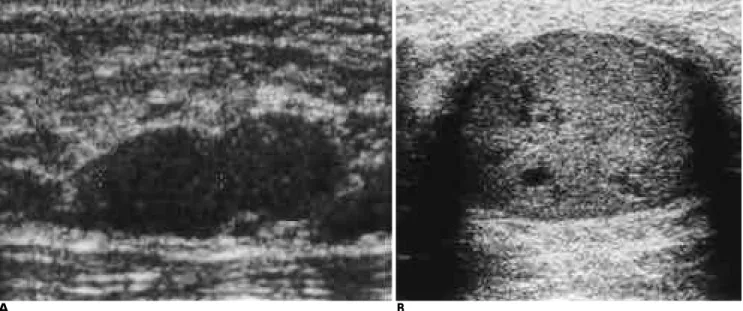
Fig. 1. Cystic or ductal typed benign and malignant papillary lesions.
Intracystic (A) and multiple intraductal papillomas (B) show smooth well-defined homogeneous mass within the cyst or ducts (white arrows; solid portion, black arrows; fluid in the duct). Intraductal papillomas show tubular in shape associated with diffuse ductal dilatation. Comparing to intracystic papilloma, intracystic papillary carcinomas show ill-defined irregular margin with inva- sion of cystic wall (C, arrows) or smoothly well-defined margin without invasion of the cyst (D, arrows), and both lesions show het- erogeneous mixed internal echo pattern that including low echoic cystic areas, and posterior enhancement of solid portion.
D
associated more with benign (80%) than malignant (20%) lesions, and three malignant lesions (60%) showed obliteration of adjacent normal structure. All the above findings were statistically significant ( p < 0 . 0 5 ) .
The sonographic findings of solid type papillary le- sions are shown in Table 5 and Figure 2. There were six malignant and 11 benign cases, with virtually no signifi- cant difference between benign and malignant lesions;
most of both these types were oval or lobulated in shape, had a smooth margin, and showed a homo- or heterogeneously hypo-echoic internal echo-pattern.
Posterior enhancement, however, was more frequently seen in malignant (three cases, 50%) than in benign le- sions (one case, 9%), a difference which was statistically significant (p<0.05).
D i s c u s s i o n
Papillary disease of the breast involves a spectrum of the entities which includes both benign and malignant lesions. The most common papillary breast neoplasm is
A B
C
Fig. 2. Solid typed benign and malignant papillary lesions.
Solid typed papilloma shows well-defined bilobed (A) homoge- neous mass without posterior change. Papillary carcinomas show well-defined margin, heterogeneous (B) or homogeneous (C) inter- nal echo pattern that similar with papilloma, but posterior en- hancement of papillary carcinomas are remarkable and character- i s t i c .
Table 5. Sonographic Findings of Solid Typed Papillary Lesions
Pathology Malignant B e n i g n
Papillary Ca IDPCa Total P a p i l l o m a
US Findings n=3 n=3 n=6(%) n = 1 1 ( % )
S h a p e
o v a l / l o b u l a t e d 3 3 6 (100) 11 (100) M a r g i n
s m o o t h 3 3 6 (100) 10 (91)0
i r r e g u l a r 01 (9)0 0
Internal echo-pattern
a) homogeneous 1 2 3 (50)0 08 (73)0
h e t e r o g e n e o u s 2 1 3 (50)0 03 (27)0
b) hypo-echoic 3 2 5 (83)0 09 (82)0
h y p e r - e c h o i c 01 (9)0 0
m i x e d - e c h o i c 1 1 (17)0 01 (9)0 0 Posterior change*
e n h a n c e m e n t 2 1 3 (50)0 01 (9)0 0
s h a d o w i n g 01 (9)0 0
no change 1 2 3 (50)0 09 (82)0
O t h e r s
thick peritumoral echo 1 1 (17)0 02 (18)0
* P < 0.05, IDPCa; Intraductal Papillary Carcinoma
the papilloma, which consists of proliferating ductal ep- ithelium on frond-forming fibrovascular stroma. A pap- illary carcinoma has similar structural characteristics but is occupied by carcinomatous epithelium (1). The term papillary carcinoma encompasses intraductal car- cinoma with a papillary configuration that involves mul- tiple ducts, solitary papillary carcinoma within a cyst, or invasive carcinoma with a papillary growth pattern characterized by microscopic frond formation (8).
Invasive papillary carcinoma is not aggressively infil- trative, and a fibrotic reaction is therefore unlikely; it is grossly well-circumscribed, often with a pseudocapsule.
In cases of papillary cancer, immunohistochemical analysis may reveal the presence of both carcinoembry- onic antigens and neurosecretory granules, both of which are absent in benign papillomas (9, 10). Histologi- cally, the absence of a myoepithelial layer differentiates carcinomas from benign papillary lesions (1, 4, 9). Mam- mography reveals that because of pseudo-infiltration caused by sclerosis and the concomitant entrapment of benign ducts within fibrous tissue at the periphery of the breast (11), some papillomas have an ill-defined or irregular margin. For these reasons, mammography can- not reliably distinguish between benign and malignant papillary breast lesions.
Most solitary papillary carcinomas are noninvasive and localized. Although the ultrasound provides more clues to the diagnosis of breast lesions, it is not always easy to differentiate benign from malignant papillary le- sions. In Han’s series (12), sonography revealed an ill- defined margin in five of eight intraductal papillomas (62.5%), and pseudo-invasion was confirmed as patho- logically. In our cases of ductal or cystic type lesions, four of five malignant papillary tumors (80%) showed an irregular ill-defined margin, while in 19 of 20 papillo- mas, the solid portion had a (90%) smooth well-defined margin. The difference was statistically significant.
Sonography revealed intracystic papillary carcinoma as a well-circumscribed, complex mass containing an ane- choic fluid component and intervening solid papillary fronds projecting from the inner wall of the mass (13). A cyst containing thick septations or solid mural nodules should suggest the possibility of intracystic papillary car- cinoma. In our study, in the case of cystic or ductal type lesions, a heterogeneous mixed-echo pattern and poste- rior enhancement of the solid portion were more char- acteristic of papillary carcinoma than benign papilloma.
A heterogeneous mixed-echoic internal echo-pattern might be related to necrosis of the tumor cells, usually
occurring as a noncomedo form of DCIS (7). In most pa- pillomas, on the other hand, a smooth well-defined ho- mogeneous hypoechoic mass is seen within a smooth- walled cyst or duct, and there is more frequent associa- tion with ductal change than in the case of malignant le- sions. Yang (14) reported the same findings.
With regard to solid type lesions, our results are the same as those of Silva (15). Papillary carcinoma mani- fests as a hypoechoic mass with lobulated smooth mar- gins, similar to benign papilloma. Posterior enhance- ment, however, was more frequent in papillary carcino- ma than in papilloma, and the difference was statistical- ly significant. Solid papillary carcinoma is frequently as- sociated with the production of mucin, and in most tu- mors, intra- and extra-cellular mucin is also found dur- ing the in situ stage. This is not, though, the finding in case of papilloma (16), and this may be why, in our study, posterior enhancement was seen to be a charac- teristic of papillary carcinomas rather than of benign pa- p i l l o m a s .
In summary, in cystic or ductal type papillary tumors, an irregular ill-defined margin, a heterogeneous mixed internal echo-pattern, and posterior enhancement of the solid portion are features, which suggest malignancy. In solid type lesions, on the other hand, posterior enhance- ment was the only sonographic finding to suggest papil- lary carcinoma rather than papilloma.
R e f e r e n c e s
1 . Rosen PP. Rosen’s breast pathology. In: Rosen PP. Benign papillary t u m o r s. Philadelphia: Lippincott-Raven, 1997:67-104
2 . Rosen PP. Rosen’s breast pathology. In: Rosen PP, Papillary carcino- m a. Philadelphia: Lippincott-Raven, 1997:335-354
3 . Gadd MA. Diseases of the breast. In: Harris JR, Lippman ME, Morrow M, Hellman S. Papillary lesions. Philadelphia: Lippincott- Raven, 1996:42-45
4 . Soo MS, Williford ME, Walsh R, Bentley RC, Korngoth PJ. Papil- lary carcinoma of the breast: imaging findings. AJR Am J Roentgenol 1995; 164:321-326
5 . Kline TS, Kannan V. Papillary carcinoma of the breast. A cytomor- phologic analysis. Arch Pathol Lab Med 1 9 8 6 ; 1 1 0 : 1 8 9 - 1 9 1
6 . Lefkowitz M, Lefkowitz W, Wargotz ES. Intraducatal (intracystic) papillary carcinoma of the breast and its variants: a clinicopatho- logical study of 77 cases. Hum Pathol 1 9 9 4 ; 2 5 : 8 0 2 - 8 0 9
7 . Powell D, Stelling C. Diagnosis and detection of breast disease. In:
Powell D, Stelling C. Papillary lesions and radial scars. St Louis, M o s b y -Year Book 1994;192-230
8 . Schneider JA. Invasive papillary breast carcinoma: mammograph- ic and sonographic appearance. Radiology 1 9 8 9 ; 1 7 1 : 3 7 7 - 3 7 9 9 . Mitnick JS, Vazquez MF, Harris MN, Schechter S, Roses DF.
Invasive papillary carcinoma of the breast: mammographic ap- pearance. R a d i o l o g y 1 9 9 0 ; 1 7 7 : 8 0 3 - 8 0 6
1 0 . Papotti M, Eusebi V, Gugliotta P, Bassolati G. Immunohistochemi-
cal analysis of benign and malignant papillary lesions of the breast.
Am J Surg Pathol 1983; 7:451-461
1 1 . Kalisher L, Rickert RR, Sharo RJ. Solitary peripheral papilloma of the breast: a radiologic-pathologic correlation of a benign lesion that may mimic breast cancer on mammography. AJR Am J Roentgenol 1998;171: 605- 609
1 2 . Han BK, Choi YH, Ko YH, Yang JH, Nam SJ. Benign papillary le- sions of the breast: sonographic-pathologic correlation. J Ultrasound M e d 1 9 9 9 ; 1 8 1 3 : 2 1 7 - 2 2 3
1 3 . Estabrook A, Asch T, Gump F, Kister SJ, Geller P. Mammographic features of intracystic papillary lesions. Surg Gynecol Obstet 1 9 9 0 ;
1 7 0 : 1 1 3 - 1 1 6
1 4 . Yang WT, Suen M, Metreweli C. Sonographic features of benign papillary neoplasms of the breast: review of 22 patients. J Ultra- sound Med 1 9 9 7 ; 1 6 : 1 6 1 - 1 6 8
1 5 . Silva R, Ferrozzi F, Paties C. Invasive papillary carcinoma in elder- ly women: sonographic and mammographic features. AJR Am J Roentgenol 1 9 9 2 ; 1 5 9 : 8 9 8 - 8 9 9
1 6 . Dickersin GR, Maluf HM, Koerner FC. Solid papillary carcinoma of breast: an ultrastructural study. Ultrastruct Pathol 1 9 9 7 ; 2 1 : 1 5 3 - 1 6 1
대한방사선의학회지 2 0 00;42:8 71- 8 7 6
유방 유두종양의 양성과 악성의 초음파 소견1
1성균관대학교 의과대학 강북삼성병원 방사선과
2한국보훈병원 방사선과
3서울대학교 의과대학 방사선과
4을지대학교 의과대학 방사선과
이창석・국신호・신현자2・문우경3・고은주4・이영욱・이영래・윤은경・정은철・박해원
목적: 유방에 발생한 유두종양의 양성 및 악성 감별에 도움이 되는 초음파 소견을 알아보고자 하였다.
대상과 방법: 병리적으로 확진된 유두종양 4 2예 (악성 1 1예; 침윤성 유두암 7예, 유관내유두암 4예, 및 양성 유두
종 3 1예)를 대상으로 초음파 소견상 병변을 낭형 또는 관형군과 고형군의 두 군으로 분류한 뒤, 각 군마다의 양 성과 악성 병변의 유방 초음파 소견상 모양, 낭 또는 관벽의 변화, 경계, 내부에코의 특징, 후방 음영 변화, 기타 동반된 소견 등을 후향적으로 분석하였다.
결과: 낭형 또는 관형 (악성 5예, 양성 2 0예)군은 2 5예로 관모양 ( 6 0 % )이 양성 병변에 많았고, 양성 2 0예 모두 뚜 렷한 경계의 부드럽고 얇은 낭 또는 관벽을 보였다. 고형부위는 악성에서 불분명하고 불규칙한 경계 ( 4예, 80%), 비균질성 혼합 내부에코 ( 3예, 60%)를 보였고, 후방 음영 증가가 2예 (40%) 있었으며, 양성의 경우 부드럽고 명 확한 경계 ( 1 9예, 95%)와 균질성의 저에코 ( 1 9예, 95%)를 보였으나 후방 음영 변화는 없었다 ( 1 9예, 95%). 고형 군은 모두 1 7예 (악성 6예, 양성 1 1예)로 양성과 악성 대부분이 부드러운 경계의 난형 또는 엽상의 저에코 종괴 를 보였으나, 후방 음영 변화는 악성 ( 3예, 50%)에서 양성 ( 1예, 9%)에 비해 현저히 많은 빈도로 관찰되었다.
결론: 낭형 또는 관형군의 경우 불분명하고 불규칙한 낭 또는 관의 벽, 고형부위의 불규칙한 경계, 비균질성 혼 합 에코, 후방 음영 증강이 악성 병변을 시사하는 소견이었고, 고형군의 경우 후방 음영 증강은 양성과 구별되 는 악성 병변의 특징적 소견이었다.