www.krspine.org
The Effect of Cervical Lordosis on Cervical Disc Degeneration in Patients with a High T1 Slope
Sung-Ha Hong, M.D., Seung-Hwan Lee, M.D., Byeong-Mun Park, M.D., Kyung-Sub Song, M.D., Sung-Tae Lim, M.D.
J Korean Soc Spine Surg 2018 Jun;25(2):54-59.
Originally published online June 30, 2018;
https://doi.org/10.4184/jkss.2018.25.2.54
Korean Society of Spine Surgery
Asan Medical Center 88, Olympic-ro 43 Gil, Songpa-gu, Seoul, 05505, Korea Tel: +82-2-483-3413 Fax: +82-2-483-3414
©Copyright 2017 Korean Society of Spine Surgery pISSN 2093-4378 eISSN 2093-4386
The online version of this article, along with updated information and services, is located on the World Wide Web at:
http://www.krspine.org/DOIx.php?id=10.4184/jkss.2018.25.2.54
This is an Open Access article distributed under the terms of the Creative Commons Attribution Non-Commercial License (http://
creativecommons.org/licenses/by-nc/4.0) which permits unrestricted non-commercial use, distribution, and reproduction in any medium, provided the original work is properly cited.
Journal of Korean Society of
Spine Surgery
is to analyze the effect of cervical compensation on cervical disc degeneration in patient with high T1 slope.
The Effect of Cervical Lordosis on Cervical Disc Degeneration in Patients with a High T1 Slope
Sung-Ha Hong, M.D.
*, Seung-Hwan Lee, M.D., Byeong-Mun Park, M.D., Kyung-Sub Song, M.D., Sung-Tae Lim, M.D.
* Department of Orthopaedic Surgery, Gwangmyeong Sungae Hospital, Gwangmyeong, Korea*Department of Orthopaedic Surgery, Sung-Ae Hospital, Seoul, Korea Study Design: Retrospective evaluation.
Objectives: To analyze the effect of cervical lordosis on cervical disc degeneration in patients with a high T1 slope.
Summary of Literature Review: The T1 slope is known to be a parameter that may be very useful in evaluating sagittal balance. We previously reported that a low T1 slope was a potential risk factor for cervical spondylosis, especially in the C6-7 cervical segment.
However, no study has analyzed the effect of cervical lordosis in patients with a high T1 slope (>25) on cervical disc degeneration.
Materials and Methods: Seventy-seven patients with a high T1 slope who underwent cervical spine MRI in our orthopedic clinic were enrolled. Patients were divided into 2 groups according to cervical compensation. The radiologic parameters obtained from radiography and cervical spine MRI were compared between the uncompensated group (cervical lordosis <25) and the compensated group (cervical lordosis ≥25).
Results: In the uncompensated group, the average degeneration grade of each segment was 2.72 (±0.70) in C2-3, 3.00 (±0.76) in C3-4, 3.02 (±0.91) in C4-5, 3.37 (±0.95) in C5-6, and 2.95 (±0.98) in C6-7. The average degeneration grade of each segment in the compensated group was 2.38 (±0.78) in C2-3, 2.38 (±0.60) in C3-4, 2.62 (±0.60) in C4-5, 2.82 (±0.72) in C5-6, and 2.41 (±0.74) in C6-7. The degeneration grade was significantly higher in the uncompensated group than in the compensated group for all cervical segments. The risk of high-grade degeneration of C3-4 was significantly higher in the uncompensated group (odds ratio = 6.268; 95% CI, 2.232-17.601; p<.001).
Conclusions: Patients with a high T1 slope without compensation of cervical lordosis had a higher grade of degeneration in all cervical segments.
Key words: Cervical disc degeneration, T1 slope, Cervical lordosis
Introduction
The T1 slope is the sagittal angle between a horizontal line and the superior end plate of T1. The T1 slope is known as a parameter that may be very useful in evaluating sagittal balance. Knott et al reported that patients with T1 tilt higher than 25° need full column radiography because of possibility of positive sagittal balance.1) Yang et al reported that patients with low T1 slope(≤25) had higher grade of degeneration regardless of age and gender and low T1 slope is a potential risk factor of cervical spondylosis especially in the C6-7 cervical segment.2)
There are many studies that evaluate risk factors of cervical disc degeneration.3-10) But there is no study that analyzed the effect of cervical compensation in patient with high T1 slope(>25) on cervical disc degeneration. Purpose of our study
Received: March 13, 2018 Revised: April 5, 2018 Accepted: June 11, 2018 Published Online: June 30, 2018
Corresponding author: Seung-Hwan Lee, M.D.
ORCID ID: Sung-Ha Hong: https://orcid.org/ 0000-0002-2913-0381 Seung-Hwan Lee: https://orcid.org/0000-0002-0432-3857 Byeong-Mun Park: https://orcid.org/ 0000-0003-2637-4257 Kyung-Sub Song: https://orcid.org/ 0000-0001-8345-5550 Sung-tae Lim: https://orcid.org/ 0000-0002-0858-8158 Department of Orthopaedic Surgery, Gwangmyeong Sungae Hospital, 36 Digital-ro, Gwangmyeong City, Gyeongido, 14241, Korea
TEL: +82-2-2680-7699, FAX: +82-2-2680-7755 E-mail: java5885@gmail.com
Compensation with Cervical Lordosis Journal of Korean Society of Spine Surgery
www.krspine.org 55
Materials and methods
This study received an exemption by the Institutional Review Board of our institute (KIRB-2018-N-004). We enrolled patient over age above 50 years old who taken cervical spine MRI from November 2015 to December 2017 who visited our orthopedic clinic consecutively. Total 120 patients were enrolled at the beginning of study. We reviewed their chart and their main symptom was neck pain, shoulder pain, arm pain and etc. We excluded patients who taken cervical MRI due to motor vehicle accident or trauma because these traumatic event can cause change of natural cervical lordosis. And we also excluded patients who were taken previous surgery on cervical spine or had acute cervical disc herniation. 43 patients were excluded and then cervical spine MRI of 77 patients were analyzed.
Patients were divided into 2 groups according to cervical compensation. Radiologic parameters obtained from radiography and cervical spine MRI were compared between uncompensated group(cervical lordosis<25) and compensated group(cervical lordosis≥25).
I. Inclusion criteria
The patients who visited our outpatient orthopedic clinic with
A. Age above 50 years old B. T1 slope more than 25 degree C. No history of surgery on spine
D. No recent History of trauma within 3months E. Patient with neck pain
were included. Average age of all patient was 61.04(±6.05) years old. Average T1 slope was 29.10(±4.19) and average cervical lordosis was 14.71(±10.35). Of 77 patients, 46 were male and 31 were female.
II. Measures analyzed
Lateral radiograph of the cervical spine was obtained in a standing position with the upper extremities attached naturally at the side of the trunk and their head facing forward for horizontal gaze.
T1 slope was measured as the angle between a horizontal line and superior endplate of T1 in standing lateral radiograph.
Cervical lordosis was measured between lower end plate of
C2 and inferior end plate of C7 in the same standing lateral radiograph.
Cervical intervertebral disc degeneration was measured based on Magnetic Resonance Imaging-based grading System.11) Grading of disc degeneration of the 60 patients was performed by 3 orthopedic doctors(observers) in a blinded fashion. Five cervical levels(C2-3, C3-4, C4-5, C5-6, C6-7) were chosen and 385 discs were assessed on T2-weighted mid sagittal images.
This classification takes into account the nucleus signal intensity, the nucleus structure, the distinction between the nucleus pulposus and the annulus fibrosus, and the disc height from Grade I to V. High grade degeneration was defined as a grade higher than GIII in which case the disc height is decreased.
Results of reliability tests (kappa statistics) were as follows:
the intra-observer reliability for cervical disc degeneration grade at different levels varied between 0.80 and 0.88. The inter-observer reliability ranged from 0.78 to 0.92, respectively.
III. Statistical analysis
Grade of disc degeneration were compared between uncompensated group and compensated group using t-test and chi-square test. And risk of high grade degeneration was analyzed using binary logistic regression test controlling age and gender. All statistical analyses were performed using the SPSS version 17.0.0 statistics package (SPSS, Inc., Chicago, IL).
A value of P < 0.05 was accepted as significant.
Results
Average age of uncompensated group was 61.72(±6.61) and that of compensated group was 60.18(±5.22). The difference of age between uncompensated group and compensated group was not statistically significant (p=
0.269
). Average T1 slope of uncompensated group was 27.14(±2.71)° and that of compensated group was 31.59(±4.43)°. The difference of T1 slope between both group was statistically significant (p<0.001).Average cervical lordosis of uncompensated group was 7.16(±
2.52)° and that of compensated group was 32.26(±6.39)°. The cervical lordosis of both group was statistically significant (p<0.001). Among both group, weight, height and body mass index were not significantly different (Table 1).
Among uncompensated group, average degeneration grade of each segment was 2.72(±0.70) in C2-3, 3.00(±0.76) in C3-4, 3.02(±0.91) in C4-5, 3.37(±0.95) in C5-6 and 2.95(±
0.98) in C6-7. And that of compensated group was 2.38(±
0.78) in C2-3, 2.38(±0.60) in C3-4, 2.62(±0.60) in C4-
5, 2.82(±0.72) in C5-6 and 2.41(±0.74) in C6-7. Grade of degeneration of uncompensated group was significantly higher compared with compensated group in all cervical segments (Table 2, Fig 1, 2).
Percentage of high grade degeneration of more than grade III in C3-4 was 79.1% in uncompensated group and 38.2%
in compensated group (p<0.001). Percentage of high grade degeneration of more than grand III in C6-7 was 62.8% in uncompensated group and 38.2% in compensated group (p=0.032) (Table 3). Risk of high grade degeneration of C3-4 was significantly higher in uncompensated group (OR 6.268, 95%CI 2.232-17.601, p<0.001) (Table 4).
Discussion
Cervical disc space narrowing, osteophytes, and disc Table 1. Comparison between compensated group and uncompensated group
Compensated group(34) Uncompensated group(43) p-value
Age (yr) 60.18(±5.22) 61.72(±6.61) 0.269
Weight (kg) 61.68(±7.44) 64.58(±6.77) 0.077
Height (cm) 162.35(±8.35) 163.37(±8.51) 0.6
BMI (kg/m2) 23.36(±1.70) 24.20(±1.82) 0.042
T1 slope 31.59(±4.43) 27.14(±2.71) <0.001
Cervical lordosis 32.26(±6.39) 7.16(±2.52) <0.001
Male:Female 17:17 29:14 0.121
Values are presented as mean±standard deviation.
Table 2. Comparison of radiologic parameter on MRI between compen- sated group and uncompensated group using t-test
Compensated group(34) Uncompensated group(43) p-value
C2-3 2.38(±0.78) 2.72(±0.70) 0.049
C3-4 2.38(±0.60) 3.00(±0.76) <0.001
C4-5 2.62(±0.60) 3.02(±0.91) 0.029
C5-6 2.82(±0.72) 3.37(±0.95) 0.007
C6-7 2.41(±0.74) 2.95(±0.98) 0.009
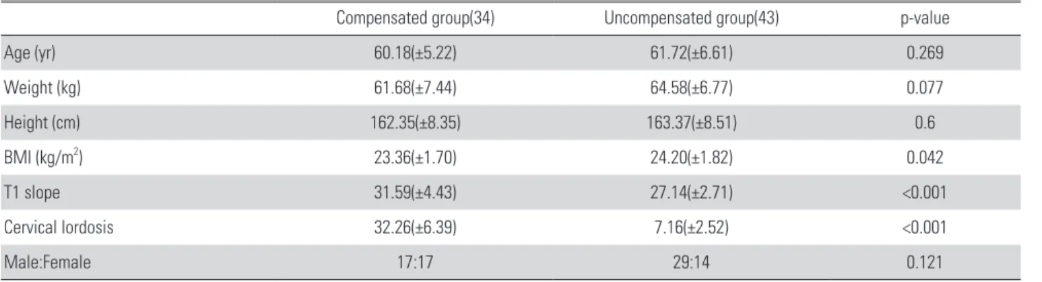
A B
Fig 1. Radiographic example of a 55-year-old female patient without cervical compensation. (A) Upright lateral X-ray. (B) T2-weighted sagittal image on cervical magnetic resonance imaging.
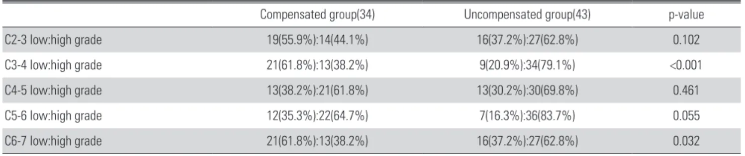
A B
Fig 2. Radiographic example of a 51-year-old female patient with cervical compensation. (A) Upright lateral X-ray. (B) T2-weighted sagittal image on cervical magnetic resonance imaging.
Compensation with Cervical Lordosis Journal of Korean Society of Spine Surgery
www.krspine.org 57 degeneration are common and increase with aging.7) The
prevalence of abnormal magnetic-resonance images of the cervical spine is related to age in asymptomatic individuals.
Clinical cervical aging studies have shown that 14% of asymptomatic subjects younger than 40 years have abnormal MRI scans with an increase to 50% by 50 years old.12) Therefore, we enrolled patient age over 50 years old to analyze disc degeneration.
Cervical lordosis in standing posture in 54 healthy volunteers was 54° (Min 36 degrees, Max 76 degrees).13) In our study, average cervical lordosis of all patient was 14.71°. Candidates of our study were patients with neck pain. Also the average age of patient was 61.04. Degenerative change with aging and underlying pathology may be a contributing factor of decrease in lordosis.
Straightened cervical lordosis causes stress concentration, so cervical lordosis may have a direct impact on cervical spondylosis.14) In our study, uncompensated group have significantly lower cervical lordosis compared with compensated group. And decreased cervical lordosis in uncompensated group may cause stress concentration on all cervical segments. This stress concentration may result in higher degree of degeneration on these segments.
The changes in sagittal alignment of the cervical spine affects the kinematics. Consequently, it may cause changes in the
segment subjected to maximum load for overall motion and accelerate its degeneration.9) The increased strain energy density and stress in the vertebral cortex over time may induce the remodeling process according to Wolff’s law, leading to the formation of osteophytes.8) Knott et al reported that patient with high T1 slope of more than 25° can have positive sagittal balance because of increased sacral vertical axis.1)
In our study, we did not evaluate whole sagittal balance of body because this study was performed retrospectively. Cervical decompensation in patient with high T1 slope may contribute to high grade of degeneration of cervical disc. Cervical disc degeneration and spondylotic change may be caused by stress concentration with decreased cervical lordosis.
The limitation of our study is whole sagittal balance of patients was not evaluated because study was performed retrospectively. And effects of upper cervical segments were not considered because cervical lordosis was measured between C2 and C7. Degree of disc degeneration of both groups was significantly different. We can know the relation between cervical decompensation and cervical disc degeneration, but we cannot conclude that the cervical decompensation causes degeneration of cervical disc. So prospective study should be performed later.
Conclusions
Patients with high T1 slope without compensation of cervical lordosis had higher grade of degeneration in all cervical segments. Cervical decompensation is related with cervical disc degeneration in patients with high T1 slope.
Table 3. Comparison of radiologic parameter between compensated group and uncompensated group using chi-square test
Compensated group(34) Uncompensated group(43) p-value
C2-3 low:high grade 19(55.9%):14(44.1%) 16(37.2%):27(62.8%) 0.102
C3-4 low:high grade 21(61.8%):13(38.2%) 9(20.9%):34(79.1%) <0.001
C4-5 low:high grade 13(38.2%):21(61.8%) 13(30.2%):30(69.8%) 0.461
C5-6 low:high grade 12(35.3%):22(64.7%) 7(16.3%):36(83.7%) 0.055
C6-7 low:high grade 21(61.8%):13(38.2%) 16(37.2%):27(62.8%) 0.032
Table 4. Odds ratio of high grade cervical degeneration of C3-4 on MRI by logistic regression analysis
Odd ratio p-value 95% CI Cervical decompensation 6.268 <0.001 2.232-17.601
Male gender 0.935 0.916 0.269-3.246
Age 0.992 0.876 0.895-1.100
REFERENCES
1. Knott PT, Mardjetko SM, Techy F. The use of the T1 sagittal angle in predicting overall sagittal balance of the spine. Spine J. 2010 Nov;10(11):994-8. DOI: 10.1016/
j.spinee.2010.08.031.
2. Yang BS, Lee SK, Song KS, et al. The Use of T1 Sagittal Angle in Predicting Cervical Disc Degeneration. Asian Spine J. 2015 Oct;9(5):757-61. DOI: 10.4184/asj.2015.9.5.757.
3. Cote P, Cassidy JD, Yong-Hing K, et al. Apophysial joint degeneration, disc degeneration, and sagittal curve of the cervical spine. Can they be measured reliably on radio- graphs? Spine (Phila Pa 1976). 1997 Apr 15;22(8):859-64.
DOI:10.1097/00007632-199704150-00007.
4. Dai L. [Disc degeneration and cervical instability]. Zhong- hua Wai Ke Za Zhi. 1999 Mar;37(3):180-2.
5. DePalma AF, Rothman RH, Levitt RL, et al.
The natural history of severe cervical disc degen- eration. Acta Orthop Scand. 1972;43(5):392-6.
DOI:10.3109/17453677208998959.
6. Friedenberg ZB. Degeneration of the Cervical Disc. West J Surg Obstet Gynecol. 1964 Jul-Aug;72:191-4.
7. Gruber HE, Phillips R, Ingram JA, et al. Spontaneous age- related cervical disc degeneration in the sand rat. Clin Or- thop Relat Res. 2014 Jun;472(6):1936-42. DOI: 10.1007/
s11999-014-3497-x.
8. Kumaresan S, Yoganandan N, Pintar FA, et al. Contribution of disc degeneration to osteophyte formation in the cervical spine: a biomechanical investigation. J Orthop Res. 2001 Sep;19(5):977-84. DOI: 10.1016/S0736-0266(01)00010- 9.
9. Miyazaki M, Hymanson HJ, Morishita Y, et al. Kine- matic analysis of the relationship between sagittal alignment and disc degeneration in the cervical spine. Spine (Phila Pa 1976). 2008 Nov 1;33(23):E870-6. DOI: 10.1097/
BRS.0b013e3181839733.
10. Sambrook PN, MacGregor AJ, Spector TD. Ge- netic influences on cervical and lumbar disc de- generation: a magnetic resonance imaging study in twins. Arthritis Rheum. 1999 Feb;42(2):366-72.
DOI: 10.1002/1529-0131(199902)42:2<366::AID- ANR20>3.0.CO;2-6.
11. Miyazaki M, Hong SW, Yoon SH, et al. Reliability of a magnetic resonance imaging-based grading sys- tem for cervical intervertebral disc degeneration. J Spinal Disord Tech. 2008 Jun;21(4):288-92. DOI: 10.1097/
BSD.0b013e31813c0e59.
12. Boden SD, McCowin PR, Davis DO, et al. Abnormal mag- netic-resonance scans of the cervical spine in asymptom- atic subjects. A prospective investigation. J Bone Joint Surg Am. 1990 Sep;72(8):1178-84. DOI:10.2106/00004623- 199072080-00008.
13. Gomez Espindola JC, Perez Viquez AF. [Cervical lordosis evaluation in asymptomatic volunteers from Navy Medical Center]. Acta Ortop Mex. 2008 Jan-Feb;22(1):7-11.
14. Wei W, Liao S, Shi S, et al. Straightened cervical lordosis causes stress concentration: a finite element model study.
Australas Phys Eng Sci Med. 2013 Mar;36(1):27-33. DOI:
10.1007/s13246-013-0182-4.
© Copyright 2018 Korean Society of Spine Surgery
Journal of Korean Society of Spine Surgery. www.krspine.org. pISSN 2093-4378 eISSN 2093-4386
This is an Open Access article distributed under the terms of the Creative Commons Attribution Non-Commercial License (http://creativecommons.org/licenses/by-nc/4.0/) which permits unrestricted non-commercial use, distribution, and reproduction in any medium, provided the original work is properly cited.
59
J Korean Soc Spine Surg. 2018 Jun;25(2):54-59. https://doi.org/10.4184/jkss.2018.25.2.59
Original Article
T1 시상각이 큰 환자에서 경추 만곡이 경추간판 퇴행에 미치는 영향
홍성하* • 이승환 • 박병문 • 송경섭 • 임성태*
광명성애병원 정형외과, *성애병원 정형외과
연구 계획: 후향적 연구
목적: T1 시상각이 큰 환자들에서 경추 만곡에 의한 보상이 경추간판의 퇴행성 변화에 미치는 효과를 분석하고자 하였다.
선행 연구문헌의 요약: T1 시상각은 시상 균형을 평가하는 유용한 지표로 알려져 있다. 저자들은 T1 시상각이 낮은 경우 제 6-7 경추간판의 퇴행성 변 화가 많다고 보고한 바 있다. 그러나 T1 시상각이 큰 환자(>25)에서 경추 만곡의 보상이 경추간판의 퇴행성 변화에 미치는 영향에 대해서는 분석하지 않았다.
대상 및 방법: 정형외과에 내원하여 경추부 MRI를 촬영한 환자들 중 T1 시상각이 25도 보다 큰 77명의 환자를 대상으로 하였다. 환자들을 경추 만곡의 보상에 따라 비보상군(경추 만곡<25)과 보상군(경추 만곡≥25)으로 분류하였다. 경추간판의 퇴행성 변화는 Miyazaki 분류에 따라 다섯 등급으로 분류하 였다. 두 군간의 방사선학적 지표와 MRI상의 추간판 퇴행성 변화를 비교하였다.
결과: 비보상군에서 경추간판의 평균 퇴행 등급은 제 2-3경추간에서 2.72, 제 3-4경추간에서 3.00, 제 4-5경추간에서 3.02, 제 5-6경추간에서 3.37, 제 6-7경추간에서 2.95로 나타났다. 보상군에서 경추간판의 평균 퇴행 등급은 제 2-3경추간에서 2.38, 제 3-4경추간에서 2.38, 제 4-5경추간에 서 2.62, 제 5-6경추간에서 2.82, 제 6-7경추간에서 2.41로 나타났다. 모든 경추 분절에서 퇴행성 변화가 비보상군에서 보상군에 비해 유의하게 높았다. 제 3-4경추 간판에서 3등급 이상의 심한 퇴행 등급의 비율 역시 비보상군에서 유의하게 높았다(OR 6.268, 95% CI 2.232-17.601, p<0.001).
결론: T1 시상각이 25도보다 큰 환자들에서 경추 만곡의 보상이 없는 경우 경추간판 전체에서 퇴행성 변화가 많은 것으로 나타났다.
색인 단어: 경추간판 퇴행, T1시상각, 경추 만곡 약칭 제목: 경추 만곡에 의한 보상
접수일: 2018년 3월 13일 수정일: 2018년 4월 5일 게재확정일: 2018년 6월 11일 교신저자: 이승환
경기도 광명시 디지털로 36 광명성애병원 정형외과
TEL: 02-2680-7699 FAX: 02-2680-7755 E-mail: java5885@gmail.com