ⓒ2018 The Korean Ophthalmological Society
This is an Open Access article distributed under the terms of the Creative Commons Attribution Non-Commercial License (http://creativecommons.org/licenses/by-nc/3.0/) which permits unrestricted non-commercial use, distribution, and reproduction in any medium, provided the original work is properly cited.
J Korean Ophthalmol Soc 2018;59(10):938-945 ISSN 0378-6471 (Print)⋅ISSN 2092-9374 (Online)
https://doi.org/10.3341/jkos.2018.59.10.938
Original Article
범망막광응고술 후 시신경유두주위 망막신경섬유층과 황반부 망막신경절세포-내망상층 두께 변화
Long-term Changes in the Peripapillary RNFL and Macular GCIPL Thicknesses after Panretinal Photocoagulation in Diabetic Retinopathy Patients
윤정현1,2⋅박동호1,2⋅김대우1,2
Jung Hyun Yoon, MD1,2, Dong Ho Park, MD, PhD1,2, Dai Woo Kim, MD, PhD1,2
경북대학교 의과대학 안과학교실1, 경북대학교병원 안과2
Department of Ophthalmology, School of Medicine, Kyungpook National University1, Daegu, Korea Department of Ophthalmology, Kyungpook National University Hospital2, Daegu, Korea
Purpose: To investigate longitudinal changes in the thicknesses of the peripapillary retinal nerve fiber layer (pRNFL) and the macular ganglion cell-inner plexiform layer (mGCIPL) in patients with diabetic retinopathy 3 years after panretinal photo- coagulation (PRP).
Methods: We retrospectively reviewed the medical records of 60 eyes of 35 patients who were diagnosed with diabetic retinop- athy and treated with PRP. The pRNFL and mGCIPL thicknesses were measured by optical coherence tomography at baseline, and then at 1, 3, 6, 9, 12, 24, and 36 months after PRP.
Results: The pRNFL and mGCIPL thicknesses (average and all sections) at 1 year after PRP increased significantly from base- line (p < 0.05, respectively). The average pRNFL and mGCIPL thicknesses showed a tendency to decrease continuously from 2 years after PRP (p < 0.05, respectively). There was no statistically significant difference in the average thicknesses of the pRNFL and the mGCIPL between pre-PRP (92.27 ± 7.76 µm, and 85.00 ± 4.80 µm, respectively) and 3 years after PRP (93.93 ± 7.49 µm, and 81.87 ± 14.00 µm, respectively) (p = 0.121, and p = 0.622, respectively).
Conclusions: Although the pRNFL and the mGCIPL thicknesses increased at 1 year after PRP, there was no statistical differ- ence in the average thicknesses of the pRNFL and the mGCIPL between pre-PRP and 3 years after PRP. These results should be considered with respect to the diagnosis and progression of glaucoma in patients with diabetic retinopathy who undergo PRP.
J Korean Ophthalmol Soc 2018;59(10):938-945
Keywords: Diabetic retinopathy, Ganglion cell-inner plexiform layer thickness, Glaucoma, Panretinal photocoagulation, Retinal nerve fiber layer thickness
■Received: 2018. 6. 14. ■ Revised: 2018. 7. 10.
■Accepted: 2018. 9. 28.
■Address reprint requests to Dai Woo Kim, MD, PhD
Department of Ophthalmology, Kyungpook National University Hospital, #130 Dongdeok-ro, Jung-gu, Daegu 41944, Korea Tel: 82-53-200-5806, Fax: 82-53-426-6552
E-mail: [email protected]
*Conflicts of Interest: The authors have no conflicts to disclose.
* Dong Ho Park was financially supported by the Basic Science Research Program of the National Research Foundation of Korea (NRF), funded by the Ministry of Education
(NRF-2017R1D1A1B03027966), and the Korea Health Technology R&D Project of the Korea Health Industry
Development Institute (KHIDI), funded by the Ministry of Health
& Welfare, Republic of Korea (HI16C1501).
* Dai Woo Kim was financially supported by the Basic Science Research Program of the National Research Foundation of Korea (NRF), funded by the Ministry of Education
(NRF-2017R1D1A1B03035483).
당뇨망막병증은 당뇨병의 가장 중요한 합병증 중 하나이 며, 20세에서 65세 성인의 시력저하와 실명의 주요 원인이 다.1 범망막광응고술(panretinal photocoagulation, PRP)은 증식당뇨망막병증 및 비증식당뇨망막병증의 고위험군에 대한 주된 치료 방법으로, 망막색소상피(retinal pigment ep- ithelium, RPE) 내에 위치한 멜라닌소체는 레이저 에너지를 흡수하여 열 손상을 일으키고 인접한 광수용체 및 망막색 소상피를 응고시킨다. 광수용체는 대사적 측면으로 볼 때 망막 내에서 산소소비율이 가장 높은 세포이기 때문에, 광 응고를 통하여 전체 대사량을 줄이고, 허혈 및 허혈로 인한 신생혈관형성물질을 감소시킨다.2
하지만 PRP 시술 후 레이저 파장이 망막내층에 직접 또 는 간접적인 영향을 줄 수 있다. RPE 내의 멜라닌소체가 레이저의 빛을 흡수하여 다시 열로 방출하는데, 이때 망막 신경절세포(retinal ganglion cells)와 시신경유두주위의 망막 신경섬유층(peripapillary retinal nerve fiber layer, pRNFL)의 손상을 유발할 수 있다.3-5
pRNFL과 황반부 망막신경절세포-내망상층(macular gan- glion cell-inner plexiform layer, mGCIPL)의 변화를 관찰하 는 것은 녹내장 진단 및 진행 여부를 파악하는 데 있어 중 요한 판단 기준이 된다. 이러한 구조적 변화를 정량적으로 관찰하기 위하여 흔히 빛간섭단층촬영(optical coherence tomography, OCT)이 이용된다.6,7 또한 전산화 부분 알고리 즘을 이용한 Cirrus OCT의 망막신경절세포분석(ganglion cell analysis, GCA)은 mGCIPL의 두께의 변화를 관찰하는 데 효과적인 것으로 알려져 있다.8,9
당뇨는 녹내장의 발병 및 진행의 위험인자이고 증식당뇨 망막병증의 경우 PRP가 주된 치료이므로, 증식당뇨망막병 증 및 비증식당뇨망막병증의 고위험군 환자에서 PRP를 시 행받은 경우 pRNFL 및 mGCIPL 두께에 대한 장기간 변화 에 대하여 관찰할 필요가 있다. 본 연구를 통하여 PRP 시 행 후 pRNFL 및 mGCIPL의 3년간의 변화에 대하여 알아 보았다.
대상과 방법
본 연구는 헬싱키선언(Declaration of Helsinki)을 준수하 여 본원의 임상연구윤리 심의위원회(institutional review board, IRB)의 승인 아래 진행되었다. 2011년 5월부터 2016 년 10월까지 본원 안과에 내원한 황반 부종을 동반하지 않 은 심한 비증식당뇨망막병증 및 비고위험 증식당뇨망막병 증 환자 중 3년간 빛간섭단층촬영기인 Cirrus HD-OCT (Carl Zeiss Meditec, Inc., Dublin, CA, USA) 검사를 시행 한 환자를 대상으로, 성별, 초기 나이, 고혈압, 초기 당뇨 이
환 기간, 초기 당화혈색소, 시력, 안압, 백내장수술 유무, 당 뇨망막병증의 정도에 대하여 후향적으로 의무 기록을 조사 하였다.
대상 선택 시 유리체절제술 등의 침습적 수술 및 유리체 내주입술과 같은 시술을 시행한 경우는 제외하였다. 당뇨 망막병증 이외 다른 망막 질환이 있는 경우, 협조도가 낮은 경우, 경과관찰 중 황반 부종이 발생한 경우, 각막 혼탁이 나 백내장 등과 같이 매질 혼탁이 심한 경우, OCT 측정 시 신호 강도가 6 미만인 경우 그리고 녹내장이 있는 경우도 대상에서 제외했다.
PRP는 아르곤 532 nm 파장의 레이저를 이용하여 1주 간 격으로 총 3회 시행하였고, 레이저의 범위는 시신경유두주 위에서 1 유두직경범위 내에는 도달하지 않게 망막의 혈관 궁 바깥 1 유두직경 떨어진 지점에서 톱니둘레까지 시행하 였다. 레이저 반흔의 크기는 200-500 µm이고, 레이저 강도 는 80 mW에서 시작하여 레이저 반흔이 회백색으로 변할 때까지 증가시켰다. 레이저 반흔 사이의 간격은 100 µm였 고, 총 레이저 조사 횟수는 약 2,000번 정도였다.
모든 OCT 검사는 1% 트로피카아마이드와 2.5% 페닐에 프린 점안제로 산동하고 Cirrus OCT system의 V.6.0 soft- ware (Carl Zeiss Meditec Inc., Dublin, CA, USA)를 이용하 여 Macular cube 200 × 200 protocol, Optic cube 200 × 200 protocol 검사를 하였다. OCT의 신호 강도는 모두 최소 6 이상이었다. pRNFL의 두께는 optic disc cube protocol을 이용하여 평균과 상측, 하측, 비측, 이측 총 4개의 구역 각 각에 대해 측정하였고, mGCIPL의 두께는 평균과 상이측, 상측, 상비측, 하비측, 하측, 하이측 총 6개의 구역 각각에 대해 the Cirrus 6.0 proprietary GCA segmentation algo- rithm을 이용해 측정하였으며 PRP 후 1, 3, 6, 9, 12개월, 2, 3년 간격으로 각각의 두께 변화를 관찰하였다(Fig. 1).
통계적 분석은 3년간 측정값의 유의한 차이가 있는지를 알아보기 위해 SPSS version 20.0 (IBM Corp., Armonk, NY, USA)을 이용하였으며, Bonferroni 방법을 이용한 반 복 측정 분석법으로 pRNFL과 mGCIPL 두께와 시력을 비 교하였다. paired t-test로 각 시기별로 이전에 방문할 때의 pRNFL과 mGCIPL의 두께와 비교하였으며, p값이 0.05 미 만일 때 통계적으로 유의하다고 판정하였다.
결 과
2011년 5월부터 2016년 10월까지 PRP를 시행한 35명 60안을 대상으로 의무 기록을 후향적으로 분석하였다. 남 성이 44명, 여성이 16명이며, 우안은 28명, 좌안은 32명, 평 균연령은 58.0 ± 8.68세, 평균 당뇨 이환 기간은 15.5 ± 5.8
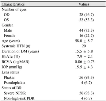
Characteristics Values Number of eyes
OD 28 (46.7)
OS 32 (53.3)
Gender
Male 44 (73.3)
Female 16 (22.7)
Age (years) 58.0 ± 8.7
Systemic HTN (n) 20
Duration of DM (years) 15.5 ± 5.8
HbA1c (%) 7.9 ± 2.1
BCVA (logMAR) 0.06 ± 0.73
IOP (mmHg) 15.5 ± 4.3
Lens status
Phakia 56 (93.3)
Pseudophakia 4 (6.7)
Status of DR
Severe NPDR 56 (93.3)
Non-high-risk PDR 4 (6.7)
Values are presented as mean ± SD or n (%) unless otherwise indicated.
OD = oculus dexter; OS = oculus sinister; HTN = hypertension;
DM = diabetes mellitus; HbA1c = hemoglobin A1c; BCVA = best-corrected visual acuity; IOP = intraocular pressure; DR = di- abetic retinopathy; NPDR = non-proliferative diabetic retinop- athy; PDR = proliferative diabetic retinopathy.
Table 1. Demographics and clinical data
A B
Figure 1. Peripapillary retinal nerve fiber layer (RNFL) and macular ganglion cell-inner plexiform layer (GCIPL) thickness in
Cirrus optical coherence tomography (OCT). (A) OCT shows the RNFL thicknesses of average and four-quadrant in both eyes. (B) OCT shows GCIPL thicknesses of average and six-sector in both eyes. OD = oculus dexter; OS = oculus sinister; GCL = ganglion cell layer; IPL = inner plexiform layer; S = superior; T = temporal; N = nasal; I = inferior; NA = non-available.년, 평균 당화혈색소(HbA1c)는 7.9 ± 2.1, 평균 안압은 15.5
± 4.2 mmHg, 수정체안은 56안, 인공수정체안은 4안, 심한 비증식당뇨망막병증은 56안, 비고위험도 증식당뇨망막병 증은 4안이었다(Table 1).
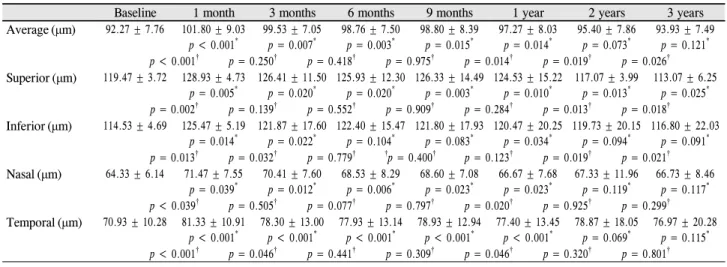
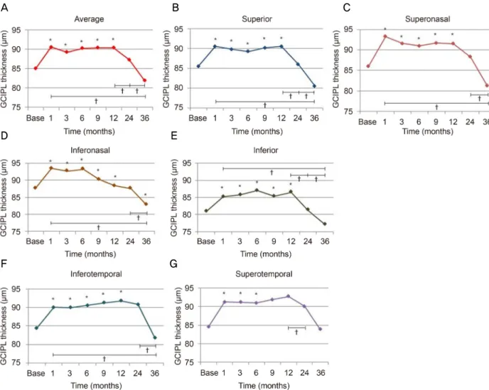
pRNFL 평균과 4개 영역 각각의 두께는 PRP 시행 전보 다 시행 1개월 후 증가하였으나(p<0.05, 모두) 이후 지속적 으로 감소하여(p<0.05, 모두), PRP 시행 1년 후 pRNFL 평 균 두께(97.27 ± 8.03 µm)는 PRP 전(92.27 ± 7.76 µm)과 비교하여 증가한 상태로 유지되었다(p=0.014) (Table 2). PRP 시행 2년 후, 상측 사분절을 제외한 나머지 RNFL 두께 및 평균 RNFL 두께는 시술 전과 비교하여 통계적으로 유의한 차이를 보이지 않았고(p>0.05, 각각), PRP 시행 3년 후의 pRNFL의 평균 및 상측 사분절을 제외한 나머지 pRNFL의 두께도 시술 전과 비교할 때 통계적으로 유의한 차이를 보 이지 않았다(p>0.05, 각각). PRP 시행 2년 후의 pRNFL 평균 두께는 PRP 시행 1년 후와 비교할 때 감소하였고(p=0.019), PRP 시행 3년 후에도 PRP 시행 2년 후에 비하여 감소하였 으나(p=0.026), PRP 시행 전에 비하여 통계적으로 유의한 차이를 보이지 않았다(p=0.121) (Fig. 2).
mGCIPL의 평균과 육분절 각각의 두께는 PRP 시행 1개월 후 증가하였고(p<0.05, 모두), PRP 시행 1년 후, mGCIPL 평균 두께(91.10 ± 7.40 µm)는 시술 전 두께(85.00 ± 4.80 µm) 에 비해 증가된 상태로 유지되었다(p=0.019) (Table 3). 이 후 3년간 mGCIPL의 평균 및 육분절 각각의 두께는 지속적 으로 감소하여, PRP 시행 3년 후, 하비측을 제외한 나머지 육분절 mGCIPL 두께와 평균 mGCIPL 두께는 시행 전과 비교할 때 통계적으로 유의한 차이를 보이지 않았다(p>0.05, 각각) (Fig. 3).
평균 최대교정시력(logMAR)은 PRP 전 0.06 ± 0.73이었
으나 3년 동안 통계적으로 유의한 수준의 변화가 없었고 (p>0.05, 모두) 시술 3년 후 0.07 ± 0.87로 시술 전과 비교 했을 때 차이가 없었다(p=0.347).
고 찰
이전 연구에서 PRP 시행 후 pRNFL 및 mGCIPL의 두께
Baseline 1 month 3 months 6 months 9 months 1 year 2 years 3 years Average(μm) 92.27 ± 7.76 101.80 ± 9.03
p < 0.001*
99.53 ± 7.05 p = 0.007*
98.76 ± 7.50 p = 0.003*
98.80 ± 8.39 p = 0.015*
97.27 ± 8.03 p = 0.014*
95.40 ± 7.86 p = 0.073*
93.93 ± 7.49 p = 0.121* p < 0.001† p = 0.250† p = 0.418† p = 0.975† p = 0.014† p = 0.019† p = 0.026† Superior(μm) 119.47 ± 3.72 128.93 ± 4.73
p = 0.005*
126.41 ± 11.50 p = 0.020*
125.93 ± 12.30 p = 0.020*
126.33 ± 14.49 p = 0.003*
124.53 ± 15.22 p = 0.010*
117.07 ± 3.99 p = 0.013*
113.07 ± 6.25 p = 0.025* p = 0.002† p = 0.139† p = 0.552† p = 0.909† p = 0.284† p = 0.013† p = 0.018† Inferior(μm) 114.53 ± 4.69 125.47 ± 5.19
p = 0.014*
121.87 ± 17.60 p = 0.022*
122.40 ± 15.47 p = 0.104*
121.80 ± 17.93 p = 0.083*
120.47 ± 20.25 p = 0.034*
119.73 ± 20.15 p = 0.094*
116.80 ± 22.03 p = 0.091* p = 0.013† p = 0.032† p = 0.779† †p = 0.400† p = 0.123† p = 0.019† p = 0.021† Nasal(μm) 64.33 ± 6.14 71.47 ± 7.55
p = 0.039*
70.41 ± 7.60 p = 0.012*
68.53 ± 8.29 p = 0.006*
68.60 ± 7.08 p = 0.023*
66.67 ± 7.68 p = 0.023*
67.33 ± 11.96 p = 0.119*
66.73 ± 8.46 p = 0.117* p < 0.039† p = 0.505† p = 0.077† p = 0.797† p = 0.020† p = 0.925† p = 0.299† Temporal(μm) 70.93 ± 10.28 81.33 ± 10.91
p < 0.001*
78.30 ± 13.00 p < 0.001*
77.93 ± 13.14 p < 0.001*
78.93 ± 12.94 p < 0.001*
77.40 ± 13.45 p < 0.001*
78.87 ± 18.05 p = 0.069*
76.97 ± 20.28 p = 0.115* p < 0.001† p = 0.046† p = 0.441† p = 0.309† p = 0.046† p = 0.320† p = 0.801† Valuesare presented as the mean ± standarddeviation.
*Repeated-measuresanalysis of variance corrected by the Bonferroni methods; †Paired t-test compared with the previous observation period.
Table 2. Longitudinal changes in peripapillary retinal nerve fiber layer thickness after panretinal photocoagulation
A B C
D E F
Figure 2. Longitudinal changes in the average and four-quadrant thicknesses (μm) of the peripapillary retinal nerve fiber layer
(RNFL) after panretinal photocoagulation (PRP). The RNFL thicknesses (average and four-quadrant) increased at 1 month after PRP and had declined continuously since then (A-E). Repeated-measures analysis of variance corrected by Bonferroni method showed that the RNFL thicknessess (average and four-quadrant) increased at 1 year after PRP (all p < 0.05), but there was no stat- istically significant difference of the average, inferior, nasal and temporal thicknesses of the RNFL between pre-PRP and 3 years af- ter PRP (A, C-E). The superior RNFL thickness (93.93 ± 7.49 μm) decreased at 3 years after PRP from the thickness (92.27 ± 7.76 μm) before PRP (p = 0.442) (B). *p < 0.05 by repeated-measures analysis of variance corrected by the Bonferroni methods; †p <0.05 by paired t-test.
변화에 대해 보고된 바 있으나,5,10 이의 장기간의 변화에 대 한 연구가 부족하기에 본 연구에서 PRP 시행 후 pRNFL과
mGCIPL의 3년간 두께 변화를 알아보았다.
Roohipoor et al11은 pRNFL 두께가 PRP 이후 초기 6개
Baseline 1 month 3 months 6 months 9 months 1 year 2 years 3 years Average (μm) 85.00 ± 4.80 90.60 ± 6.90
p < 0.001*
89.30 ± 7.54 p = 0.010*
90.30 ± 6.74 p = 0.004*
90.40 ± 7.25 p = 0.027*
91.10 ± 7.40 p = 0.037*
87.33 ± 7.31 p = 0.096*
81.87 ± 14.00 p = 0.622* p < 0.001† p = 0.195† p = 0.215† p = 0.341† p = 0.245† p = 0.036† p = 0.045† Superior (μm) 85.53 ± 4.21 90.53 ± 5.71
p < 0.001*
89.80 ± 6.37 p < 0.001*
89.21 ± 6.75 p < 0.001*
90.10 ± 6.63 p = 0.019*
90.53 ± 7.25 p = 0.017*
86.03 ± 7.34 p = 0.671*
80.53 ± 10.21 p = 0.588* p < 0.001† p = 0.377† p = 0.417† p = 0.265† p = 0.725† p = 0.038† p = 0.040† Superonasal (μm) 86.00 ± 5.77 93.33 ± 7.72
p = 0.005*
91.60 ± 7.28 p < 0.001*
91.04 ± 7.34 p < 0.001*
91.73 ± 7.88 p < 0.001*
91.53 ± 8.65 p < 0.001*
88.27 ± 13.71 p = 0.208*
81.27 ± 15.46 p = 0.937* p = 0.002† p = 0.073† p = 0.196† p = 0.750† p = 0.719† p = 0.752† p = 0.040† Inferonasal (μm) 87.67 ± 15.10 93.53 ± 18.87
p < 0.001*
92.73 ± 15.47 p < 0.001*
93.30 ± 17.25 p < 0.001*
90.27 ± 11.27 p < 0.001*
88.40 ± 14.29 p = 0.023*
87.61 ± 16.45 p = 0.308*
82.80 ± 15.48 p < 0.001* p < 0.001† p = 0.591† p = 0.572† p = 0.027† p = 0.943† p = 0.655† p = 0.039† Inferior (μm) 80.87 ± 1.73 85.20 ± 2.06
p < 0.001*
85.73 ± 2.16 p < 0.001*
87.01 ± 2.70 p = 0.020*
85.30 ± 2.60 p = 0.017*
86.53 ± 2.84 p = 0.017*
81.33 ± 3.80 p = 0.242*
77.13 ± 5.52 p = 0.592* p < 0.001† p = 0.320† p = 0.251† p = 0.090† p = 0.256† p = 0.027† p = 0.049† Inferotemporal (μm) 84.46 ± 1.22 90.07 ± 1.47
p < 0.001*
90.00 ± 1.46 p = 0.005*
90.60 ± 1.54 p = 0.006*
91.29 ± 2.03 p = 0.038*
91.80 ± 1.96 p = 0.011*
90.87 ± 4.95 p = 0.120*
81.73 ± 7.50 p = 0.071* p < 0.001† p = 0.418† p = 0.719† p = 0.541† p = 0.135† p = 0.188† p = 0.034† Superotemporal
(μm)
84.60 ± 3.16 91.27 ± 4.01 p = 0.007*
91.20 ± 4.18 p = 0.035*
91.00 ± 3.93 p = 0.020*
91.93 ± 5.93 p = 0.179*
92.73 ± 7.33 p = 0.463*
90.07 ± 7.44 p = 0.851*
83.93 ± 17.17 p = 0.679* p < 0.001† p = 0.849† p = 0.642† p = 0.590† p = 0.230† p = 0.037† p = 0.160† Values are presented as the mean ± standard deviation.
*Repeated-measures analysis of variance corrected by the Bonferroni methods; †Paired t-test compared with the previous observation period.
Table 3. Longitudinal changes in macular ganglion cell-inner plexiform layer thickness after panretinal photocoagluation
월까지 증가한다고 보고하였으며, Kim et al10은 PRP 시행후 1년간 pRNFL과 mGCIPL 두께는 시행 전보다 증가되었 다고 보고하였다. Park and Jee12는 532 nm 파장의 Pattern Scanning Laser (PASCAL)를 사용하여 PRP 시행 1년 후 pRNFL 두께가 시술 전과 차이가 없었다고 보고했지만, Kim et al5은 PRP 시행 후 초기에는 광응고가 망막의 염증 과 부종을 발생시키지만, 이후 망막신경섬유를 퇴행시키므 로 pRNFL 두께는 PRP 이후 처음 3개월까지 증가하지만 시행 2년 후에는 그 두께가 통계적으로 유의한 수준으로 감소한다고 보고했다. Shin and Lee13는 577 nm 파장의 PASCAL을 사용하여 PRP 시행 1년 후 pRNFL 두께는 시 술 전과 차이가 없으나 황반부 망막층 각각의 두께는 증가 하였다고 보고했다.
PRP는 증식당뇨망막병증 환자에서 병의 진행을 예방하 는 데 중요한 치료이지만, 광응고에 의한 레이저 반흔이 염 증 반응으로 인해 망막에 손상을 주어 망막신경섬유와 망 막신경절세포에 영향을 줄 수 있다.14 레이저 반흔을 입은 망막 조직에서 분비된 화학 물질들이 망막 모세혈관계에 혈역학적인 문제를 초래할 수 있고15 림프구들을 자극하여 혈관의 투과성을 증가시킬 수 있다.16 Itaya et al17은 PRP가 신경망막과 망막색소상피-맥락막에서 혈관내피성장인자를 증가시키고, 혈액-망막 장벽을 붕괴시켜 망막 각층의 부종 을 일으킬 수 있다고 설명하며, Jampol et al18은 항혈관내 피성장인자를 안구 내 주입한 경우 망막신경섬유층의 부종
을 완화시키고, 혈관내피세포성장인자의 신경방어 효과를 차단함으로써 망막신경절세포를 손상시키기 때문에 pRNFL 두께가 감소할 수 있다고 하였다. 본 연구에서도 PRP 시행 후 1년 동안 pRNFL 및 mGCIPL의 두께가 시행 전보다 증 가한 것은 염증 반응과 그에 따른 혈관내피성장인자 증가 로 인한 것으로 생각된다.
당뇨망막병증은 신경절세포나 뮬러세포와 같은 세포층 의 손상19과 신경교세포의 손상으로 망막신경섬유층의 손 상에 영향을 주어20 pRNFL과 mGCIPL의 두께 감소를 초 래할 수 있다. Kim et al21은 비증식당뇨망막병증 환자들의 눈에서 pRNFL 평균 두께 및 코쪽 사분원의 두께가 당뇨 이환 기간이 증가함에 따라 감소한다고 하였다. 한편 Apple et al22은 동물을 이용한 실험에서 망막에 레이저를 조사하 면 그 강도에 따라 가역적 또는 비가역적인 망막신경섬유 와 신경절세포의 손상을 일으킬 수 있다고 하였다. Yang et al23은 당뇨망막병증 환자에서 PRP를 시행하면 레이저 광 선이 망막신경섬유층의 방사상으로 배열된 다발에 직접 손 상을 주어 pRNFL의 상측, 하측, 비측 사분원의 두께가 감 소했다고 보고하였다. 또한 Kim et al5은 당뇨망막병증 환 자에서 PRP를 시행한 후 pRNFL의 상측, 하측 사분원만 통 계적으로 유의한 수준의 두께 감소가 있었다고 보고했다. Tso et al24은 고강도의 레이저 광선이 망막신경절세포를 포 함한 망막 각층을 파괴시키기 때문에 비록 PRP 후 초기에 pRNFL과 mGCIPL의 두께가 증가되더라도 결국 그 두께는
A B C
D E □
F G □
Figure 3. Longitudinal changes in the average and six-sector thicknesses (μm) of the macular ganglion cell-inner plexiform layer
(GCIPL) after panretinal photocoagulation (PRP). Repeated-measures analysis of variance corrected by Bonferroni method showed that macular GCIPL thicknesses (average and 6-sector) increased in 1 year after PRP (all p < 0.05). The average GCIPL thickness showed a tendency to increase until 1 year, but to decrease continuously from 2 year after PRP. There was no statistically significant difference of the average, superior, superonasal, inferior, inferotemporal and superotemporal GCIPL thicknesses between pre-PRP and 3 years after PRP (A-C, E-G). The inferonasal GCIPL thickness (81.87 ± 19.0 μm) decreased at 3 years after PRP from the thickness (85.00 ± 4.80 μm) before PRP (p = 0.544) (D). *p < 0.05 by repeated-measures analysis of variance corrected by the Bonferroni methods; †p < 0.05 by by paired t-test.감소할 것이라고 설명하였다. Van et al25은 당뇨가 진행함 에 따라 시신경과 황반 주변의 혈관 변화와 신경적 퇴행을 초래하여 pRNFL과 망막신경절세포층의 두께가 얇아진다 고 하였으며, Lee et al26은 당뇨망막병증 환자에서 PRP를 시행하면 신경조직을 더욱 감소시켜 결국 pRNFL 두께가 시술을 하지 않은 경우보다 더 감소한다고 하였다. 본 연구 에서는 앞선 연구와 달리, PRP 시행 3년 후 pRNFL과 mGCIPL의 평균 두께는 시술 전과 비교할 때 통계적으로 의미있는 차이를 보이지 않았다. 하지만 Maeshima et al27 은 PRP 시행 후 4년을 기준으로 시행 4년 후 레이저 반흔 의 크기 증가율이 시행 4년 이전에 비하여 크다고 하였고, 본 연구에서도 pRNFL의 평균 두께는 PRP 후 1개월 시점
으로부터 지속적으로 통계적으로 유의한 수준으로 감소하 고 있음을 고려할 때, 추후 pRNFL 두께는 지속적으로 감 소하여 결국 PRP 시행 전보다 감소할 것으로 예상된다.
Shin et al28은 당뇨망막병증에서 pRNFL 결손을 상측과 하측으로 나눌 때 상측이 더 많았다고 보고했으며 이는 일 반적으로 알려진 정상안압녹내장의 특징에 비해 망막의 허 혈이 망막신경섬유층의 결손에 주요인자이기 때문이라고 설명한다. 본 연구에서도 경과관찰 기간 중 pRNFL의 다른 영역보다 상측 사분원의 두께 감소가 두드러졌는데, 이는 당뇨망막병증의 경과에서 망막의 허혈이 상측에 더 많이 발생하는 것과 관련이 있다고 생각된다. 일반적으로 당뇨 망막병증이 있는 눈에서의 시신경유두는 창백한 경향을 보
이기 때문에 녹내장성 시신경유두를 판별하는 것에 어려움 이 있다. 따라서 시신경유두의 녹내장성 변화가 의심되는 부위의 pRNFL 및 mGCIPL의 변화를 유심히 살펴볼 필요 가 있는데, PRP 이후의 기간별 pRNFL 및 mGCIPL의 변화 에 대한 정보가 경과관찰에 유용한 판단 기준이 될 것으로 보인다.
본 연구는 몇 가지 제한점이 있다. 후향적이고 비무작위 선택에 의한 선택편위가 있을 수 있고, mGCIPL 이외의 다 른 각 망막 단층의 두께 및 병리적 변화에 대하여도 연구가 필요할 것으로 보인다. 또한 PRP를 시행받은 당뇨망막병증 환자를 녹내장 유무에 따른 군으로 비교 분석하여 PRP 후 pRNFL과 mGCIPL의 시간에 따른 두께 변화에 대하여 알 아보는 것도 필요할 것이다.
결론적으로, pRNFL과 mGCIPL의 두께는 PRP 후 1년까 지 증가하였지만, 3년 후에는 시술 전과 큰 차이를 보이지 않았다. 이를 통하여, 당뇨망막병증으로 PRP를 시행받은 환자를 경과관찰 함에 있어서 경과 기간에 따른 pRNFL 및 mGCIPL의 두께 변화를 감안하여 녹내장의 진단과 진행 여부를 판단하는 것이 필요할 것으로 보인다.
REFERENCES
1) Barber AJ. A new view of diabetic retinopathy: a neurodegenerative disease of the eye. Prog Neuropsychopharmacol Biol Psychiatry 2003;27:283-90.
2) Stefanson E. Oxygen and diabetic eye disease. Graefes Arch Clin Exp Ophthalmol 1990;228:120-3.
3) Frisch GD, Shawaluk PD, Adams DO. Remote nerve fibre bundle alterations in the retina as caused by argon laser photocoagulation.
Nature 1974;248:433-5.
4) Retineour RJ, Kozousek V, Chauhan BC. The effect of panretinal photocoagulation for diabetic retinopathy on retinal nerve fiber layer thickness and optic disc topography. Br J Ophthalmol 2009;
93:838-9.
5) Kim J, Woo SJ, Ahn J, et al. Long-term temporal changes of peri- papillary retinal nerve fiber layer thickness before and after pan- retinal photocoagulation in severe diabetic retinopathy. Retina 2012;32:2052-60.
6) Mwanza JC, Budenz DL, Godfrey DG, et al. Diagnostic performance of optical coherence tomography ganglion cell--inner plexiform layer thickness measurements in early glaucoma. J Ophthalmology 2014;121:849-54.
7) Sihota R, Sony P, Gupta V, et al. Diagnostic capability of optical co- herence tomography in evaluating the degree of glaumcomatous reti- nal nerve fiber damage. Invest Ophthalmo Vis Sci 2006;47:2006-10.
8) Mwanza JC, Durbin MK, Budenz DL, et al. Profile and predictors of normal ganglion cell-inner plexiform layer thickness measured with frequency-domain optical coherence tomography. Invest Ophthalmol Vis Sci 2011;52:7872-9.
9) Mwanza JC, Oakley JD, Budenz DL, et al. Macular ganglion cell-in- ner plexiform layer: automated detection and thickness reproduci-
bility with spectral domain-optical coherence tomography in glaucoma. Invest Ophthalmol Vis Sci ophthalmol 2011;52:8323-9.
10) Kim JJ, Im JC, Shin JP, et al. One-year follow-up of macular gan- glion cell layer and peripapillary retinal nerve fiber layer thickness changes after panretinal photocoagulation. Br J ophthalmol 2014;
98:213-7.
11) Roohipoor R, Dantism S, Ahmadrji A, et al. Subfoveal choroidal thickness after panretinal photocoagulation with red and green lase in bilateral proliferative diabetic retinopathy patients: short term results. J Ophthalmol 2016;2016:9364861.
12) Park YR, Jee D. Changes in peripapillary retinal nerve fiber layer thickness after pattern scanning laser photocoagulation in patients with diabetic retinopathy. Korean J Ophthalmol 2014;28:220-5.
13) Shin JS, Lee YH. Changes in macular retinal layers and peripapil- lary nerve fiber layer thickness after 577-nm pattern scanning laser in patients with diabetic retinopathy. Korean J Ophthalmol 2017;
31:497-507.
14) Mendívil A, Cuartero V, Mendívil MP. Ocular blood flow velocities in patients with proliferative diabetic retinopathy before and after scatter photocoagulation: a prospective study. Eur J Ophthalmol 1995;5:259-64.
15) Hiroshiba N, Ogura Y, Nishiwaki H, et al. Alterations of retinal mi- crocirculation in response to scatter photocoagulation. Invest Ophthalmol Vis Sci 1998;39:769-76.
16) Nonaka A, Kiryu J, Tsujikawa A, et al. Inflammatory response af- ter scatter laser photocoagulated retina. Invest Ophthalmol Vis Sci 2002;43:1204-9.
17) Itaya M, Sakurai E, Nozaki M, et al. Upregulation of VEGF in mur- ine retina via monocyte recruitment after retinal scatter laser photocoagulation. Invest Ophthalmol Vis Sci 2007;48:5677-83.
18) Jampol LM, Odia I, Glassman AR, et al. Panretinal photocoagulation versus ranibizumab for proliferative diabetic retinopathy comparison of peripapillary retinal nerve fiber layer thickness in a randomized clinical trial. Retina 2017;10 doi:10.1097/IAE.0000000000001909.
19) Gillies MC, Su T, Stayt J, et al. Effect of high glucose on perme- ability of retinal capillary endothelium in vitro. Invest Ophthalmol Vis Sci 1997;38:635-42.
20) Rungger-Brändle E, Dosso AA, Leuenberger PM. Glial reactivity, an early feature of diabetic retinopathy. Invest Ophthalmol Vis Sci 2000;41:1971-80.
21) Kim JT, Lee JK, Moon NJ, Cho HK. Analysis of the optic nerve head and RNFL thickness using optical coherence tomography in diabetes. J Korean Ophthalmol Soc 2008;49:935-41.
22) Apple DJ, Wyhinny GJ, Goldenberg MF, et al. Experimental argon laser photocoagulation. I. Effects on retinal nerve fiber layer. Arch Ophthalmol 1976;94:137-44.
23) Yang HS, Kim JG, Cha JB, et al. Quantitative analysis of neural tis- sues around the optic disc after panretinal photocoagulation in pa- tients with diabetic retinopathy. Plos one 2017;12:e0186229.
eCollection 2017.
24) Tso MO, Wallow IH, Elgin S. Experimental photocoagulation of the human retina: I. Correlation of physical, clinical, and patho- logic data. Arch Ophthalmol 1977;95:1035-40.
25) Van Dijk HW, Verbraak FD, Stehouwer M, et al. Association of visual function and ganglion cell layer thickness in patients with diabetes mellitus type 1 and no or minimal diabetic retinopathy.
Vision Res 2011;51:224-8.
26) Lee SB, Kwag JY, Lee HJ, et al. The longitudinal changes of retinal
= 국문초록 =
범망막광응고술 후 시신경유두주위 망막신경섬유층과 황반부 망막신경절세포-내망상층 두께 변화
목적: 본 연구에서는 당뇨망막병증 환자를 대상으로 범망막광응고술(panretinal photocoagulation, PRP) 후 시신경유두주위 망막신경 섬유층(peripapillary retinal nerve fiber layer, pRNFL)과 황반부 망막신경절세포-내망상층(macular ganglion cell-inner plexiform layer, mGCIPL)의 장기간 두께 변화를 추적관찰 하고자 하였다.
대상과 방법: PRP를 시행한 당뇨망막병증 환자 35명, 60안의 의무기록을 후향적으로 분석하였다. PRP 전과 이후 1, 3, 6, 9, 12, 24, 36개월 간격으로 pRNFL과 mGCIPL 두께 변화에 대해 살펴보았다.
결과: PRP 시행 1년 후 pRNFL과 mGCIPL의 평균과 각 영역의 두께는 시술 시행 전과 비교할 때 증가하였다(p<0.05, 모두). 이후 평균 두께는 지속적으로 감소하는 경향을 보였고(p<0.05, 모두), 시행 후 3년째 pRNFL과 mGCIPL의 평균 두께(93.93 ± 7.49 µm, 81.87 ± 14.00 µm, 각각)는 시술 전의 두께(92.27 ± 7.76 µm, 85.00 ± 4.80 µm, 각각)와 비교할 때 통계적으로 유의한 차이를 보이지 않았다(p=0.121, p=0.622, 각각).
결론: pRNFL과 mGCIPL의 두께는 PRP 후 1년까지 증가하였지만 3년 후에는 시술 전과 큰 차이를 보이지 않았다. PRP를 시행한 당뇨망막병증 환자의 녹내장 진단과 진행 여부를 판단함에 있어 이 점을 고려해야 할 것으로 생각된다.
<대한안과학회지 2018;59(10):938-945>
윤정현 / Jung Hyun Yoon
경북대학교병원 안과 Department of Ophthalmology, Kyungpook
National University Hospital nerve fiber layer thickness after panretinal photocoagulation in dia-
betic retinopathy patients. Retina 2013;33:188-93.
27) Maeshima K, Utsugi-Sutah N, Otani T, Kishi S. Progressive enlarge- ment of scattered photocoagulation scars in diabetic retinopathy.
Retina 2004;24:507-11.
28) Shin YJ, Kyoung HS, Park KH, Yu HG. The analysis of retinal nerve fiber layer in the patients with nonproliferative diabetic retinopathy. J Korean Ophthalmol Soc 2003;44:2010-5.